Korean J Thorac Cardiovasc Surg 2012;45:334-337 □ Case Report □ http://dx.doi.org/10.5090/kjtcs.2012.45.5.334 ISSN: 2233-601X (Print) ISSN: 2093-6516 (Online)
− 334 −
Department of Thoracic and Cardiovascular Surgery, Pusan National University School of Medicine Received: February 1, 2012, Revised: February 27, 2012, Accepted: March 3, 2012
Corresponding author: Seunghwan Song, Department of Thoracic and Cardiovascular Surgery, Pusan National University Hospital, Pusan National University School of Medicine, 179 Gudeok-ro, Seo-gu, Busan 602-739, Korea
(Tel) 82-51-240-7267 (Fax) 82-51-243-9389 (E-mail) [email protected]
C
The Korean Society for Thoracic and Cardiovascular Surgery. 2012. All right reserved.
CC
This is an open access article distributed under the terms of the Creative Commons Attribution Non-Commercial License (http://creative- commons.org/licenses/by-nc/3.0) which permits unrestricted non-commercial use, distribution, and reproduction in any medium, provided the original work is properly cited.
Application of Percutaneous Cardiopulmonary Support for Cardiac Tamponade Following Blunt Chest Trauma: Two Case Reports
Seon Hee Kim, M.D., Seunghwan Song, M.D., Yeong Dae Kim, M.D., Jeong Su Cho, M.D., Chung Won Lee, M.D., Jong Geun Lee, M.D.
Since the advent of percutaneous cardiopulmonary support (PCPS), its application has been extended to massively injured patient. Cardiac injury following blunt chest trauma brings out high mortality and morbidity. In our cases, patients had high injury severity score by blunt trauma and presented sudden hemodynamic collapse in emergency room. We quickly detected cardiac tamponade by focused assessment with sonography for trauma and implemented PCPS. As PCPS established, their vital sign restored and then, they were transferred to the operation room (OR) securely. After all injured lesion repaired, PCPS weaned successfully in OR. They were discharged without compli- cation on day 26 and 55, retrospectively.
Key words: 1. Extracorporeal circulation 2. Trauma, blunt
3. Cardiac tamponade 4. Ultrasonic diagnosis 5. Cardiac rupture
CASE REPORT 1) Case 1
A 34-year-old woman involved in car accident was admit- ted to the emergency department (ED) in a drowsy, hemody- namically unstable. Her clinical data presented in Table 1.
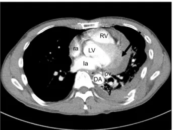
There was no evidence of external wound except skin lacer- ation on nose. Initial chest X-ray showed enlargement of car- diac shadow only (Fig. 1). Focused assessment with sonog- raphy for trauma (FAST) revealed the hemopericardium com- pressing the right ventricle (RV). At that time, sudden cardiac arrest occurred. We started cardiopulmonary resuscitation (CPR) and performed subxiphoid drainage. We also set per- cutaneous cardiopulmonary support (PCPS) by using Capioxe-
mergency bypass system (CapioxEBS; Terumo Inc., Tokyo, Japan) within few minutes because the vital sign did not restore. Following that, the patient was rushed into the oper- ation room (OR). We performed median sternotomy and iden- tified rupture of the right atrium appendage (2×1 cm) and su- perior vena cava (1 cm). After repairing all injured sites, PCPS was weaned smoothly. Total 73 units of blood compo- nents were given to correct circulation and coagulopathy.
Postoperatively, the patient required ventilatory support for 14
hours due to pulmonary edema. Visual disturbance appeared
due to multifocal embolic infarctions, but recovered during
follow-up. She discharged on postoperative day (POD) 22 af-
ter closed reduction of nasal bone.
PCPS for Cardiac Tamponade
− 335 − Table 1. Clinical characteristics and surgical details
Characteristic Case 1 Case 2 Age/sex
Injury mechanism Cardiac lesion
Associated injuries
Arrival (hr)
a)Initial BP, HR, SpO2 CPR
GCS ISS RTS TRISS (%) First diagnostic tool Diagnose to surgery (hr) PCPS
Catheter size (Fr.) Flow (L/min/m
2) Running time (hr) ACT (sec) ICU satys (day) Intubation time (hr) Hospital stay (day)
34/female High speed MVA RAA rupture (2×1 cm) SVC laceration 1 cm Sternal fracture Nasal bone fracture
0.67
70/50, 133, 92 Yes
15 26 5.4388 85.5 FAST 0.67
Artery: 16, vein: 20 1−2
3 220 4 14 26
45/male Fall-down
Left pericardial laceration (10 cm) Bronchial artery rupture
Left hemothorax with multiple rib fractures 2nd cervical spine fracture
Left pelvic bone fracture Left humerus fracture 6
100/60, 90, 89 No
9 22 7.8408 98.3 FAST 3.5
Artery: 16, vein: 20 2−2.5
3 150 6 30 55
MVA, motor-vehicle accident; RAA, right atrium appendage; SVC, superior vena cava; BP, blood pressure; HR, heart rate; SpO2, peripheral oxygen saturation; CPR, cardiopulmonary resuscitation; GCS, Glasgow Coma Scale; ISS, injury severity score; RTS, re- vised trauma score; TRISS, survival probability of trauma score-injury severity score; FAST, focused assessment with sonography for trauma; PCPS, percutaneous cardiopulmonary support; Fr., French; ACT, activated clotting time; ICU, intensive care unit.
a)