http://dx.doi.org/10.5090/kjtcs.2012.45.1.1 ISSN: 2233-601X (Print) ISSN: 2093-6516 (Online)
Department of Thoracic and Cardiovascular Surgery, Gospel Hospital, Kosin University College of Medicine Received: September 30, 2010, Revised: November 25, 2011, Accepted: November 25, 2011
Corresponding author: Sung-Rae Cho, Department of Thoracic and Cardiovascular Surgery, Gospel Hospital, Kosin University College of Medicine, 34 Amnam-dong, Seo-gu, Busan 602-702, Korea
(Tel) 82-51-990-6466 (Fax) 82-51-990-3099 (E-mail) srcho@kosinmed.or.kr
C The Korean Society for Thoracic and Cardiovascular Surgery. 2012. All right reserved.
CC This is an open access article distributed under the terms of the Creative Commons Attribution Non-Commercial License (http://creative- commons.org/licenses/by-nc/3.0) which permits unrestricted non-commercial use, distribution, and reproduction in any medium, provided the original work is properly cited.
Induction of ER Stress-Mediated Apoptosis by α-Lipoic Acid in A549 Cell Lines
Jong In Kim, M.D., Sung-Rae Cho, M.D., Chang Min Lee, M.D., Eok-Sung Park, M.D., Ki Nyun Kim, M.D., Hyung Chul Kim, M.D., Hae Young Lee, M.D.
Background: α-Lipoic acid (α-LA) has been studied as an anticancer agent as well as a therapeutic agent for diabetes and obesity. We performed this study to evaluate the anticancer effects and mechanisms of α-LA in a lung cancer cell line, A549. Materials and Methods: α-LA-induced apoptosis of A549 cells was detected by fluo- rescence-activated cell sorting analysis and a DNA fragmentation assay. Expression of apoptosis-related genes was analyzed by western blot and reverse transcription–polymerase chain reaction analyses. Results: α-LA induced apoptosis and DNA fragmentation in A549 cells in a dose- and time-dependent manner. α-LA increased caspase activity and the degradation of poly (ADP-ribose) polymerase. It induced expression of endoplasmic reticulum (ER) stress-related genes, such as glucose-regulated protein 78, C/EBP-homologous protein, and the short form of X-box binding protein-1, and decreased expression of the anti-apoptotic protein, X-linked inhibitor of apoptosis protein.
Reactive oxygen species (ROS) production was induced by α-LA, and the antioxidant N-acetyl-L-cysteine de- creased the α-LA-induced increase in expression of apoptosis and ER stress-related proteins. Conclusion: α-LA induced ER stress-mediated apoptosis in A549 cells via ROS. α-LA may therefore be clinically useful for treating lung cancer.
Key words: 1. Lung neoplasms 2. Cell death 3. Neoplasm marker
INTRODUCTION
Oxidative stress plays a key role in the development of de- generative diseases and tumors [1,2], and endogenous anti- oxidative agents have been shown to protect against these disorders [3,4]. α-Lipoic acid (α-LA), which is produced in small quantities in the human body, is an intermediate fatty acid that functions as a cofactor for mitochondrial respiratory enzymes. α-LA has been shown to have anticancer effects by activating glutathione peroxidase and decreasing oxidative
stress in cancer patients [5,6].
In addition, α-LA has been shown to have a prophylactic and therapeutic effect on diabetic neuropathy and alcohol-re- lated or toxic hepatic injury [7,8]. However, it can act as a pro-oxidant as well as an antioxidant depending on the dose applied and the length of application [9,10]. Further studies are therefore required to determine under what conditions α- LA protects or promotes oxidative stress.
Apoptosis describes the process of active, controlled cell death that occurs during embryonic development, pre-
developmental stages, and in response to hormones and dif- ferent types of chemical agents. Because the induction of apoptosis is closely related to cancer therapy, several studies at the molecular level have investigated the effects of natural products such as resveratrol, curcumin, and genistein on tu- mors [11-13].
Unlike other compounds, α-LA does not induce apoptosis in normal cells such as neurons and hepatocytes [14,15], but has been shown to induce apoptosis of various human cancer cell lines. Consequently, because α-LA is innocuous to nor- mal cells but inhibits the growth or promotes apoptosis of cancer cells, it has great potential therapeutic benefit as an anticancer agent.
An understanding of the pathways and mechanisms of apoptosis induced by an anti-carcinogen is crucial to the de- velopment of an effective anticancer therapy [16]. Apoptosis pathways can be initiated via membrane proteins, mitochon- dria, or the endoplasmic reticulum (ER). By clarifying the apoptosis pathway induced by a particular anticancer agent, the anticancer effect can be maximized using combination chemotherapy that activates several apoptosis pathways [17,18].
For example, the anticancer agent tumor necrosis factor-re- lated apoptosis-inducing ligand (TRAIL) activates tumor ne- crosis factor (TNF) receptors (or death receptors) in the cel- lular membrane, resulting in the apoptosis of tumor cells.
However, many tumor cells are resistant to TRAIL, and com- bination chemotherapy is required to increase the suscepti- bility of these tumor cells to TRAIL [19,20]. Therefore, a ba- sic knowledge of the mechanisms underlying the anticancer effect of a compound should improve the effectiveness of an- ticancer therapy.
In this study, we investigated the anticancer effects and mechanism of apoptosis of α-LA in lung cancer cells. Lung cancer is one of the most common causes of cancer-related death in Korea and in contrast to other cancers, most cases are treated by anticancer chemotherapy because this cancer is rarely diagnosed at an early stage. Despite the development of several novel anticancer agents, their therapeutic effective- ness is limited.
In this study, we investigated whether α-LA induces apoptosis in A549 lung cancer cells, whether α-LA-induced
apoptosis is induced by an antioxidant or pro-oxidant effect, and the relationship between α-LA-induced apoptosis and the ER stress response.
MATERIALS AND METHODS 1) Materials
α-LA ((+)-1,2-dithiolane-3-pentanoic acid) was purchased from Thiocaid Inc. (Germany) and cisplatin was obtained from Sigma Inc. (St. Louis, MO, USA). The antioxidant, N-acetyl-L-cysteine (NAC) and the caspase inhibitor, z-VAD, were purchased from Carbiochem Inc. (Darmstadt, Germany) and 2‘7’-dichlorodihydrofluorescein diacetate (DCFH-DA) for cell staining was purchased from Invitrogen, Inc. (Karlsro, Germany).
Western blotting antibodies against heat shock cognate pro- tein 70, C/EBP-homologous protein (CHOP), and myeloid cell leukemia 1 (Mcl-1) were obtained from Santa Cruz Inc.
(Santa Cruz, CA, USA), while antibodies targeting glucose-re- gulated protein 78 (GRP78) and caspase 3 were obtained from Stressgen Inc. (USA). X-linked inhibitor of apoptosis protein (XIAP) and Bcl2-associated X protein (BAX) anti- bodies were purchased from BD Biosciences Inc. (Franklin Lakes, NJ, USA), extracellular signal-regulated kinase (ERK) antibodies were obtained from Transduction Laboratory Inc.
(Lexington, KY, USA), and poly (ADP-ribose) polymerase (PARP) antibodies were obtained from Cell Signaling Inc.
(Beverly, MA, USA).
2) Methods
(1) A549 cell culture: A549 cells (lung cancer cell line) were cultured in RPMI 1640 media supplemented with 10%
fetal bovine serum, penicillin (100 U/mL), streptomycin (100 mg/mL), and gentamicin (10 mg/mL).
(2) Analysis of apoptosis: Cells cultured in α-LA-con- taining media were detached with 2.5 X trypsin-EDTA (GIBCO BRL, Grand Island, NY, USA), harvested, and then rinsed using phosphate buffered saline (PBS). Cells were then resuspended in 100 μL PBS and fixed at 4°C for more than one hour by the addition of 200 μL 100% ethanol. After centrifugation, the upper aqueous layer was removed and the cells were resuspended in 100 μL PBS.
We added 250 μL of 50 μg/mL RNase A (Sigma Inc.) to the cells and incubated them at 37oC for 30 minutes. We then cultured the cells at 37oC for 20 minutes in the dark af- ter the addition of 250 μL of 50 μg/mL propidium iodide (Sigma Inc.). We measured cell cycle progression and apopto- sis (%) using a fluorescence activated cell sorting Calibur-A flow cytometer (BD Bioscience Inc.). Averages and standard deviations were calculated by analysis of triplicate assays.
(3) Observation of DNA fragmentation: We added 500 μL cytosolic DNA extraction buffer containing 5 mM Tris (pH 7.4), 20 mM EDTA, 0.5% Triton X-100, and 1 mM of phenylmethylsulfonyl fluoride (Amresco Inc., Solon, OH, USA) to cultured A549 cells, and the cells were then incubated in ice water for 30 minutes with shaking at ten-minute intervals.
The cell solution was centrifuged at 12,000 rpm at 4oC for 20 minutes. A 500-μL aliquot of the upper layer after cen- trifugation was transferred to a new test tube and then mixed with the same amount of Tris-saturated phenol.
After centrifugation at 12,000 rpm at room temperature for ten minutes, 400 μL of the upper layer was collected in a new tube and centrifuged at 12,000 rpm for ten minutes after the addition of 400 μL chloroform. The collection and cen- trifugation steps were repeated two more times. A 200 μL aliquot of the final upper layer was collected in a new tube and 20 μL of 3 M sodium acetate and 1 mL of 100% etha- nol was added followed by centrifugation at 12,000 rpm at 4oC for 20 minutes. After removal of the upper layer, the pellet was washed with 70% ethanol by centrifugation at 12,000 rpm at 4°C for 15 minutes.
After removal of the upper layer, the dried pellet was dis- solved in 10 μL of sterile distilled water and incubated at 37oC for 30 minutes after the addition of 2 μL RNase A (10 mg/mL). We added gel loading buffer to this solution and loaded it on a 2% agarose gel. We electrophoresed the DNA through the gel at 50 V for one hour and then ob- served the DNA using a UV-transilluminator.
(4) Western blot analysis: Approximately 0.8×10⁶ cul- tured cells were lysed by adding 50−100 μL of lysis buffer to the harvested cells (137 mM NaCl, 15 mM EGTA, 0.1 mM sodium orthovanadate, 15 mM MgCl2, 0.1% Triton X-100, 25 mM MOPS, 100 μM phenylmethylsulfonylflu- oride, 20 μM leupeptin, pH 7.2). After centrifugation of the
cells at 12,000 rpm for 30 minutes, the upper protein-contain- ing layer was collected.
After measuring the protein concentration using a Bradford Protein Assay Reagent Kit (Pierce Inc., Rockford, IL, USA), equal amounts of protein extracts were loaded on a poly- acrylamide gel and electrophoresed. Proteins were transferred to Immobilon-P membranes (Millipore Co., Bedford, MA, USA) and probed with antibodies to caspase 3, PLC-r1, PARP, cIAP2, Mcl-1, and HSP70. Specific protein binding was detected using an ECL Western Blotting Kit (Millipore Co.).
(5) Reverse transcription-polymerase chain reaction: We used TRIzol for RNA isolation. In brief, cells were lysed by the addition of TRIzol after washing in PBS containing 0.1%
diethyl pyrocarbamate (DEPC) and chloroform. The upper layer after centrifugation was collected. Finally, RNA was sedimented and dried.
To reverse transcribe the RNA, we prepared an RT mixture containing 2 mL of 5× RT buffer, 1 mL of 10 mM dNTPs, 0.25 mL of Moloney murine leukemia virus reverse tran- scriptase (200 U/mL, Life Technologies Inc., Gaithersburg, MD, USA), 0.25 mL of RNase inhibitor, 0.5 mL of 50 mM oligo-dT primer, 4 mL of DEPC-water, and 2 mg of total RNA, and then performed cDNA synthesis at 42°C for one hour. After electrophoresis, PCR products were detected on 1.2% agarose gels using a UV transilluminator. The primer sequences used for the PCR were as follows: XBP-1 sense primer 5'-CCTTGTAGTTGAGAACCAGG-3' and antisense primer 5'-GGGGCTTGGTATATATGTGG-3'.
RESULTS 1) Effects of α-LA on A549 cells
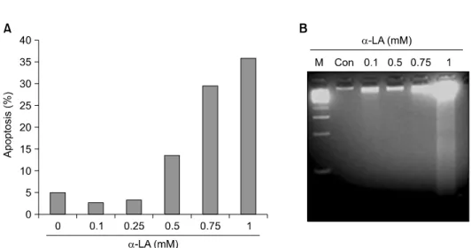
To determine whether α-LA induces apoptosis in A549 cells, cells were cultured for 18 hours in media containing 0, 0.1, 0.25, 0.5, or 1 mM of α-LA. The collected cells were stained with propidium iodide and cells in the sub-G1 phase were counted using a flow cytometer (Fig. 1A). Apoptosis was detected in groups treated with 0.5 mM or higher α-LA, and the higher the concentration of α-LA, the greater the extent of apoptosis. To determine whether cell death was di- rectly induced by α-LA treatment, we treated A549 cells
Fig. 1. α-Lipoic acid (α-LA) induces apoptosis in lung carcinoma A549 cells. (A) α-LA induced apoptosis in a dose-dependent manner.
A549 cells were treated with various concentrations of α-LA for 18 hours. Apoptosis was analyzed by determining the sub-G1 fraction of cells by fluorescence activated cell sorting (FACS). (B) α-LA-induced DNA fragmentation. A549 cells were treated with various concen- trations of evodiamine for 18 hours. DNA was extracted and was subjected to electrophoresis.
Fig. 2. α-Lipoic acid (α-LA) induces caspase 3 activation and poly ADP-ribose polymerase (PARP) degradation. A549 cells were treated with various concentrations of α-LA for 18 hours. Equal amounts of cell lysates were subjected to electrophoresis and ex- pression of PARP and caspase 3 was evaluated by western blot analysis. Heat shock cognate protein 70 (Hsc 70) was used as a loading control.
with various concentrations of α-LA; we observed DNA fragmentation in cells incubated with α-LA concentrations
≥0.75 mM (Fig. 1B).
2) Effects of α-LA on caspase-3 activation and PARP cleavage in A549 cells
Western blot analysis of protein extracts of A549 cells cul- tured for 18 hours with various concentrations of α-LA re- vealed a decrease in levels of inactivated caspase-3 in the group treated with 0.25 mM α-LA and an increase in PARP
cleavage at α-LA concentrations of 0.75 mM and higher (Fig. 2).
3) Analysis of apoptosis and protein expression according to α-LA treatment time
A549 cells treated with 1 mM α-LA were collected at 3, 6, 12, 18, and 24 hours. Cells were stained with propidium iodide and cells in the sub-G1 phase were counted using a flow cytometer. Apoptosis was detected after 12 hours or more of treatment with α-LA, and the apoptosis increased in a time-dependent manner (Fig. 3A).
We used western blot analysis to determine the pathway of apoptosis (Fig. 3B). The expression of the ER stress-related proteins, GRP78 and CHOP, increased with α-LA treatment time. Additionally, levels of inactive caspase-3 and PARP cleavage decreased, and levels of caspase-3 substrate in- creased with α-LA treatment time. Levels of XIAP, a pro- tein known to inhibit apoptosis, decreased.
4) ER stress-related response and α-LA-induced X-box binding protein splicing
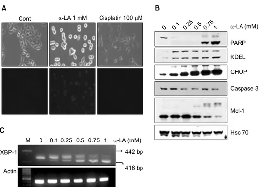
To clarify the relationship between apoptosis and the α- LA-induced ER stress-related response in A549 cells, we tre- ated A549 cells with α-LA (1 mM) or cisplatin (100 μM) (Fig. 4A). Cells treated with α-LA had a rounder morphol-
Fig. 3. Time kinetics of α-lipoic acid (α-LA)-induced apoptosis in A549 cells. (A) A549 cells were treated with 1 mM of α-LA for the designated time periods, and apoptosis was analyzed by determining the sub-G1 fraction of cells by fluorescence activated cell sorting (FACS). (B) Equal amounts of cell lysates were subjected to electrophoresis and expression of glucose regulated protein 78 (GRP78), C/EBP-homologous protein (CHOP), poly ADP-ribose polymerase (PARP), X-linked inhibitor of apoptosis protein (XIAP), and caspase 3 was evaluated by western blot analysis. Heat shock cognate protein 70 (Hsc 70) was used as a protein loading control.
ogy than the control or cisplatin-treated cells and blue fluo- rescence was detected only in the α-LA group after cell staining using an ER tracking dye.
To investigate changes in levels of ER stress-related pro- teins in response to α-LA treatment, we performed western blot analysis of cells treated with α-LA for 18 hours (Fig.
4B). As the concentration of α-LA increased, the levels of inactive caspase-3 decreased, but the levels of its substrate, PARP 3, decreased to an even greater extent. In addition, ex- pression of ER stress-related proteins such as KDEL, CHOP, and Mcl-1 increased in an α-LA concentration-dependent manner.
We measured transcript levels of the short form (416 bp) of the ER stress-related protein X-box binding protein (XBP)-1 using reverse transcription-polymerase chain reaction (RT-PCR) (Fig. 4C). At low concentrations of α-LA, the in-
active long form (422 bp) of XBP-1 was detected. However, as the concentration of α-LA increased, more of the active short form (416 bp) was detected.
5) Changes in levels of reactive oxygen species in A549 cells treated with α-LA
To determine whether α-LA induces the production of re- active oxygen species (ROS) and to determine the effect of ROS on apoptosis, we treated A549 cells with 1 mM α-LA for the following periods of time: 30 minutes, 1 hour, 3 hours, and 6 hours. Collected cells were stained with DCFH-DA to measure their levels of H2O2. Production of H2O2, measured by a flow cytometer, reached a maximum 30 minutes after α-LA treatment (Fig. 5A). We also detected the maximum degree of fluorescence in the group treated with α-LA for 30 minutes by direct observation of intra-
Fig. 4. Induction of the endoplasmic reticulum (ER) stress-related response and X-box binding protein (XBP) splicing by α-lipoic acid (α-LA).
(A) A549 cells treated with 1 mM α-LA or 100 μM cisplatin were stained with an ER tracking dye. (B) α-LA induced proteins related to ER stress. A549 cells were treated with various concentrations of α-LA for 18 hours. Equal amounts of cell lysates were subjected to electrophoresis and the expression of poly ADP-ribose polymerase (PARP), KDEL, C/EBP-homologous protein (CHOP), caspase 3, and myeloid cell leukemia 1 (Mcl-1) was determined by western blot analysis. Heat shock cognate protein 70 (Hsc 70) was used as a protein loading control. (C) α-LA induced X-box binding protein (XBP) mRNA splicing. mRNA from A549 cells treated with α-LA was isolated, and XBP-1 transcripts were detected by reverse transcription-polymerase chain reaction.
cellular changes in H2O2 levels using a fluorescence micro- scope (Fig. 5B).
6) The role of ROS in α-LA-induced apoptosis To determine whether ROS in A549 cells was directly in- volved in α-LA-induced apoptosis, we treated A549 cells with α-LA (1 mM), the antioxidant NAC (10 μM), or α-LA + NAC, and then after culturing the cells for 18 hours, we measured the proportion of cells in the sub-G1 phase using a flow cytometer. In contrast to the group treated with NAC-only, there was a remarkable increase in cells in the sub-G1 phase in the α-LA-only treatment group.
In contrast, the proportion of cells in the sub-G1 phase de- creased in both the NAC and α-LA+NAC-treated groups (Fig. 6A). Cells treated with α-LA had a circular morphology and condensed shape, but cells treated with both α-LA and NAC were similar in shape to control group cells (Fig. 6B).
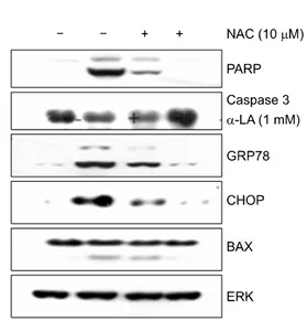
7) Effects of NAC pretreatment on the expression of apoptosis-related proteins induced by α-LA We treated A549 cells with α-LA (1 mM), NAC (10 μM), or α-LA (1 mM)+NAC (10 μM) and then investigated protein expression by western blot analysis (Fig. 7). Consi- stent with our previous results, α-LA treatment increased PARP cleavage, decreased levels of inactive caspase 3, and induced the expression of the ER stress-related proteins GRP78 and CHOP. However, in the group treated with both α-LA and NAC, cleavage of PARP and the expression of GRP78 and CHOP were inhibited. There were no changes in the expression levels of BAX or ERK.
DISCUSSION
Apoptosis, in contrast to necrosis, is initiated in response
Fig. 5. Induction of reactive oxygen species production following α-lipoic acid (α-LA) treatment of A549 cells. A549 cells were treated with 1 mM α-LA for 30 minutes, 1 hour, 3 hours, or 6 hours and then harvested and stained with 2‘7’-dichlorodihydro- fluorescein diacetate (DCFM-DA). H202 production was measured by fluorescence-activated cell sorting (A), and cell fluorescence was detected using a fluorescence microscope (B).
Fig. 6. Effect of pretreatment of A549 cells with N-acetyl-L-cysteine (NAC) on α-LA-induced apoptosis. A549 cells were treated with 1 mM α-lipoic acid (α-LA) or 10 μM NAC alone or in combination for 18 hours. The sub G1 fraction was measured by fluo- rescence-activated cell sorting (A), and cell morphology was observed using a microscope (B).
to various signals that activate cell death signaling pathways.
Caspase is one of the key proteins in apoptosis. Activated caspases induce cells to undergo apoptosis by activating or degrading proteins involved in DNA replication and gene ex- pression, as well as structural proteins and those involved in cellular homeostasis [21].
Three types of signals activate caspases: membrane protein signals, mitochondrial signals, and ER signals [17,18]. ER stress-related apoptosis develops due to impaired protein folding. ER-mediated apoptosis is activated by a failed un- folded protein response (UPR) related to pancreatic ER kinase (PKR)-like ER kinase, activating transcription factor 6, or in- ositol-requiring enzyme 1, and reflects the effort of the cell to prevent further misfolding of proteins when the ER is sa- turated with unfolded proteins [22-24].
In this study, we investigated the signaling pathways and mechanisms of α-LA-induced mediated apoptosis using the experimental lung cancer cell line, A549. First, we treated lung cancer cells with various concentrations of α-LA and counted cells in the sub-GI phase (i.e., apoptotic cells) by flow cytometry to assess the anticancer effects of α-LA. We found that apoptosis increased in a concentration- and time-dependent manner in groups treated with α-LA.
However, it was not clear whether this phenomenon was
Fig. 7. Effect of N-acetyl-L-cysteine (NAC) pretreatment of A549 on α-lipoic acid (α-LA)-induced protein expression. A549 cells were treated with 1 mM of α-LA or 10 μM of NAC alone or in combination for 18 hours. Equal amounts of cell lysates were subjected to electrophoresis and the expression of poly ADP-ri- bose polymerase (PARP), caspase 3, glucose-regulated protein 78 (GRP78), C/EBP-homologous protein (CHOP), Bcl2-associated X protein (BAX), P21, and extracellular signal-regulated kinase (ERK) was analyzed by western blot analysis.
due to cell necrosis or α-LA-induced apoptosis. We there- fore performed DNA electrophoresis and detected fragmented DNA, confirming apoptosis. In addition, western blot analysis revealed fragmentation of PARP induced by an increase in activated caspase-3 after α-LA treatment. Western blot anal- ysis also revealed an increase in expression of GRP78 and CHOP, which are ER stress-related proteins, after different periods of α-LA treatment. GRP78 is an ER stress-respon- sive protein and CHOP, by increasing the expression of DR5, is involved in ER mediated-apoptosis [25,26].
Together, our results suggest that α-LA induced apoptosis in the lung cancer cells via the ER stress pathway. The sup- pression of XIAP, a protein that inhibits apoptosis, was in- ferred from a decrease in XIAP levels [27] and ER stress was also detected by morphological changes in the cancer cells after α-LA treatment.
The ER stress-induced transcript factor, XBP-1, can be spliced in two different forms; the short form induces apopto- sis [28]. In our study, levels of the long form decreased whereas levels of the short form increased as the concen- tration of α-LA increased. These changes in morphology and
expression of specific molecules imply that treatment of cells with α-LA increases ER stress.
ROS is a well-known ER stress-inducing factor [29,30].
We observed an increase in intracellular production of H2O2
after α-LA treatment, which suggests a correlation between ROS and ER stress-mediated apoptosis in α-LA-treated lung cancer cells. Treatment of cells with the antioxidant, NAC, decreased apoptosis and restored the cell morphology to normal. To analyze the mechanism of action of ROS, we per- formed western blot analysis. We observed no changes in the activity of caspase 3 upon NAC treatment, but found that levels of GRP78 and CHOP decreased. These results imply that ROS acts as a mediator in ER stress-mediated apoptosis induced by α-LA treatment.
In light of our results, we suggest that α-LA may be a potent anti-lung cancer agent as it appears to be able to in- duce ER stress-mediated apoptosis in cancer cells by increas- ing levels of GRP78, CHOP, and the short form of XBP-1.
Furthermore, our experimental results show that ROS is cor- related with ER stress. Consequently, we hypothesize that α- LA acts as a pro-oxidant and apoptosis inducer in A549 can- cer cells.
CONCLUSION
We investigated the effects of α-LA on the induction of apoptosis, DNA fragmentation, and changes in the expression of apoptosis-related proteins in A549 lung cancer cells. Our major findings are as follows:
α-LA treatment induced apoptosis and DNA fragmenta- tion in A549 cells in a concentration- and time-dependent manner.
α-LA treatment increased levels of active caspase 3 and cleavage of PARP as well as expression of the ER stress re- sponse-related proteins GRP78 and CHOP and the tran- scription of the active form of XBP-1. Levels of the anti- apoptosis protein, XIAP, decreased in response to α-LA treatment.
α-LA treatment increased ROS levels, while NAC (antioxidant) treatment inhibited the α-LA-induced increase in expression of apoptosis- and ER stress response-related proteins.
These results provide a rationale for the clinical use of α- LA as an anticancer agent for treating lung cancer.
REFERENCES
1. Glaab WE, Hill RB, Skopek TR. Suppression of sponta- neous and hydrogen peroxide-induced mutagenesis by the antioxidant ascorbate in mismatch repair-deficient human colon cancer cells. Carcinogenesis 2001;22:1709-13.
2. Forsberg L, de Faire U, Morgenstern R. Oxidative stress, human genetic variation, and disease. Arch Biochem Bio- phys 2001;389:84-93.
3. Moghadasian MH, Freeman HJ, Godin DV. Endogenous an- tioxidant status in neoplastic and adjacent tissues in 1,2-di- methylhydrazine-induced colon cancer in rats: effects of olsalazine. Carcinogenesis 1996;17:983-7.
4. Packer L, Witt EH, Tritschler HJ. alpha-Lipoic acid as a bi- ological antioxidant. Free Radic Biol Med 1995;19:227-50.
5. Mantovani G, Maccio A, Madeddu C, et al. The impact of different antioxidant agents alone or in combination on re- active oxygen species, antioxidant enzymes and cytokines in a series of advanced cancer patients at different sites: cor- relation with disease progression. Free Radic Res 2003;37:
213-23.
6. Mantovani G, Maccio A, Madeddu C, et al. Reactive oxygen species, antioxidant mechanisms, and serum cytokine levels in cancer patients: impact of an antioxidant treatment. J Environ Pathol Toxicol Oncol 2003;22:17-28.
7. Bludovska M, Kotyzova D, Koutensky J, Eybl V. The influ- ence of alpha-lipoic acid on the toxicity of cadmium. Gen Physiol Biophys 1999;18 Spec No:28-32.
8. Bustamante J, Lodge JK, Marcocci L, Tritschler HJ, Packer L, Rihn BH. Alpha-lipoic acid in liver metabolism and disease. Free Radic Biol Med 1998;24:1023-39.
9. Biewenga GP, Haenen GR, Bast A. The pharmacology of the antioxidant lipoic acid. Gen Pharmacol 1997;29:315-31.
10. Packer L, Roy S, Sen CK. Alpha-lipoic acid: a metabolic antioxidant and potential redox modulator of transcription.
Adv Pharmacol 1997;38:79-101.
11. Kuo PL, Chiang LC, Lin CC. Resveratrol-induced apoptosis is mediated by p53-dependent pathway in Hep G2 cells. Life Sci 2002;72:23-34.
12. Rao CV, Rivenson A, Simi B, Reddy BS. Chemoprevention of colon carcinogenesis by dietary curcumin, a naturally oc- curring plant phenolic compound. Cancer Res 1995;55:259- 66.
13. Li M, Zhang Z, Hill DL, Chen X, Wang H, Zhang R.
Genistein, a dietary isoflavone, down-regulates the MDM2 oncogene at both transcriptional and posttranslational levels.
Cancer Res 2005;65:8200-8.
14. Piotrowski P, Wierzbicka K, Smialek M. Neuronal death in
the rat hippocampus in experimental diabetes and cerebral ischaemia treated with antioxidants. Folia Neuropathol 2001;
39:147-54.
15. Pierce RH, Campbell JS, Stephenson AB, et al. Disruption of redox homeostasis in tumor necrosis factor-induced apop- tosis in a murine hepatocyte cell line. Am J Pathol 2000;
157:221-36.
16. Lopez-Beltran A, Maclennan GT, de la Haba-Rodriguez J, Montironi R, Cheng L. Research advances in apoptosis-me- diated cancer therapy: a review. Anal Quant Cytol Histol 2007;29:71-8.
17. Gupta S. Molecular signaling in death receptor and mitoch- ondrial pathways of apoptosis (Review). Int J Oncol 2003;
22:15-20.
18. Kadowaki H, Nishitoh H, Ichijo H. Survival and apoptosis signals in ER stress: the role of protein kinases. J Chem Neuroanat 2004;28:93-100.
19. Lee TJ, Jung EM, Lee JT, et al. Mithramycin A sensitizes cancer cells to TRAIL-mediated apoptosis by down-regu- lation of XIAP gene promoter through Sp1 sites. Mol Cancer Ther 2006;5:2737-46.
20. Jung EM, Park JW, Choi KS, et al. Curcumin sensitizes tu- mor necrosis factor-related apoptosis-inducing ligand (TRAIL)- mediated apoptosis through CHOP-independent DR5 upregu- lation. Carcinogenesis 2006;27:2008-17.
21. Kim R, Emi M, Tanabe K. Caspase-dependent and -inde- pendent cell death pathways after DNA damage (Review).
Oncol Rep 2005;14:595-9.
22. Gaut JR, Hendershot LM. The modification and assembly of proteins in the endoplasmic reticulum. Curr Opin Cell Biol 1993;5:589-95.
23. Kaufman RJ. Orchestrating the unfolded protein response in health and disease. J Clin Invest 2002;110:1389-98.
24. Schroder M, Kaufman RJ. The mammalian unfolded protein response. Annu Rev Biochem 2005;74:739-89.
25. Yu Z, Luo H, Fu W, Mattson MP. The endoplasmic retic- ulum stress-responsive protein GRP78 protects neurons aga- inst excitotoxicity and apoptosis: suppression of oxidative stress and stabilization of calcium homeostasis. Exp Neurol 1999;155:302-14.
26. Yamaguchi H, Wang HG. CHOP is involved in endoplasmic reticulum stress-induced apoptosis by enhancing DR5 expre- ssion in human carcinoma cells. J Biol Chem 2004;279:
45495-502.
27. Devi GR. XIAP as target for therapeutic apoptosis in pros- tate cancer. Drug News Perspect 2004;17:127-34.
28. Yoshida H. Unconventional splicing of XBP-1 mRNA in the unfolded protein response. Antioxid Redox Signal 2007;9:
2323-33.
29. England K, Driscoll CO, Cotter TG. ROS and protein oxida- tion in early stages of cytotoxic drug induced apoptosis.
Free Radic Res 2006;40:1124-37.
30. Xue X, Piao JH, Nakajima A, et al. Tumor necrosis factor alpha (TNFalpha) induces the unfolded protein response (UPR) in a reactive oxygen species (ROS)-dependent fashion,
and the UPR counteracts ROS accumulation by TNFalpha. J Biol Chem 2005;280:33917-25.