DOI:10.5090/kjtcs.2011.44.4.273 ISSN: 2233-601X (Print) ISSN: 2093-6516 (Online)
*Department of Thoracic and Cardiovascular Surgery, Sanggye Paik Hospital, Inje University
**Department of Thoracic and Cardiovascular Surgery, Seoul National University Bundang Hospital
***Department of Thoracic and Cardiovascular Surgery, Kosin University Gospel Hospital
****Department of Thoracic and Cardiovascular Surgery, Eulji University Hospital
*****Department of Thoracic and Cardiovascular Surgery, Seoul Metropolitan Boramae Hospital, Seoul National University Received: September 30, 2010, Revised: March 17, 2011, Accepted: March 30, 2011
Corresponding author: Cheong Lim, Department of Thoracic and Cardiovascular Surgery, Seoul National University Bundang Hospital, 300, Gumi-dong, Bundang-gu, Seongnam 461-822, Korea
(Tel) 82-31-787-7140 (Fax) 82-31-787-7141 (E-mail) [email protected]
C
The Korean Society for Thoracic and Cardiovascular Surgery. 2011. All right reserved.
CC
This is an open access article distributed under the terms of the Creative Commons Attribution Non-Commercial License (http://creative- commons.org/licenses/by-nc/3.0) which permits unrestricted non-commercial use, distribution, and reproduction in any medium, provided the original work is properly cited.
Results of Extracorporeal Membrane Oxygenation (ECMO) Support before Coronary Reperfusion in Cardiogenic Shock
with Acute Myocardial Infarction
Eui Suk Chung, M.D.*, Cheong Lim, M.D.**, Hae-Young Lee, M.D.***, Jin-Ho Choi, M.D.****, Jeong-Sang Lee, M.D.*****, Kay-Hyun Park, M.D.**
Background: Despite aggressive treatment, the mortality rate of cardiogenic shock with acute myocardial infarction (AMI) is high. We performed extracorporeal membrane oxygenation (ECMO) prior to coronary reperfusion, and eval- uated the early clinical results and risk factors. Materials and Methods: From May 2006 to November 2009, we reviewed the medical records of 20 patients in cardiogenic shock with AMI (mean age 67.7±11.7 yrs, M : F 14 : 6).
After initially performing ECMO using the CAPIOX emergency bypass system (EBS
ⓇTerumo, Tokyo, Japan), pa- tients underwent coronary reperfusion (coronary artery bypass grafting, 13; percutaneous coronary intervention, 7).
Results: All patients were in a cardiogenic shock state, cardiopulmonary resuscitations (CPR) were performed for fourteen patients (mean CPR time 20.8±26.0 min). The mean time from vascular access to the initiation of ECMO was 17.2±9.4 min and mean support time was 3.8±4.0 days. Fourteen patients were able to be weaned from ECMO and ten patients were discharged (mean admission duration 50.1±31.6 days). Patients survived on average 476.6±374.6 days of follow-up. Longer CPR and support time, increased cardiac enzyme, lower ejection fraction, lower albumin, and major complications were the risk factors of mortality (p<0.05). Conclusion: The early applica- tion of ECMO prior to coronary reperfusion and control of risk factors allowed for good clinical results in cardio- genic shock with AMI.
Key words: 1. Extracorporeal membrane oxygenation (ECMO) 2. Coronary reperfusion
3. Acute myocardial infarction 4. Cardiogenic shock
INTRODUCTION
The mortality rate of cardiogenic shock due to acute my- ocardial infarction (AMI) is as high as 60∼80% and may be
higher without aggressive treatment. Besides the classical
medical treatment, circulatory supportive devices such as the
Intra Aortic Balloon Pump (IABP) and Ventricular Assistance
Device (VAD) may be helpful [1-5]. Recently ECMO has
been adapted to the percutaneous approach and the self-charg- ing system, so that it can support the whole body and coro- nary circulation fast, and has shown good clinical results in patients experiencing cardiogenic shock due to AMI [6-9]. On the other hand, there is controversy about which procedure should be first, coronary reperfusion or ECMO, for cardio- genic shock due to AMI. Therefore, the authors have re- viewed the short-term results of patients who had received ECMO just before coronary reperfusion in cases of cardio- genic shock due to AMI.
MATERIALS AND METHODS
We retrospectively reviewed 20 cases (median age 67.7±11.7, male : female 13 : 7) of coronary reperfusion after application of EBS (Capiox
ⓇEmergency Bypass System [Terumo Inc., Tokyo, Japan]) due to postinfarct cardiogenic shock among 106 cases of ECMO from February 2005 to November 2009. We defined a state of cardiac arrest, systolic blood pressure below 70 mmHg for over 30 minutes and clinical symptoms such as oliguria, confusion, and ventricular tachycardia as cardiogenic shock. We applied ECMO to those patients experiencing cardiogenic shock and performed CPR in 14 of the cases (70%) (mean time 20.8±26.0 minutes). In all cases, we underwent coronary artery bypass under the support of ECMO and chose the method of coronary re- perfusion according to the results. After using ECMO sup- port, arterial pulse wave was disappeared in 14 cases, and in those cases we additionally inserted an IABP. Immediately after coronary reperfusion, we started Aspirin and Clopidogrel in all patients.
To perform ECMO, we inserted a 17 Fr cannula into the artery and 21 Fr cannula into the vein (Medtronic Inc.
Ⓡ, DLP, MN, USA) percutaneously using the Seldinger method and adjusted the size of cannulae according to the patient’s body weight and body surface area. We infused with as much as 5,000 IU heparin before cannulation, and maintained an ACT (Activated Coagulation Time) in the range of 180 to 200 minutes. We maintained cardiopulmonary circulatory blood flow in the range of 3.0∼3.5 L/min/m
2and stopped or lowered the vasopressor to the minimum dosage after ECMO support. During ECMO support, we performed echocardiog-
raphy daily to check heart function and to check for any ven- tricular dilation or ventricular thrombus. We maintained plate- let and hemoglobin counts over 50,000/μ and 10.0 gm/dL, respectively. During ECMO support, we employed continuous renal replacement therapy (CRRT) in cases of acute renal failure (ARF) by connecting CRRT unit to the ECMO circuit directly.
The weaning from ECMO was begun after the initial 48 hours from the application, raising the dosage of vasopressors and decreasing supportive circulatory blood flow. After wean- ing from ECMO, the cannula insertion sites were sutured af- ter identification of the femoral artery intima in the Intensive Care Unit (ICU), and there were no complications from the cannula insertion site sutures.
We analyzed the risk factors using a 2×2×2 K Pearson's chi-square test and Fisher’s exact test. We compared the mean values of the clinical results using a nonparametric Mann-Whitney U-test.
RESULTS
The mean cannular insertion time for ECMO application was 17.2±9.4 minutes, we performed peripheral circulation at the same time to prevent lower extremity from necrosis in two cases. The cannular insertions were performed in the cor- onary catheterization room in most cases (17 cases). Two cannular insertions were performed in the Emergency Room and one in the ICU.
Coronary angiography showed 11 cases of three-vessel dis-
eases, 6 cases of two-vessel diseases, one case of single coro-
nary disease and 6 cases of left main branch lesions. The
main lesion was the left main branch in 5 cases, the left an-
terior descending artery in 4, the left circumflex artery in 6,
and the right coronary artery in 5 cases. We decided the
method of coronary reperfusion according to each patient’s
coronary angiography results and state. We performed 13
cases of coronary bypass graft surgery and 7 cases of
Percutaneous Coronary Intervention (PCI). There were surge-
ries combined with coronary bypass graft surgery in 5 cases
(mitral valvuloplasty in 2 cases, repair of a left ventricle rup-
ture in one case, Dor procedure in one case, and repair of
postinfarct ventricular septal defect in one case).
Table 1. Clinical results
Total Survivors (n=10) Death (n=10) p-value
ECMO support (hrs) ICU stay (days)*
Admission duration (days)
3.8±4.3 11.05±6.8 28.75±31.1
2.1±1.4 14.8±6.2 50.1±31.6
5.6±5.4 7.3±5.4 7.4±5.3
0.06 0.00 0.01
*ICU=Intensive care unit.
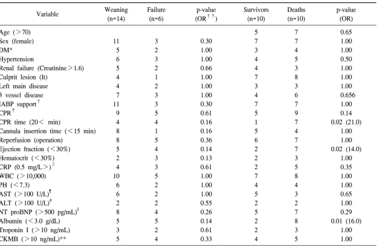
Table 2. Pre ECMO risk factors
Variable Weaning
(n=14)
Failure (n=6)
p-value (OR
††)
Survivors (n=10)
Deaths (n=10)
p-value (OR) Age (>70)
Sex (female) DM*
Hypertension
Renal failure (Creatinine>1.6) Culprit lesion (It)
Left main disease 3 vessel disease IABP support
†CPR
‡CPR time (20< min)
Cannula insertion time (<15 min) Reperfusion (operation)
Ejection fraction (<30%) Hematocrit (<30%) CRP (0.5 mg/L>)
∥WBC (>10,000) PH (<7.3) AST (>100 U/L)
¶ALT (>100 U/L)
#NT proBNP (>500 pg/mL)
§Albumin (<3.0 g/dL) Troponin I (>10 ng/mL) CKMB (>10 ng/mL)**
11 5 6 5 4 4 7 11 9 4 8 8 5 2 4 10 6 6 2 8 5 3 5
3 2 3 2 1 2 3 3 5 4 1 5 4 3 3 5 2 2 2 4 5 2 4
0.30 1.00 1.00 0.66 1.00 1.00 1.00 0.30 0.61 0.16 0.16 0.36 0.14 0.13 0.61 1.00 1.00 1.00 0.55 0.26 0.14 0.61 0.33
5 7 3 4 4 7 3 4 7 5 1 5 6 2 2 2 7 4 5 2 5 2 2 4
7 7 4 5 3 8 3 6 7 9 7 4 7 7 3 5 8 4 3 2 7 8 3 5
0.65 1.00 1.00 0.50 1.00 1.00 1.00 0.656
1.00 0.14 0.02 (21.0)
1.00 1.00 0.02 (14.0)
1.00 0.35 1.00 1.00 0.65 1.00 0.29 0.01 (16.0)
1.00 1.00
*=Diabetes mellitus;
†=Cardiopulmonary resuscitation;
‡=Intra aortic balloon pump;
§=N-Terminal fragment of the prohormone, Brain-Type natriuretic peptide;
∥=C reactive peptide;
¶=Aspartate aminotransferase;
#=Alanine aminotransferase; **=Creatin kinase MB;
††=Odd ratio.
Mean ECMO support time was 3.8±4.3 days (0.5 to 19.2 days). Fourteen cases (70%) were weaned from ECMO and 10 patients (50%) were alive and discharged from the hospital. The mean length of hospitalization was 28.8±31.1 days (1 to 58 days) and mean time in ICU was 11.1±6.8 days (1 to 25 days) (Table 1).
The cause of death was failure of ECMO weaning in 6 cases, brain injury in 2 cases, restenosis of coronary artery in
one case, and sepsis in one case. The causes of ECMO
weaning failure were failure of left ventricular decompression
in 2 cases, decrease of circulatory blood flow due to bleed-
ing, and sepsis in 3 cases and heart dysfunction before
ECMO support in one case. The main complications were
sepsis and Disseminated Intravascular Coagulation (DIC) in 9
cases, acute renal failure in 9 cases, bleeding in 8 cases, and
brain injury in 4 cases. We applied CRRT to 8 of the 9 cas-
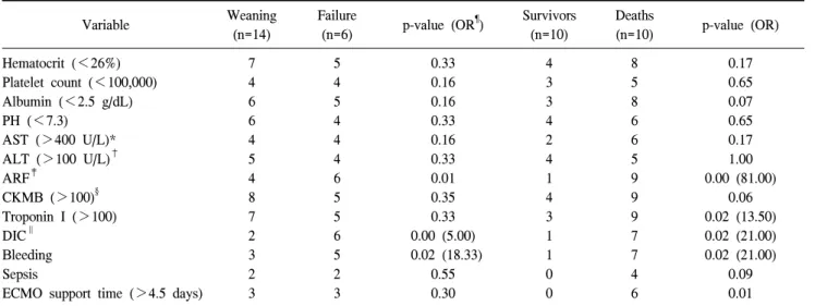
Table 3. Risk factors during ECMO
Variable Weaning
(n=14)
Failure
(n=6) p-value (OR
¶) Survivors (n=10)
Deaths
(n=10) p-value (OR) Hematocrit (<26%)
Platelet count (<100,000) Albumin (<2.5 g/dL) PH (<7.3)
AST (>400 U/L)*
ALT (>100 U/L)
†ARF
‡CKMB (>100)
§Troponin I (>100) DIC
∥Bleeding Sepsis
ECMO support time (>4.5 days) 7 4 6 6 4 5 4 8 7 2 3 2 3
5 4 5 4 4 4 6 5 5 6 5 2 3
0.33 0.16 0.16 0.33 0.16 0.33 0.01 0.35 0.33 0.00 (5.00) 0.02 (18.33)
0.55 0.30
4 3 3 4 2 4 1 4 3 1 1 0 0
8 5 8 6 6 5 9 9 9 7 7 4 6
0.17 0.65 0.07 0.65 0.17 1.00 0.00 (81.00)
0.06 0.02 (13.50) 0.02 (21.00) 0.02 (21.00)
0.09 0.01
*=Aspartate aminotransferase;
†=Alanine aminotransferase;
‡=Incase creatininin over 50% of normal range;
§=Creatin Kinase;
∥