서 론
인공와우 이식기는 심장박동기 및 뇌심부자극계와 더 불어 신경보형물(neural prosthesis) 중 질병 치료에 있 어서 유용한 것으로 알려져 있다. 1990년대 이후 인공와 우 기기의 신호처리전략과 코딩기술이 빠르게 발전하여
술 후 언어수행능력의 큰 향상을 가져오게 되었고, 인공 와우 이식은 고도이상의 난청환자들에게 유용한 청각을 제공함으로써 효과적인 청각재활방법으로 인정되고 있 어 중요성이 더욱 크다고 할 수 있다. 그런데 만성중이 염수술 등으로 인한 근치유양돌절제술 상태인 환자들에 게 인공와우 이식수술을 시행하는 것은 복잡한 과정이 다. 유양동 내의 얇은 상피내막은 탄력이 있는 전극을
개방형 유양동을 가진 환자에서의 인공와우 이식술
중앙보훈병원 이비인후과
최전하
·
백훈희·
조현상·
김춘동Cochlear Implantation in the Patient with Open Mastoid Cavity
Jeon Ha Choi, MD, Hun Hee Baek, MD, Hyun Sang Cho, MD and Choon Dong Kim, MD, PhD Department of Otolaryngology-Head and Neck Surgery, Veterans Health Service Medical Center, Seoul, Korea
- ABSTRACT -
Background and Objectives:Performing cochlear implantation is a difficult process for those patients who
have received radical mastoidectomy due to chronic otitis media. However, we could perform a single stage operation successfully without external auditory meatus and mastoid complete obliteration. Materials andMethods:The authors performed 13 cases of cochlear implantation in open cavity mastoidectomy state pa-
tients without obliteration of mastoid or with the partial obliteration of mastoid from July 2011 to July 2015. Of these thirteen patients, eight patients received cochlear implantation in the site of open mastoid cavity and oth- ers in opposite site. The medical records and radiological findings were reviewed retrospectively. Results:Three of eight patients who had undergone open cavity mastoidectomy did not perform mastoid cavity oblitera- tion during cochlear implantation. Another five patients with an open cavity mastoid had undergone the partial obliteration of mastoid during cochlear implantation. There were no postoperative medical complications such as infections of mastoid cavity, recurrence of cholesteatoma and necrosis of skin flap during following up peri- ods except for 1 case of delayed electrode extrusion. Conclusions:The advantages for this procedure as the following. First, we could overcome the difficulty of radiological diagnosis for primary or recurrent cholestea- toma due to mastoid obliteration. Second, a single-stage procedure allowed the implantation as safe as two- stage procedures. At last, non-obliterative or partial obliterative procedures may replace the typical mastoid obliteration process during cochlear implantation for it allows a safe procedure for open cavity patients reduc- ing complications such as electrode extrusion or infection.
(J Clinical Otolaryngol 2016;27:92-97)
KEY WORDS
:Cochlear ImplantationㆍOtitis Media.논문접수일 :2016년 3월 18일 / 논문수정일 :2016년 4월 27일 / 심사완료일 :2016년 5월 20일 교신저자 :김춘동, 05368 서울 강동구 진황도로61길 53 중앙보훈병원 이비인후과
전화 :(02) 2225-1384・전송 :(02) 2225-1385・E-mail:[email protected]
덮기에는 외부로 전극의 노출 위험이 있어 불충분하다.
개방형 유양동에 인공와우 이식을 할 경우 중이염이나 진주종의 재발의 위험성, 개방동의 감염, 전극의 노출 등 의 문제가 발생할 수 있어 이를 해결하기 위하여 지금까 지 다양한 방법들이 시도되어왔다.1,2) 많은 연구 보고서 에서 개방형 유양동을 가진 귀의 인공와우 이식은 병의 재발방지와 인공와우 이식물의 노출 가능성을 줄이기 위해 이관 및 외이도 폐쇄를 함께하는 추체아전절제술 을 권장하고 있다.3) 그러나 기존의 연구들에서 권장하는 추체아전절제술로서 외이도를 완전히 폐쇄하였을 때 수 술 후 염증이나 진주종의 재발을 추적하기 어렵고 넓은 이도성형술이 되어 있는 경우 외이도 입구의 폐쇄도 기 술적으로 어렵다는 문제점이 있다.4)
유양동을 폐쇄하기 위한 여러 방법들이 고안되었는 데, 지방을 이용하는 것, tricalcium phosphate와 hydr- oxyapatite의 혼합물을 이용하는 것, 골분 반죽과 조직 접합체를 이용하는 것, 골막-연조직 피판 또는 피부이 식물을 이용하는 것 등이 있다.5-7) 유양동을 폐쇄하는 목 적은 자가 정화되고 감염없는 공동를 만들어 유돌동 문 제를 해결하는 것에 있고 이는 인공와우를 이식받으려 는 환자들에 있어서 필요하다고 알려져 있다. 또한 여러 연구에서 진주종이나 만성중이염이 있는 환자들에게 인 공와우를 시행할 때 2단계 수술을 하는 것이 바람직하다
고 추천하고 있다.1)
그렇지만 본 저자들은 개방형 유양동을 가진 환자들 에게 인공와우수술을 시행함에 있어서 기존의 방법과 달리 외이도를 폐쇄 하지 않고, 유양동 폐쇄술을 시행하 지 않을 뿐만 아니라 1단계만의 수술로도 큰 합병증 없 이 수술이 가능하다는 것을 보여주고자 한다.
재료 및 방법
2011년 7월부터 2015년 7월까지 중앙보훈병원 이비 인후·두경부외과에서 시행한 인공와우이식술 중 개방 동 유양돌기 수술병력이 있는 13예를 대상으로 의무기 록, 수술기록, 방사선기록을 후향적으로 고찰하였다.
연령대는 35세에서 84세까지로 평균연령은 67세였고 남자 12명, 여자 1명이었다. 인공와우이식술 후 추적관 찰 기간은 28개월에서 42개월까지였으며, 평균 31.9개월 이었다. 개방동 유양돌기 수술 후의 경과 기간은 3년에 서 58년까지로, 개방동 유양돌기 수술을 받은 곳에 인공 와우를 이식한 환자는 9명이고(Table 1. Participant 1~9), 반대쪽에 인공와우를 이식한 환자는 5명이었다(Table 1.
Participant 10~13). 기존의 개방형 유양동 부위에 인공 와우 이식을 한 경우 평균청력은 표준순음청력검사상 4 분법으로 공기전도 115 dB이었으며 반대쪽(정상귀)의
Table 1. Changes in postoperative PTA
Parti-
cipant Sex Age (years)
Duration after canal wall down mastoidectoy (years)
Follow up period (months)
Pre OP PTA (4freq) Post OP PTA (4freq) CI side
(dB) Contralateral
side (dB) CI side (dB)
1 M 65 7 30 120/76 80/76 35
2 M 70 20 31 120/76 72/71 40
3 M 72 55 26 120/76 65/64 44
4 M 35 6 19 120/76 59/56 34
5 M 73 53 18 120/76 70/65 48
6 M 61 13 15 108/72 36/36 40
7 M 84 1 16 111/76 95/70 51
8 M 68 5 29 116/64 85/51 51
9 M 79 10 22 106/75 82/75 30
10 M 68 0.3 27 95/61 92/66 35
11 M 68 7 25 105/76 81/60 40
12 M 68 12 20 95/76 80/71 48
13 M 68 13 23 116/76 120/76 45
14 F 65 35 33 120/76 120/76 48
평균 공기전도는 72 dB였다. 개방형 유양동이 아닌 부위 에 인공와우 이식을 한 경우 수술을 한 쪽(정상귀)의 평 균 공기전도는 106 dB였으며 반대쪽(개방동)의 평균 공 기전도는 99 dB였다. 이 경우에는 잔청이 있는 쪽에 보 청기를 착용하기 위해(bimodal hearing) 정상귀에 인공 와우이식을 시행하였다. 개방동 유양돌기 수술을 받은 곳에 인공와우 이식을 한 경우 모든 경우에서 외이도 폐 쇄술을 시행하지 않았고 유양동의 완전폐쇄도 시행하지 않았다. 그 중 유양동은 4예에서 유양동 폐쇄술을 전혀 시행하지 않았고, 5예에서 부분적 폐쇄술을 시행하였다.
결 과
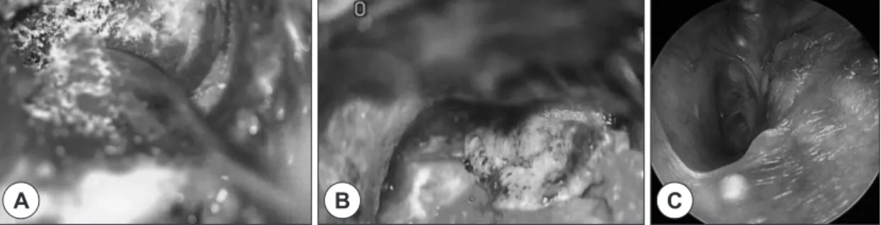
유양동 폐쇄술을 시행하지 않은 4예는 안면신경관 후 방부와 개방동 내에 홈(groove)을 만들고(Fig. 1) 전극을 위치시킨 후 원래의 상피내막과 측두근막으로 덮었으며 그 중 1예에서 6개월 후에 전극이 노출되었다(Fig. 2).
또 다른 3예에서는 기존의 개방동이 일부 폐쇄되어있는 상태로서 전극을 보호하기에 충분히 두꺼운 상피내막과 피하조직을 가지고 있어 그 자체 만으로 전극을 덮었다.
부분폐쇄술을 시행한 5예 중 2예에서는 유양동 내 홈을 만들어 전극을 위치시키고 골분 반죽(bone pa′te)과 피부 전층 유리피판 이식술을 시행하였다(Fig. 3). 부분 폐쇄 술을 시행한 다른 2예에서는 유양동을 덮는 피부층이 충 분히 두꺼워 홈을 만들지 않았고 유양돌기 부분에는 골 분 반죽과 피부 전층 유리피판 이식술을 시행하여 부분 폐쇄하였다. 1예에서는 생착률을 높이기 위해 피부 전층 유리피판 이식술을 하지 않고 하부 기저 피판을 이용하 여 전극을 덮었다(Fig. 4). 전극의 노출이 있었던 1예를 제외한 8예에서는 추적기간 동안에 모두 유양동의 감염,
진주종의 발생 및 피부피판의 괴사 등의 합병증은 없었 으며 없었다. 수술 후 시행된 전산화단층촬영상 전극의 위치는 잘 유지되었다(Fig. 5). 외래 추적관찰시 기저 피 판을 이용하여 전극을 덮는 경우가 피부 전층 유리피판 이식을 시행하는 경우보다 빨리 호전되는 양상을 보였다.
고 찰
만성중이염 또는 진주종성중이염 환자들에서 개방동 중이염 수술은 고도 난청이나 심지어는 전농까지 초래 할 수 있으며 어떤 이유로든 양측 고도 난청이 될 경우 인공와우 전극을 삽입하는 것만이 다시 청력을 되살리 는 유일한 방법이다.8) 기존 중이수술로 인한 개방형 유 양동을 가진 환자에서 인공와우 이식을 할 경우 대부분 은 이전에 수술을 받지 않은 쪽(정상측두골)에 와우이식
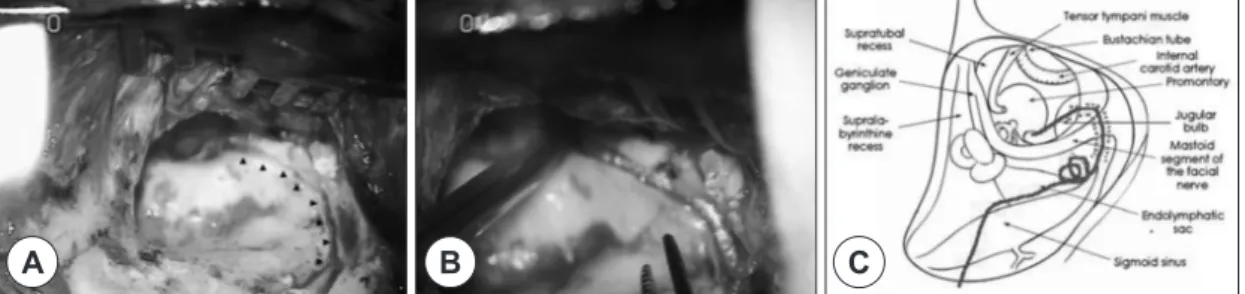
Fig. 1. Operative findings. A : Groove for electrode in mastoid cavity (arrowheads). B : The electrode was inserted in the groove. C : Design for groove for electrode. Redline is a electrode. dotted line shows the groove.
C B
A
Fig. 2. Postperative findings. The eletrode lead was ex-
posed (asterisk).
을 시행하게 된다.21) 그 이유는 개방동 상태에서는 이식 기 전극의 노출이나 감염, 기존질환의 재발 등이 병발할 수 있기 때문이다.9) 하지만 개방형 유양동 절제술을 시 행한 귀는 그렇지 않은 귀보다 잔청이 없는 경우가 많아 추후 양이청취(bimodal hearing)를 계획할 때 잔청이 있 는 쪽에 보청기를 착용하기 때문에 어쩔 수 없이 개방형
유양동이 있는 쪽에 인공와우 이식을 하게 되는 경우가 있다.10) 개방형 유양동 측에 인공와우의 전극을 보호하 기 위해 특별한 방법들이 소개되었는데, 인공와우 이식 전에 유양동의 해부학적 재건을 주장하기도 하고,5) 유양 동폐쇄술도 알려져 있다. 유양동폐쇄술은 추체아전절제 술이나 근치유양돌기절제술에서 잘 알려진 바이나, 인
Fig. 5. Temporal CT : showing the lead electrode in the mastoid cavity after partial obliteration with bone páte and full thickness skin graft (arrow).
Fig. 4. A, B : The partial obliteration of mastoid during cochlear implantation with inferior based flap (dotted line). C : Post-operative otoscopic findings. No specific event has occurred in 1 year since implantation.
B C
A
Fig. 3. A, B : The partial obliteration of mastoid during cochlear implantation with bone páte and full thickness skin graft. C : Post-operative otoscopic findings. No specific event has occurred in 1 year since implantation.
C B
A
공와우 이식술에서는 그 중요성이 더욱 강조되고 있 다.11) 유양동폐쇄술에는 복부지방과 측두근피판이 이용 되고, 개방동 유양돌기절제술시에는 이개연골이나 이주 연골, 측두근막, 골막-연조직 피판을 이용하여 와우개창 부위를 포함한 중이강과 유양동을 폐쇄하고 전극을 매 몰할 수 있다.12) 완전폐쇄의 경우에는 이관을 잘 막은 후 유양동을 골분, hydroxyapatite, tricalcium phosphate 같은 물질로 채우고, 외이도를 폐쇄하여 맹낭폐쇄(blind sac closure)를 하게 된다. 그러나 이렇게 되면 해부학적 지표가 모호지고 인공와우 이식을 위한 2차 수술 시에 드릴링을 해야하기 때문에 단점이 될 수 있다. 13)
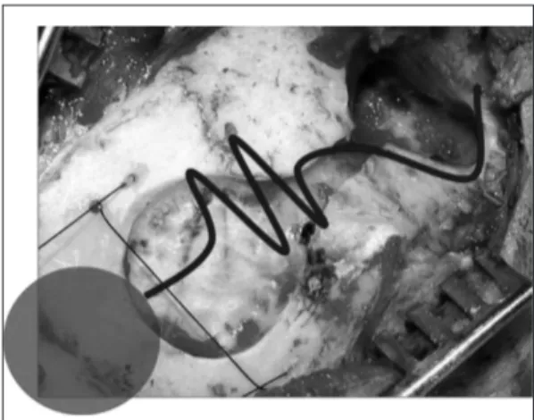
지금까지는 개방형 유양동 상태인 경우에 앞서 언급 된 것과 같이 외이도와 이관, 유양동을 모두 폐쇄한 후 인공와우 이식을 하는 것이 권장되어 왔다.10) 그 이유는 유양동을 폐쇄하지 않은 인공와우 이식 환자는 감염의 재발이나 전극의 노출같은 위험성이 증가될 것을 우려 해 유양동을 폐쇄하고 와우개창술(cochleostomy)을 폐 쇄하며 외이도 피부에 맹낭폐쇄(blind sac closure)를 하 면 그 위험성이 최소화될 것으로 생각되었기 때문이 다.10) 하지만 개방형 유양동 상태의 귀에 인공와우 이식 을 시행한 최근의 논문들을 보건대 몇가지 이유로 이것 에 대한 반론이 제기되었다. 첫째, 유양동을 폐쇄하지 않 는 것이 방사선학적으로 유양동을 잘 볼 수 있어 진주종 의 발생을 용이하게 추적이 가능하고,14,15) 둘째, 단계적 수술이나 재수술시에 수술적 접근을 용이하게 하며,13) 셋째, 이관을 통한 감염의 이론적인 위험도가 그 동안의 경험상 상당한 기간에 걸친 추적관찰을 통해 임상적으 로 문제가 되지 않는다는 것이 증명되었기 때문이다.16) 따라서 외이도와 이관의 폐쇄 없이 유양동 내의 전극만 을 보호하기 위한 부분적인 유양동 폐쇄술로서 측두근 유경피판, 피부 전층 유리피판 이식, 골분 반죽과 조직 접합제, 측두근 유리피판이나 골막 피판 또는 지방조직 그리고 이들을 혼합하는 방법들이 제안되었다.14) 그 밖 에 유양동을 폐쇄하지 않는 또 다른 방법으로 수용자극 기의 두개골 홈(well)을 일반적인 위치보다 더욱 후방에 만들고 전극의 경로 홈(tract groove)을 물결 모양으로 만들어 전극의 많은 부분을 유양동 바깥쪽에 유지시킴 으로써 피판이나 상피내막의 궤양과 괴사를 감소시키는 방법이 있다(Fig. 6).17-19) 또한 유양동 내부에도 홈
(groove)을 만들어 전극을 홈에 위치 시킨 후 골분 반죽 이나 골 시멘트 또는 조직 접합제로 경로(tract)를 덮어 평평한 표면을 만든 후, 연골이나 측두근막, 골막 피판 등과 그것들의 조합으로 그 표면을 다시 덮을 수 있
다.12,17-19) 결과적으로 이러한 방법은 비폐쇄 또는 부분적
폐쇄 유양동을 만들 수 있고, 이후에 이 유양동은 표면 이 상피내막으로 덮히게 된다.1,5,20) 위에 언급한 수술방법 들은 전극을 받아들일 수 있는 각각의 유양동의 상태에 따라 적절한 선택을 하였다.
그러므로 본 저자들은 경험상 이러한 술기의 장점을 다음과 같이 정리하였다. 첫째, 유양동을 폐쇄함으로 인 해 영상학적으로 진주종의 발생 또는 재발을 진단하는 데 어려움이 있던 것을 해결할 수 있다. 둘째, 2단계 수 술로 진행하지 않고 1단계 수술로 진행하여도 안전한 인 공와우 이식을 할 수 있다. 마지막으로 전극의 탈출, 감 염 등의 수술 후 합병증이 거의 없어 인공와우 이식수술 시 유양동을 폐쇄하는 수술을 대체할 수 있는 안전한 술 식이 될 수 있기 때문에 개방형 유양동 수술을 받은 환 자들에 있어서 인공와우 이식술 시에 적합한 수술이 될 수 있다.
중심 단어:인공와우・개방형 유양동.
REFERENCES
1) Hamzavi J, Baumgartner W, Franz P, Plenk H. Radical
cavities and cochlear implantation. Acta Otolaryngol 2001;
Fig. 6. Design for well and electrode : The well is located
in the rear position of the typical position and the elec-
trode groove has a zigzag-shape.
121:607-9.
2) Harada T, Ishida K, Endo M, Takahashi M, Sakai M. Recur-
rent extrusion of cochlear implant at an interval of 5 years.
Otol Neurotol 2003;24:83-5.
3) Bendet E, Cerenko D, Linder TE, Fisch U. Cochlear implan-
tation after subtotal petrosectomies. Eur Arch Otorhino- laryngol 1998;255:169-74.
4) Kim CS, Shim WS, Chang SO, Oh SH, Kim YH, Lee HJ,
et al. Cochlear implantation in chronic otitis media. Kore- an J Otolaryngol 2004;47:730-5.
5) Axon PR, Mawman DJ, Upile T, Ramsden RT. Cochlear
implantation in the presence of chronic suppurative otitis media. J Laryngol Otol 1997;111:228-32.
6) Decher H. Reduction of radical cavities by homologous car-
tilage chips. Laryngorhinootologie 1985;64:423-6.
7) Decher H, Migdal H. Partial obliteration of radical cavities
with ceramic granules. Laryngorhinootologie 1992;71:106-9.
8) Basavarij S, Shanks M, Sivaji N, Allan AA. Cochlear im-
plantation and management of chronic suppurative otitis media: single stage procedure? Eur Arch Otorhinolaryngol 2005;262:852-5.
9) Ching TY, van Wanrooy E, Dillon H. Binaural-bimodal fit-
ting or bilateral implantation for managing severe to pro- found deafness: a review. Trends Amplif 2007;11(3):161-92.
10) Meyerhoff WL, Stringer SP, Roland PS. Ramdo procedure:
Modification and application. Layngoscope 1988;98:795-6.
11) Bendet E, Cerenko D, Linder TE, Fisch U. Cochlear implan-
tation after subtotal petrosectomies. Eur Arch Otorhinolar- yngol 1998;255:169-74.
12) Manrique M, Cervera Paz FJ, Espinosa JM, Perez N, Garcia Tapia. Cochlear implantation in radical cavities of mastoid-
ectomy. Laryngoscope 1996;106:1562-5.
13) Gray RF, Ray J. McFerran DJ. Further experience with fat
graft obliteration of mastoid cavities for cochlear implants.
J Laryngol Otol 1999;113:881-4.
14) El-Kashlan HK, Arts HA, Telian SA. Cochlear implanta-
tion in chronic suppurative otitis media. Otol Neurotol 2002;
23:53-5.
15) El-Kashlan HK, Arts HA, Telian SA. External auditory ca-
nal closure in cochlear implant surgery. Otol Neurotol 2003;
24:404-8.
16) Hussam K, El-kashlan, Steven A Telian. Cochlear implan-
tation in the chronically diseased ear. Current opinion in Otolaryngology & Head and Neck Surgery 2004;12:384-6.
17) Himi T, Harabuchi Y, Shintani T, Yamaguchi T, Yoshio- ka I, Kataura A. Well Audiol Neurootol 1997;2:410-7.
18) Marangos N, Laszig R. Cochlear implant surgery and rad-
ical cavities. Adv Otorhinolaryngol 1997;52:147-50.
19) Kiefer J, von Ilberg C. Special surgical problems in cochle-
ar implant patients. Adv Otorhinolaryngol 1997;52:135-9.
20) Gray RF, Irving RM. Cochlear implants in chronic suppu-
rative otitis media. Am J Otol 1995;16:682-6.
21) Oh SH, Kim SY. Hearing preservation surgery in cochlear