76
KISEP Case Reports 臨床耳鼻:臨床耳鼻:第••••••••••••••••••••••••••••••••••••••••••••••••••••••••••••••••••• 臨床耳鼻:臨床耳鼻:第第 第10 卷卷卷卷 第第第第 1 號號號號 1999
J Clinical Otolaryngol 1999;;;;10::::76-78
상악동내 전위분출치 1례
인천기독병원 이비인후과,1 치과2
유용원1·서준영1·최호순1·김영철1·김상균1·최덕준2
Ectopic Third Molar in Maxillary Sinus
Yong-Weon Yoo, MD
1, Jun-Young Seo, MD
1, Ho-Soon Choi, MD
1, Young-Chul Kim, MD
1, Sang-Gyoon Kim, MD
1and Duc-Jun Choi, MD
21Department of Otorhinolaryngology-Head and Neck Surgery, 2Dentistry, Inchon Christian Hospital, Inchon, Korea
- -
-- ABSTRACT ----
Ectopic teeth eruption into the dental environ are a common developmental abnormality. However, ectopic teeth eruption in other sites are rare. We experienced a case of ectopic third molar in the left maxillary sinus that is thought to evoke maxillary sinusitis. The tooth was removed through Caldwell-Luc approach under local anesthesia without any problem. ((((J Clinical Otolaryngol 1999;10:76-78))))
KEY WORDS:Ectopic third molar·Maxillary sinus.
서 론
치아의 전위분출은 비교적 흔한 발생학적 질환이다.1)2) 그러나 치조골 이외의 전위분출은 드문 것으로 되어 있 으며, 그 장소로는 하악과, 오훼돌기, 구개, 비강, 그리고 상악동 등이다.3)4)
저자는 2년간에 걸쳐 재발과 호전을 반복하는 만성 부 비동염 환자에 있어 좌측 상악동내에서 전위분출치를 발 견, 제거 하였기에 문헌 고찰과 함께 보고하는 바이다.
증 례
28세된 외국인 남자근로자로서 2개월간의 좌측 협골
부의 간헐적인 종창 및 압통을 주소로 내원하였다. 과 거력상 특이 사항은 없었으며 2년전부터 간헐적으로 종 창 및 압통이 있을 때마다 자가로 증상치료만 받아왔다.
환자 내원시 이학적 검사상 좌측 협골부위에 점증하는 종괴가 촉지되고 압통을 동반하며 국소소견상 중비도에 약간의 농성 분비물을 볼 수 있었다.
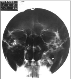
단순 부비동 X-선상 좌측 상악동기저부에 치아처럼 보이는 골밀도 이물의 음영을 나타내며, 좌측 상악동의 전반적인 점막 비후의 음영이 보이나 골파괴의 소견은 없어 보였다(Fig. 1).
파노라마방사선촬영상 좌측 상악 제3대구치의 결손을 보였으나, 그외 치아의 숫자나 모양의 이상은 없었다 (Fig. 2).
1998년 4월 8일 국소마취로 Caldwell-Luc씨 접근 방법을 통하여 좌측 상악동 치조와(alveolar recess)에 있는 15×20 mm크기의 전위분출치를 발견하고 제거 하였다.
수술소견상 좌측 상악동 점막은 전반적인 부종 및 농 논문접수일:1998년 11월 28일
심사완료일:1999년 1월 22일
교신저자:서준영, 400-130 인천 광역시 중구 율목동 237 인천기독병원 이비인후과
전화:(032) 761-9745・전송:(032) 763-9409
유용원 외:상악동내 전위분출치 1례
77 성분비물이 관찰되었으며 상악동 외하벽쪽에 15×20 mm 정도의 골 손실이 있었으나 구강으로의 교통은 없었다 (Figs. 3 and 4).
술후경과는 특이한 합병증 없이 양호하였다.
고 찰
치아의 전위분출은 비교적 흔한 발생학적 질환으로 서1)2) 치조골내의 전위분출시에는 50명의 소아당 1명 의 유병률을 보인다.2) 그러나, 치조골 이외의 전위분출
은 드물며 그 장소로는 하악과, 오훼돌기, 구개, 비강, 그 리고 상악동으로의 빈도순으로3)4) 특히 상악동내로 제 3대구치의 전위는 극히 드문 것으로 되어있다.2)
전위분출치아의 원인으로는 외상, 감염, 치아군집, 치판 발생시의 이상등이며3)4) 상악동내의 낭종이나 종양도 한 원인이 될 수 있다.2) 이 환자의 경우는 외상, 감염의 과 거력이 없으며, 파노라마 방사선상 치아 군집의 양상을 보이지는 않아, 치판발생시의 이상을 원인으로 보는 것 이 합당하다 하겠다.
증상은 치아의 전위분출장소에 따라 다양하며1) 하악 과나 오훼돌기에 발생한 경우에는 개구장애, 저작장애, 이전부의 종창등이,2) 비강내 존재시는 비폐색이 나타날 수 있다.3) 하지만 대부분이 골막으로 전위분출치가 주위 와 분리되기 때문에 무증상이 많은 것으로 되어 있으 며,2) 본 환자의 경우 전위분출치(Fig. 2, black arrow) 가 골막을 뚫고 부비동 내로 나와있었기 때문에 부비동 Fig. 1. Waters’view showing tooth in the left maxillary
sinus.
Fig. 2. Panoramic radiograph revealing the tooth in the left maxillary sinus(black arrow:tooth root).
Fig. 3. Endoscopic view of the left maxillary sinus shows ectopic tooth.
Fig. 4. The extracted tooth shows a relatively normal sh- ape and size of crown with underdeveloped roots.
J Clinical Otolaryngol 1999;10:76-78
78 염을 발생시킨 것으로 생각된다.
그 기전으로는 점막 섬모운동방향의 결손이나 이물작 용으로 생각되어지는데, 이때 Ferris에 의하면 부비동 이물이 증상을 나타내는 경우는 이물이 점막이나 골벽에 손상을 주는 경우, 부비동내 분비물의 배출을 차단하는 경우, 이물이 결석의 핵으로 작용하여 부비동 벽을 손상 하는 경우라고 한다.6)
이러한 전위분출치로 거의 대부분이 골밀도 음영을 보 이기 때문에 단순 부비동 X-선이나 파노라마방사선 촬 영으로 충분하며 컴퓨터단층촬영은 불필요한 것으로 되 어 있다.4)5)
치료는 외과적 적출이며, 증상이 없는 경우는 주기적 방사선 검사로 충분하다.2-4)
적출된 치아는, 치근의 발달은 미숙해 보였으나 치아 관의 발달은 정상소견을 보였다. 이는 치아의 발생시 여 러 단계를 거쳐 치아관이 완성된 뒤에 치근초(root sh- eath)를 따라 상아질 형성이 되면서 치근을 형성하기 때문에, 발생단계에 문제가 있으면 치아관의 모양이나
크기가 정상이더라도 치근은 미성숙 될 수 있다고 생각 된다.
저자들은 부비동염을 동반한 좌측 상악동내로의 제3 대구치의 전위분출을 치험하였기에 문헌고찰과 함께 보 고하는 바이다.
중심 단어:상악동 전위분출치.
REFERENCES
1) Yusuf H, Quayle AA. Intracondylar tooth. Int J Oral Ma- xillofac Surg 1989;8:323.
2) Elango S, Palaniappan SP. Ectopic tooth in the roof of the maxillary sinus. Ear, Nose and Throat J 1991;70:365-6.
3) Pracy JPM, Williams HOL, Montgomery PQ. Nasal teeth.
J Laryngol Otol 1992;106:366-7.
4) Difelice R, Lombardi T. Ectopic third molar in the maxi- llary sinus. Australian Dental J 1995;40:236-7.
5) Mckenna JD, Chidyllo SA. Localization of a maxillary third molar immobilized within an infected maxillary si- nus. J An Dent Assoc 1984;108:208-9.
6) Ferris KP. Foreign body in the sphenoid sinus. J Laryngo 1961;78:697-701.