INTRODUCTION
Oral antiviral agents are currently available and are the main option for the treatment of patients with chronic hepatitis B.
In a large proportion of patients, antiviral therapy can slow down the disease progression by hepatitis B virus (HBV) DNA suppression and hepatitis ‘e’ antigen (HBeAg) seroconversion.
However, the potential emergence of drug resistant mutations remains a caveat of oral antivirals; mutations increase as the treatment duration extends (1, 2). In addition, mutations to one drug may lead to resistance to other drugs. Patients with lamivudine (LMV)-resistant rtM204I/V (±rtL180M) muta- tions in the catalytic tyrosine-methionine-aspartate-aspartate (YMDD) motif are less responsive to entecavir (ETV) (1, 2).
Among adefovir (ADV) treated patients, rtA181T or rtA181V mutations have been associated with a 2-6 fold decrease in sensitivity to LMV (2).
Mutation of the HBV polymerase can alter the amino acid codon of the surface genes and vice versa, because the poly- merase gene, the main target of antiviral drug resistant muta- tions, has overlapping reading frames with the surface region (1, 3). In LMV treated patients, the P120A mutation in the S region results in hepatitis B surface antigen (HBsAg) detec-
tion failure (4). Lee et al. (5) reported overlapping surface gene mutations of HBV in an LMV-treated patient who finally lost HBsAg. In this patient, they detected deletions of nucleo- tides 23 to 55 (amino acids 12 to 22) in the Pre-S2 region, and amino acid substitutions at I126S, T131N, M133T, and S136Y in the ‘a’ determinant region of HBsAg.
Overlap of surface gene mutations, such as sW172stop in patients carrying rtA181T, and sL173F in patients carrying rtA181V, have been reported in chronic hepatitis B patients receiving ADV treatment. However, the clinical consequences of these changes have not been determined (1, 2). The goal of this study was to evaluate the surface gene sequence in ADV- resistant patients carrying A181T/V mutations and to deter- mine the associated clinical significance.
MATERIALS AND METHODS Patients
Twenty-two patients with chronic hepatitis B were includ- ed in this study. Nine patients had not been previously treat- ed with antiviral agents and thus formed the control group
257
Jeong Han Kim, Young Kul Jung, Moon Kyung Joo, Ji Hoon Kim, Hyung Joon Yim, Jong-Jae Park, Jae Seon Kim, Young-Tae Bak, Jong Eun Yeon, and Kwan Soo Byun
Division of Gastroenterology and Hepatology, Department of Internal Medicine, Korea University Guro Hospital, Korea University College of Medicine, Seoul, Korea
Address for Correspondence Jong Eun Yeon, M.D.
Division of Gastroenterology and Hepatology, Department of Internal Medicine, Korea University Guro Hospital, Korea University College of Medicine, 97 Gurodong-gil, Guro-gu, Seoul 152-703, Korea Tel : +82.2-2626-1030, Fax : +82.2-838-6591 E-mail : 93haan@hanmail.net
Hepatitis B Viral Surface Mutations in Patients with Adefovir Resistant Chronic Hepatitis B with A181T/V Polymerase Mutations
The hepatitis B virus (HBV) polymerase gene has overlapping reading frames with surface genes, which allows to alter the amino acid codon of the surface genes. In adefovir (ADV) treated chronic hepatitis B patients carrying rtA181T/rtA181V muta- tions, overlap with surface gene mutations such as sW172stop/sL173F has been reported. However, the clinical consequences of such surface mutations have not been determined. The aim of this study was to determine the surface gene sequence in ADV-resistant patients carrying the A181T/V mutation and to describe the clini- cal significance. Of the 22 patients included in this study, 13 were ADV-resistant with rtA181T/V mutations (polymerase mutation group, Group P) and nine were antiviral treatment-naive (control group, Group C). The Pre-S1 gene mutation, V60A, was detected in 11 patients (Group P=8, Group C=3). A start codon mutation in the Pre-S2 gene was found in five patients (Group P=3, Group C=2). An S gene mutation, sA184V, was found in nine patients, all of whom were in group P. Although sW172stop and sL173F mutations were detected, reduced HBsAg titer was not observed. Fur- ther study of these mutations and their clinical implications are needed.
Key Words : Hepatitis B, Chronic; Adefovir; Hepatitis B Surface Antigens
Received : 27 November 2008 Accepted : 25 March 2009
ⓒ 2010 The Korean Academy of Medical Sciences.
This is an Open Access article distributed under the terms of the Creative Commons Attribution Non-Commercial License (http://creativecommons.org/licenses/by-nc/3.0) which permits unrestricted non-commercial use, distribution, and reproduction in any medium, provided the original work is properly cited.
. .
(nucleos[-t]ide naive patients, Group C). The remaining thir- teen patients were ADV-resistant, having an rtA181T muta- tion, rtA181V mutation, or both mutations (ADV polymerase mutation group, Group P). These patients developed ADV resistance while undergoing ADV monotherapy as rescue ther- apy for LMV-resistant HBV (rtM204I/V±rtL180M). The ADV-resistant mutations were detected using restriction frag- ment mass polymorphism (RFMP) method as previously des- cribed (6). Serum was collected from the patients every two to three months during treatment and was kept at -20℃until the mutation analyses were performed. The serum collected at the time of ADV-resistant rtA181T/V mutation detection was used for sequencing analysis. Informed consent was ob- tained from each patient and the experimental protocol was approved by the Korea University Guro Hospital Human Research Committee.
Biochemical and virology monitoring
At the time of serum collection, we conducted routine full blood examinations and liver function tests, including tests for aspartate aminotransferase (AST), alanine aminotransferase (ALT), total bilirubin, albumin, prothrombin time, blood urea nitrogen (BUN), and creatinine. HBsAg and antibody to the HBs antigen were measured using the commercially available radioimmunoassay kits (Abbot Laboratories, North Chicago, IL, USA). The HBsAg titer was measured using the chemiluminescent microparticle immunoassay (CMIA) ARCHITECT�HBsAg assay (Abbot Laboratories). HBeAg and antibody to the HBe antigen were measured using com- mercially available radioimmunoassay kits (Shin Jin Medics Inc., Goyang, Korea). HBV-DNA was quantified using the Hybrid Capture II assay (Digene Diagnostics Inc., Beltsville, MD, USA) or the VERSANT�HBV DNA 3.0 assay (Sie- mens Healthcare Diagnostics Inc., Tarrytown, NY, USA) and data showed ‘pg/mL’ were converted to ‘copies/mL’ (1 pg=
283,000 copies).
Polymerase chain reaction
Hepatitis B viral DNA was extracted from 200 mL of serum using the QIAamp�MinElute�Virus Spin kit (Qiagen Inc., Vanencia, CA, USA) according to the manufacturer’s instruc- tions. The S coding region of HBV was amplified using HBS- 1S (the forward primer) and HBS-1AS (the backward primer) as previously described (5). The amplification conditions con- sisted of 94℃for 3 min, 35 cycles of 94℃for 30 sec, and 55℃for 30 sec. This was followed by a final primer exten- sion at 72℃for 1 min using the Taq PCR Core Kit (Qiagen Inc.). The polymerase chain reaction (PCR) amplified HBV DNA was purified using the QIAquick�PCR Purification Kit (Qiagen Inc.) according to the manufacturer’s instructions.
Sequencing
The primers used for direct sequencing were HBS-1S, HBS- 1AS, HBS-2S, and HBS-3S as previously described (5). Sequ- encing reactions were performed in an MJ Research PTC- 225 Peltier Thermal Cycler using an ABI PRISM BigDyeTM Terminator Cycle Sequencing Kit with AmpliTaq DNA poly- merase (Applied Biosystems Inc., Foster, CA, USA) accord- ing to the manufacturer’s instructions. Single-pass sequenc- ing was performed on each template using the aforemention- ed primers. The fluorescent-labeled fragments were purified from the unincorporated terminators with an ethanol precipi- tation protocol. The samples were resuspended in distilled water and subjected to electrophoresis in an ABI PRISM 3730XL Analyzer (Applied Biosystems Inc.).
Analysis
The nucleotide sequences were compared with those of the HBV genotype C subtype ‘adr’ registered at the Nation- al Center for Biotechnology Information (NCBI, nucleotide LOCUS AF286594). The genotype of the obtained HBV DNA sequences was determined by a web-based genotyp- ing tool for viral sequences at the NCBI (7).
Statistical analysis
We conducted the Fisher’s exact tests for categorical vari- ables and Mann-Whitney’s U-tests for continuous variables to compare patients with the rtA181T/V mutation to those without the rtA181T/V mutation. Values are expressed as median (range). A P value <0.05 was considered statistical- ly significant.
RESULTS
Demographic and biochemical characteristics
Demographic and biochemical data are summarized in Table 1. Among the 13 patients in Group P, the majority were males (n=10). All patients in Group C were males. Bio- chemical data are represented as median (range) (median of Group P vs. Group C, P value). Group P was significantly older than Group C, 45 (28-75) (47 vs. 39, P=0.025), had higher levels of albumin 4.2 (2.5-5) (4.2 vs. 3.6, P>0.05).
Group P also had lower levels of AST 100.5 (23-385) (65 vs. 193, P=0.001), ALT 155 (22-385) (78 vs. 262, P<0.001), and total bilirubin 1.1 (0.4-33.6) (1.0 vs. 1.3, P>0.05). Ten of the 22 patients had liver cirrhosis (LC) (Group P=7, Group C=3). Thirteen of the 22 patients were HBeAg positive (Group P=7, Group C=6). Two cases of hepatocellular carcinoma (HCC) were included (Group P=1, Group C=1).
. .
ADV, adefovir; HBeAg, hepatitis B e antigen; HBV, hepatitis B virus; AST, aspartate aminotransferase; ALT, alanine aminotransferase; CH, chronic hepatitis; LC, liver cirrhosis; HCC, hepatocellular carcinoma.
ADV resistant
mutation Gender Age
(yr) HBeAg HBV DNA
(log copies/mL)
AST/ALT (IU/L)
Bilirubin (mg/dL)
Albumin
(g/dL) Clinical status Group-
number
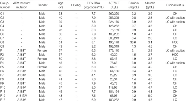
C1 - Male 33 - 7.7 260/290 2.2 3.6 CH
C2 - Male 40 + 7.9 253/325 0.8 2.5 LC with ascites
C3 - Male 39 + 7.8 224/170 3.9 2.5 LC with ascites
C4 - Male 34 + 8.0 125/262 0.7 4.5 CH
C5 - Male 28 + 8.3 75/169 0.9 4.4 CH
C6 - Male 30 + 7.9 103/262 1.0 4.7 CH
C7 - Male 75 - 8.6 385/249 3.4 2.6 LC
C8 - Male 51 - 8.1 190/178 33.6 3.1 HCC
C9 - Male 43 + 9.2 193/319 1.3 4.5 CH
P1 A181T Female 57 - 6.3 273/110 3.1 2.8 LC with ascites
P2 A181T Male 57 + 6.2 40/30 1.5 3.7 HCC
P3 A181T Female 50 + 5.8 67/47 1.9 3.3 LC
P4 A181T Male 45 + 7.9 75/63 3.0 3.3 LC with ascites
P5 A181T Female 70 + 6.3 64/73 0.4 4.2 CH
P6 A181V Male 40 - 5.8 64/156 0.4 4.7 LC
P7 A181V Male 46 - 4.1 29/22 0.9 3.0 LC
P8 A181T Male 41 + 7.0 23/34 1.4 4.8 CH
P9 A181T Male 45 - 7.9 40/78 1.0 4.7 CH
P10 A181V Male 57 + 8.0 118/96 1.0 4.7 LC
P11 A181T Male 49 - 7.7 101/154 0.9 4.1 CH
P12 A181T/V Male 43 + 7.5 65/96 1.2 5.0 CH
P13 A181V Male 47 - 6.9 100/232 0.9 4.8 LC
Table 1. Patients’ demographic, biochemical and clinical characteristics
Number of point mutations
Pre-S1 region Pre-S2 region S region Entire region Group-
number Genotype HBsAg
subtype
HBsAg titer (IU/mL)
C1 C adr 2,075 6 3 2 11
C2 C adr 12,335 2 8 8 18
C3 C adr 24,205 2 0 3 5
C4 C adr 8,850 2 2 1 5
C5 C adr 50,660 4 5 1 10
C6 C adr 15,090 2 4 0 6
C7 C adr 18,250 3 2 6 11
C8 C adr 2,490 4 4 2 10
C9 C adr 24,770 3 0 3 6
Median (range) of Group C 15,090 (2,075-50,660) 3 (2-6) 3 (0-8) 2 (0-8) 10 (5-18)
P1 C adr 2,010 5 1 6 12
P2 C adr 4,530 3 6 9 18
P3 C adr 960 4 1 1 6
P4 C adr 6,330 4 2 2 8
P5 C adr 6,055 0 1 1 2
P6 C adr 5,605 3 0 4 7
P7 C adr 2,340 1 0 4 5
P8 C adr 98,440 2 0 2 4
P9 C ayr 37,590 3 0 3 6
P10 C adr 21,385 4 1 6 11
P11 C adr 5,175 3 2 5 10
P12 C adr 14,280 4 0 4 8
P13 C adr 5,950 2 2 4 8
Median (range) of Group P 5,950 (960-98,440) 3 (0-5) 1 (0-6) 4 (1-9) 8 (2-18)
Total 7,590 (960-98,440) 3 (0-6) 1.5 (0-8) 3 (0-9) 8 (2-18)
Group C vs. Group P P=0.235 P=0.487 P=0.133 P=0.413 P=0.583
Table 2. Genotype, HBsAg subtype, HBsAg titer and point mutations
HBsAg, hepatitis B surface antigen.
HBV genotype, subtype, and point mutations
All patients in our study were infected with HBV geno- type C. Twenty-one of the 22 patients had the ‘adr’ subtype and one patient had the ‘ayr’ subtype. For the Pre-S1, Pre- S2, and S genes, the incidence of mutations in Group P and Group C were 3 vs. 3 (P>0.05), 1 vs. 3 (P>0.05), and 4 vs.
2 (P>0.05), respectively. The median number of point muta- tions in the entire region was 8 (Group P=8, Group C=10, P>0.05). Most cases had mutations in the S gene except for one case in Group C. One patient in Group P did not have a mutation in the Pre-S1 gene and seven patients (Group P=5, Group C=2) had no mutations in the Pre-S2 gene (Table 2).
Changes in the serum hepatitis B surface antigen titer
We conducted serum HBsAg titers to evaluate the clini- cal consequences of the surface gene mutation. The median HBsAg titer was 7,590 IU/mL (Group P=5,950, Group C=15,090, P>0.05). There was no significant difference, regardless of whether they harbored the rtA181T/V muta- tion (Table 2).
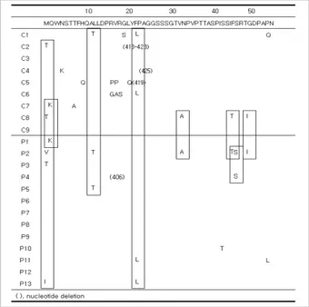
Pre-S1 region
In the Pre-S1 region, 12 patients (six cases in each group) had nucleotide deletions at position ‘21’ (amino acid [a.a.]
7) and 18 patients (nine cases in each group) had nucleotide deletions at position ‘30’ (a.a. 10). Nucleotide deletions at position ‘59’ (a.a. 20) were detected in 5 of 13 patients in Group P and 2 of 9 patients in Group C. An H51Q muta- tion was detected in two patients (one case in each group).
Eight of 13 patients in Group P and 3 of 9 patients in Group C had a V60A mutation. G2A (n=1), G73D (n=1), L74I (n=2), A91T (n=1), and A95T (n=1) mutations were detect- ed in Group P. In Group C, A81T (n=1), T86A (n=2), and A95V (n=1) mutations were detected. Three cases of I84T mutations were found in Group C, compared to only one in Group P (Fig. 1).
Pre-S2 region
A start codon mutation was identified in five patients (Group P=3, Group C=2) in the Pre-S2 gene. All five pati- ents with this mutation had cirrhosis, and two of these pati- ents also had HCC (Group P=1, Group C=1). V32A, I45T, and T49I mutations were detected in the patients with HCC, both groups equally. An F46S mutation was observed in Group P (n=2); a Q2K mutation in Groups P (n=1) and C (n=1);
an A11T mutation in Groups P (n=2) and C (n=1); and an F22L mutation in Groups P (n=2) and C (n=2).
In Group C, there was one case of each of the following
Fig. 1. Sequence changes in the Pre-S1 regions. Twelve patients (six cases in each group) and 18 patients (nine cases in each group) had nucleotide deletions at positions ‘21’ and ‘30’. Nucleo- tide deletions at position ‘59’ were detected in 5 of 13 patients in Group P and in 2 of 9 patients in Group C. An H51Q mutation was detected in two patients (one case in each group). A V60A muta- tion was detected in 8 of 13 patients in Group P and 3 of 9 patients in Group C. L74I mutations in Group P (n=2) and T86A mutations in Group C (n=2) were detected. Three cases of an I84T mutation were found in Group C and one case in Group P. G2A, G73D, A91T, and A95T mutations were detected in Group P. A81T and A95V mutations were detected in Group C.
Fig. 2. Sequence changes in the Pre-S2 regions. Five start codon mutations were identified in the Pre-S2 region. V32A, I45T and T49I mutations were detected in HCC cases. Q2K (Group P=1, Group C=1), A11T (Group P=2, Group C=1), F22L (Group P=2, Group C=2), and F46S (Group P=2) mutations were observed. In Group C, there were following mutations: N4K, T7A, H9Q, R16P, R16G, V17P, V17A, R18S, G19S, L20Q, P54Q, nucleotide ‘418-423’ dele- tion, ‘419’ deletion, and ‘425’ deletion. In Group P, one case each of I42T, P54L mutation and nucleotide 406 deletion were detected.
mutations: N4K, T7A, H9Q, R16P, R16G, V17P, V17A, R18S, G19S, L20Q, P54Q, nucleotide 418-423 deletion (a.a.
21-22), 419 deletion (a.a. 21), and 425 deletion (a.a. 23). In Group P, mutations I42T, P54L, and nucleotide 406 dele- tion (a.a. 17) were detected (Fig. 2).
S region
Among the patients in Group P, sW172stop (n=5) and sL173F (n=5) mutations were detected. In the S gene, an A184V mutation was found in 9 of 13 patients in Group P.
However, it was not detected in Group C (P=0.002). Other mutations that we detected in the S gene include S3N (Group P=4, Group C=2), I68T (Group P=3, Group C=1), I126T (Group P=4, Group C=1), and L213I (Group P=6, Group C=1). These mutations were most frequent in Group P.
In Group C, there was one case of each of the following mutations: G10stop, L15V, I28V, W36L, F41S, T47A, Q51L, N59H, C69stop, L87V, F93L, V96G, L97P, G102C, T131P, Y200F, and C221Y. Similarly, in Group P, there was one case of each of the following mutations: F41C, A45V, P49L, Q54L, P62L, I92T, L98V, T140S, A157V, W182stop, and S204R.
An L21S mutation was detected in Groups P (n=2) and C (n=
2), and R24Q and P203R mutations were found in Groups P (n=1) and C (n=1) (Figs. 3, 4).
Entire amino acid changes in both groups
Among these mutations, only the W172stop/L173F (P<
0.001) and A184V (P=0.002) mutation were significantly different between the groups (data not shown).
Comparison of demographic, biochemical and clinical characteristics between patients with and without the W172stop/L173F mutation
We also compared patients both with and without the W172stop/L173F mutation (Table 3). AST (P=0.035) and ALT (P=0.009) levels were significantly lower in patients
Fig. 3. Sequence changes in the S regions up to the front of ‘a’
determinant. An S3N mutation in Groups P (n=4) and C (n=2), and an I68T mutation in Groups P (n=3) and C (n=1) were detect- ed. G10stop, L15V, I28V, W36L, F41S, T47A, Q51L, N59H, C69- stop, L87V, F93L, V96G, L97P, and G102C mutations were detect- ed in Group C (one case each). F41C, A45V, P49L, Q54L, P62L, I92T, and L98V mutations were identified in Group P (one case each). An L21S mutation in Groups P (n=2) and C (n=2) and an R24Q mutation in Groups P (n=1) and C (n=1) were detected.
Fig. 4. Sequence changes in the S regions from ‘a’ determinant. In 10 of 13 patients in Group P, sW172stop (n=5) and sL173F (n=5) mutations were detected. A184V mutations were found in 9 of 13 patients in Group P. I126T mutations in groups P (n=4) and C (n=
1) and L213I mutations in Groups P (n=6) and C (n=1) were detect- ed. T131P, Y200F, and C221Y mutations were detected in Group C (one case each). T140S, A157V, W182stop, and S204R muta- tions were identified in Group P (one case each). One P203R muta- tion was detected in each group.
AST, aspartate aminotransferase; ALT, alanine aminotransferase; HBV, hepatitis B virus; HBsAg, hepatitis B surface antigen; HCC, hepatocellu- lar carcinoma.
W172stop/
L173F (n=10)
Non-W172stop/
L173F (n=12) P value
Male 9 (90%) 10 (83.3%) 1.0
Age (yr) 46.5 (40-57) 41.5 (28-75) 0.155 AST (IU/L) 69.5 (23-273) 157.5 (64-385) 0.035 ALT (IU/L) 87 (22-232) 213.5 (47-325) 0.009 Bilirubin (mg/dL) 1.0 (0.4-3.1) 1.3 (0.4-33.6) 0.644 Albumin (g/dL) 4.4 (2.8-4.8) 3.9 (2.5-5.0) 0.291 HBV DNA 7.0 (4.1-8.0) 7.9 (5.8-9.2) 0.052
(log copies/mL)
HBsAg (IU/mL) 5,777 13,307 0.582
(2,010-98,440) (960-50,660)
Cirrhosis 6 (60%) 4 (33.3%) 0.231
HCC 1 (10%) 1 (8.3%) 1.0
Table 3. Comparison of demographic, biochemical and clinical characteristics between patients with and without W172stop/
L173F mutation
with W172stop/L173F mutation. However, there was no sig- nificant difference when the same comparison was done in Group P alone.
Comparison of demographic, biochemical and clinical characteristics between patients with and without the sA184V mutation
We compared patients both with and without the sA184V mutation (Table 4). Only the ALT level was significantly lower in patients with the sA184V mutation (P=0.012). However, there was no significant difference when the same compari- son was done in Group P alone.
DISCUSSION
HBV is an enveloped virus and contains a partially double- stranded DNA genome of approximately 3,200 base pairs (1, 3, 8). The major envelope protein of HBsAg consists of 226 amino acids and is coded by the S gene (9). The polymerase gene overlaps all six other genes, including the three enve- lope genes, Pre-S1, Pre-S2, and S, which encode for the large (by the Pre-S1/S2/S gene), middle (by the Pre-S2/S gene), and small (the S gene) envelope proteins, respectively (1, 10). Due to the overlap between the polymerase and surface genes, the selection of a drug-resistant HBV polymerase gene mutant may have important clinical and diagnostic implications (1, 3).
Deletions and mutations of overlapping surface genes have been reported in LMV treated patients who loose HBsAg (3).
In addition to surface gene deletions and mutations, deletions of nucleotides in the Pre-S2 region and amino acid substitu- tions in the ‘a’ determinant region of HBsAg have been report- ed (5). The LMV-resistant polymerase mutation, rtV173L
(sE164D), has been reported to reduce the antigenicity of HBsAg (11).
Drug resistant mutations to ADV have been reported main- ly in the HBV polymerase domain D rtN236T or the domain B rtA181V/T (1, 12). Whereas a domain D rtN236T muta- tion does not overlap with the envelope gene, a mutation at rtA181T can result in a stop mutation in the envelope region of the S gene (sW172stop). In addition, an ADV-resistant mutation at rtA181V results in a concomitant change at sL- 173F (1). Therefore, we selected LMV-resistant chronic hep- atitis B patients who had developed ADV-resistant A181T/V mutations during ADV rescue therapy. We sought to detect other mutations caused by the ADV-resistant mutation and its clinical implications. Patients who had not received antivi- ral treatment were selected for the control group.
In the present study, biochemical data showed that AST, ALT and total bilirubin levels were higher in Group C. And it can be considered that Group C has relatively worse bio- chemical level. One limitation should be noted: we used indi- rect markers or surrogates of liver function in the biochemi- cal tests and fibrosis, proliferation, apoptosis, and other patho- genic processes were not measured. The patients should be followed for a long period of time and the clinical impact of the rtA181T/V mutation should be evaluated.
The results of our study showed that treatment induced or naturally occurring mutations of the Pre-S1, Pre-S2, and surface antigen regions were detected in most of the patients studied. In Group P, although statistically insignificant, Pre- S1 region nucleotide deletions at position ‘59’, and mutations V60A and L74I were the primary changes detected. Five pati- ents carrying Pre-S2 start codon mutations had either cirrho- sis or HCC. In addition, V32A, I45T, and T49I mutations at Pre-S2 were detected in the HCC patients only. An F46S mutation in the Pre-S2 region was detected only in patients with the ADV-resistant rtA181T/V mutation. These results are consistent with a number of previous reports indicating that Pre-S mutations are associated with advanced liver dis- ease and active viral replication (13-19). In Group C, a num- ber of point mutations were detected. A similar pattern of mutations was also detected in Group P. There were no spe- cific mutations confined to the Pre-S2 region in Group C.
Nucleotide 21 deletions were frequently observed in both Groups P and C, and nucleotide 30 deletions were observed in all but four cases. We are uncertain as to what caused the sequence changes. It is difficult to suggest that the sequence changes were caused by resistant mutations because these dele- tions were more common in Group C. The development of Pre-S deletion mutants depends on HBV genotypes (20).
Recently, one report showed that Pre-S deletions were more common in Korean HBV genotype strains and correlated with genotype C and advanced liver disease (21). The muta- tions in the Pre-S region, particularly the deletions, may affect the ratio between the small and large envelope proteins, result- ing in endoplasmic reticulum stress associated with the aggra-
AST, aspartate aminotransferase; ALT, alanine aminotransferase; HBV, hepatitis B virus; HBsAg, hepatitis B surface antigen; HCC, hepatocel- lular carcinoma.
sA184V (n=9)
Non-sA184V
(n=13) P value
Male 8 (88.9%) 11 (84.6%) 1.0
Age 45 (40-57) 43 (28-75) 0.525
AST (IU/L) 65 (23-273) 125 (40-385) 0.061 ALT (IU/L) 96 (22-156) 232 (30-325) 0.012 Bilirubin (mg/dL) 1.0 (0.4-3.1) 1.3 (0.4-33.6) 0.664 Albumin (g/dL) 4.7 (2.8-5.0) 3.7 (2.5-4.8) 0.204 HBV DNA 7.5 (4.1-8.0) 7.9 (5.8-9.2) 0.171
(log copies/mL)
HBsAg (IU/mL) 6,330 8,850 0.948
(2,010-98,440) (960-50,660)
Cirrhosis 5 (55.6%) 5 (38.5%) 1.0
HCC 0 2 (15.4%) 0.494
Table 4. Comparison of demographic, biochemical and clinical characteristics between patients with and without sA184V muta- tion
vation of liver disease (21, 22). These patients should be ob- served closely.
The S gene mutation, A184V, was detected in the ADV- resistant rtA181T/V polymerase mutation group alone. This mutation site overlaps with the rt192 region in the polymerase gene. However, the A184V mutation did not result in amino acid changes in the polymerase region. There is one publish- ed report that the A184V mutation was related to reduced or negative HBsAg signal (23). In our study, however, changes in HBsAg titer were not observed. When compared with patients who have not A184V mutation including Group C, ALT level alone was significantly lower in patients with A184V mutation. But it is difficult to conclude that it is caused by A184V mutation, because this difference was no longer significant when the analysis was done in pure Group P and patient’s number was not enough. The clinical impli- cations of this remain to be determined. Of all mutations within the ‘a’ determinant region of HBsAg, the most com- mon antigenic determinant mutations are in amino acids 124 to 147 (24). Substitutions within the ‘a’ loop can create a puta- tive glycosylation site in mutant HBVs that result in unde- tectable HBsAg antigenicity (25). In our study, an I126T mutation was detected in 4 of 13 ADV mutants and one of nine control cases. One case of a T131P mutation in Group C and one case of a T140S mutation in Group P were also detected. Some investigators have reported that s126 threo- nine is a wild type variant and nucleotide substitution of thre- onine has no specific effect (26-29). To determine the clini- cal significance of I126T, T131P, and T140S mutations, fur- ther studies are required. It is possible that these amino acid substitutions were caused by a remaining LMV-resistant muta- tion, though a rtM204I/V mutation was not detected in se- quencing analysis.
Although surface gene mutations were common in Group P, serum HBsAg titers were not significantly different from those of Group C. Despite the fact that we did not measure in vitro hepatitis B surface antigen production, the impact of coexisting wild type virus, viral replication, fitness, pack- aging, and antigenic targets for immunoassay should be con- sidered. Full-length HBV genomes should be cloned and the S gene should be sequenced and analyzed. It should be noted that, in this study, patients who were nucleoside treatment- naive controls had a variety of Pre-S gene mutations as well as surface gene mutations.
We detected sW172stop and sL173F mutations in 10 of the 13 patients with ADV-resistant rtA181T/V polymerase mutations. A large percentage of the cases with rtA181T mutations developed sW172stop mutations. In cases with ADV treated LMV-resistant mutations, the rtA181T muta- tion was reported at the ADV treatment baseline with low HBV DNA titers (30).
In conclusion, the results of this study showed that treat- ment-induced or naturally occurring mutations of the Pre- S1, Pre-S2 and surface antigen regions were frequent. The
sA184V mutation was detected in the A181T/V polymerase mutation group alone. Although sW172stop and sL173F mutations were detected in the polymerase mutation group, a reduction in HBsAg titer was not found. Further study is needed to determine the clinical implications of viral repli- cation, topographic alteration, fitness, envelope gene produc- tion, and interaction with wild type HBV DNA.
REFERENCES
1. Bartholomeusz A, Locarnini S. Hepatitis B virus mutations associ- ated with antiviral therapy. J Med Virol 2006; 78 (Suppl 1): S52-5.
2. Locarnini S, Mason WS. Cellular and virological mechanisms of HBV drug resistance. J Hepatol 2006; 44: 422-31.
3. Torresi J. The virological and clinical significance of mutations in the overlapping envelope and polymerase genes of hepatitis B virus.
J Clin Virol 2002; 25: 97-106.
4. Hsu CW, Yeh CT, Chang ML, Liaw YF. Identification of a hepati- tis B virus S gene mutant in lamivudine-treated patients experienc- ing HBsAg seroclearance. Gastroenterology 2007; 132: 543-50.
5. Lee SY, Choi MS, Lee D, Lee JH, Koh KC, Paik SW, Yoo BC. Over- lapping gene mutations of hepatitis B virus in a chronic hepatitis B patient with hepatitis B surface antigen loss during lamivudine ther- apy. J Korean Med Sci 2005; 20: 433-7.
6. Yeon JE, Yoo W, Hong SP, Chang YJ, Yu SK, Kim JH, Seo YS, Chung HJ, Moon MS, Kim SO, Byun KS, Lee CH. Resistance to adefovir dipivoxil in lamivudine resistant chronic hepatitis B patients treated with adefovir dipivoxil. Gut 2006; 55: 1488-95.
7. Rozanov M, Plikat U, Chappey C, Kochergin A, Tatusova T. A web- based genotyping resource for viral sequences. Nucleic Acids Res 2004; 32: W654-9.
8. Summers J, O’Connell A, Millman I. Genome of hepatitis B virus:
restriction enzyme cleavage and structure of DNA extracted from Dane particles. Proc Natl Acad Sci USA 1975; 72: 4597-601.
9. Michel ML, Tiollais P. Structure and expression of the hepatitis B virus genome. Hepatology 1987; 7 (1 Suppl): 61S-3S.
10. Tiollais P, Pourcel C, Dejean A. The hepatitis B virus. Nature 1985;
317: 489-95.
11. Torresi J, Earnest-Silveira L, Deliyannis G, Edgtton K, Zhuang H, Locarnini SA, Fyfe J, Sozzi T, Jackson DC. Reduced antigenicity of the hepatitis B virus HBsAg protein arising as a consequence of sequence changes in the overlapping polymerase gene that are select- ed by lamivudine therapy. Virology 2002; 293: 305-13.
12. Angus P, Vaughan R, Xiong S, Yang H, Delaney W, Gibbs C, Bros- gart C, Colledge D, Edwards R, Ayres A, Bartholomeusz A, Locarni- ni S. Resistance to adefovir dipivoxil therapy associated with the selec- tion of a novel mutation in the HBV polymerase. Gastroenterology 2003; 125: 292-7.
13. Choi MS, Kim DY, Lee DH, Lee JH, Koh KC, Paik SW, Rhee JC, Yoo BC. Clinical significance of pre-S mutations in patients with geno- type C hepatitis B virus infection. J Viral Hepat 2007; 14: 161-8.
14. Chen CH, Hung CH, Lee CM, Hu TH, Wang JH, Wang JC, Lu SN, Changchien CS. Pre-S deletion and complex mutations of hepatitis
. .
B virus related to advanced liver disease in HBeAg-negative patients.
Gastroenterology 2007; 133: 1466-74.
15. Raimondo G, Costantino L, Caccamo G, Pollicino T, Squadrito G, Cacciola I, Brancatelli S. Non-sequencing molecular approaches to identify preS2-defective hepatitis B virus variants proved to be asso- ciated with severe liver diseases. J Hepatol 2004; 40: 515-9.
16. Preikschat P, Gunther S, Reinhold S, Will H, Budde K, Neumayer HH, Kruger DH, Meisel H. Complex HBV populations with muta- tions in core promoter, C gene, and pre-S region are associated with development of cirrhosis in long-term renal transplant recipients.
Hepatology 2002; 35: 466-77.
17. Fan YF, Lu CC, Chen WC, Yao WJ, Wang HC, Chang TT, Lei HY, Shiau AL, Su IJ. Prevalence and significance of hepatitis B virus (HBV) pre-S mutants in serum and liver at different replicative stages of chronic HBV infection. Hepatology 2001; 33: 277-86.
18. Pollicino T, Zanetti AR, Cacciola I, Petit MA, Smedile A, Campo S, Sagliocca L, Pasquali M, Tanzi E, Longo G, Raimondo G. Pre-S2 defective hepatitis B virus infection in patients with fulminant hep- atitis. Hepatology 1997; 26: 495-9.
19. Chen BF, Liu CJ, Jow GM, Chen PJ, Kao JH, Chen DS. High preva- lence and mapping of pre-S deletion in hepatitis B virus carriers with progressive liver diseases. Gastroenterology 2006; 130: 1153-68.
20. Sugauchi F, Ohno T, Orito E, Sakugawa H, Ichida T, Komatsu M, Kuramitsu T, Ueda R, Miyakawa Y, Mizokami M. Influence of hep- atitis B virus genotypes on the development of preS deletions and advanced liver disease. J Med Virol 2003; 70: 537-44.
21. Mun HS, Lee SA, Jee Y, Kim H, Park JH, Song BC, Yoon JH, Kim YJ, Lee HS, Hyun JW, Hwang ES, Kook YH, Kim BJ. The preva- lence of hepatitis B virus preS deletions occurring naturally in Kore- an patients infected chronically with genotype C. J Med Virol 2008;
80: 1189-94.
22. Caselmann WH, Meyer M, Kekule AS, Lauer U, Hofschneider PH, Koshy R. A trans-activator function is generated by integration of hepatitis B virus preS/S sequences in human hepatocellular carci-
noma DNA. Proc Natl Acad Sci USA 1990; 87: 2970-4.
23. Olinger CM, Weber B, Otegbayo JA, Ammerlaan W, van der Taelem- Brule N, Muller CP. Hepatitis B virus genotype E surface antigen detection with different immunoassays and diagnostic impact of muta- tions in the preS/S gene. Med Microbiol Immunol 2007; 196: 247-52.
24. Carman WF, Zanetti AR, Karayiannis P, Waters J, Manzillo G, Tanzi E, Zuckerman AJ, Thomas HC. Vaccine-induced escape mutant of hepatitis B virus. Lancet 1990; 336: 325-9.
25. Koyanagi T, Nakamuta M, Sakai H, Sugimoto R, Enjoji M, Koto K, Iwamoto H, Kumazawa T, Mukaide M, Nawata H. Analysis of HBs antigen negative variant of hepatitis B virus: unique substitutions, Glu129 to Asp and Gly145 to Ala in the surface antigen gene. Med Sci Monit 2000; 6: 1165-9.
26. Ohnuma H, Machida A, Okamoto H, Tsuda F, Sakamoto M, Tanaka T, Miyakawa Y, Mayumi M. Allelic subtypic determinants of hep- atitis B surface antigen (i and t) that are distinct from d/y or w/r. J Virol 1993; 67: 927-32.
27. Yamamoto K, Horikita M, Tsuda F, Itoh K, Akahane Y, Yotsumoto S, Okamoto H, Miyakawa Y, Mayumi M. Naturally occurring escape mutants of hepatitis B virus with various mutations in the S gene in carriers seropositive for antibody to hepatitis B surface antigen. J Virol 1994; 68: 2671-6.
28. Okamoto H, Yano K, Nozaki Y, Matsui A, Miyazaki H, Yamamoto K, Tsuda F, Machida A, Mishiro S. Mutations within the S gene of hepatitis B virus transmitted from mothers to babies immunized with hepatitis B immune globulin and vaccine. Pediatr Res 1992; 32: 264-8.
29. Kohno H, Inoue T, Tsuda F, Okamoto H, Akahane Y. Mutations in the envelope gene of hepatitis B virus variants co-occurring with anti- body to surface antigen in sera from patients with chronic hepatitis B. J Gen Virol 1996; 77 (Pt 8): 1825-31.
30. Tatti KM, Korba BE, Stang HL, Peek S, Gerin JL, Tennant BC, Schi- nazi RF. Mutations in the conserved woodchuck hepatitis virus poly- merase FLLA and YMDD regions conferring resistance to lamivu- dine. Antiviral Res 2002; 55: 141-50.