Pregabalin as a Neuroprotector after Spinal Cord Injury in Rats:
Biochemical Analysis and Effect on Glial Cells
As one of trials on neuroprotection after spinal cord injury, we used pregabalin. After spinal cord injury (SCI) in rats using contusion model, we observed the effect of pregabalin compared to that of the control and the methylprednisolone treated rats. We observed locomotor improvement of paralyzed hindlimb and body weight changes for clinical evaluation and caspase-3, bcl-2, and p38 MAPK expressions using western blotting. On histopathological analysis, we also evaluated reactive proliferation of glial cells. We were able to observe pregabalin’s effectiveness as a neuroprotector after SCI in terms of the clinical indicators and the laboratory findings. The caspase-3 and phosphorylated p38 MAPK expressions of the pregabalin group were lower than those of the control group (statistically significant with caspase-3). Bcl-2 showed no significant difference between the control group and the treated groups. On the histopathological analysis, pregabalin treatment demonstrated less proliferation of the microglia and astrocytes. With this animal study, we were able to demonstrate reproducible results of pregabalin’s neuroprotection effect. Diminished production of caspase-3 and phosphorylated p38 MAPK and as well as decreased proliferation of astrocytes were seen with the administration of pregabalin. This influence on spinal cord injury might be a possible approach for achieving neuroprotection following central nervous system trauma including spinal cord injury.
Key Words: Spinal Cord Injuries; Pregabalin; Apoptosis; Astrocytes; Microglia Kee-Yong Ha1, Eugene Carragee2,
Ivan Cheng2, Soon-Eok Kwon1, and Young-Hoon Kim1
1Department of Orthopaedic Surgery, College of Medicine, The Catholic University of Korea, Seoul, Korea; 2Department of Orthopaedic Surgery, Stanford University, Stanford, CA, USA Received: 11 October 2010
Accepted: 10 January 2011 Address for Correspondence:
Young-Hoon Kim, MD
Department of Orthopedic Surgery, Seoul St. Mary’s Hospital, The Catholic University of Korea, 641 Banpo-ro, Seocho-gu, Seoul 137-701, Korea
Tel: +82.2-2258-2837, Fax: +82.2-535-9834 E-mail: [email protected]
This work was supported by the Korea Research Foundation Grant funded by the Korean Government (KRF-311-2008- E00193). No other funds or benefits in any form have been or will be received from a commercial party related directly or indirectly to the subject of this article.
DOI: 10.3346/jkms.2011.26.3.404 • J Korean Med Sci 2011; 26: 404-411
INTRODUCTION
Numerous efforts to overcome the neurologic deficits after spi- nal cord injury have been attempted, and have advanced our understandings of the pathophysiologic mechanisms after spi- nal cord injury (SCI) (1-4). However, few clinical trials have been completed. Contemporary research on spinal cord injury has been performed in two ways. One is neuro-protection that is fo- cused on limiting or ameliorating the secondary pathophysio- logic mechanisms. The other is neuro-regeneration that is fo- cused on overcoming an established SCI such as gliosis. We have focused on excitotoxicity as one of these efforts to reduce the secondary mechanism of SCI. Excitatory neurotransmitters such as glutamate and asparate rapidly increase and then fall follow- ing SCI. Presynaptic release, disrupted neurons, axons and/or glial cells, and especially astrocytes and oligodendrocytes, have been suggested as sources of glutamate in the gray matter and white matter. These over-expressed excitatory amino acids can subsequently cause apoptosis of neurons and glia cells, and es- pecially oligodendrocytes (2, 5-8). Alteration of the intracellular sodium and calcium, which is mediated by the N-methyl-D-as- parate (NMDA) and α-amino-3-hydroxy-5-methylisoxazole-4-
propionate-kainate (AMPA-kainate) receptors subsequently produces cytotoxic edema, acidosis and activation of a number of enzymes, including phospholipase, protease and nuclease.
These enzymes subsequently elicit the destruction of cellular structures and they initiate apoptosis (9).
Pregabalin is a drug that is used for controlling neurogenic pain in various clinical conditions, including diabetic neuropa- thy, neuralgia and complex regional pain syndrome. Its chemical structure is similar to that of gamma-aminobutyric acid (GABA).
However, it does not act like GABA and it does not bind to the GABA receptors. It is known that it binds at the α2δ subunit of the voltage-controlled calcium channels. Its potent binding at this site reduces Ca2+ influx at the presynaptic nerve endings and therefore, it reduces the release of several neurotransmitters, in- cluding glutamate and noradrenalin (10-12). In vivo and in vitro studies have demonstrated the effect of pregabalin on the release of glutamate (13). However, most of these studies were designed for assessing the effect on neuropathic pain and no experimen- tal trials have been completed in traumatic SCI. Thus, we de- signed an experiment to study the effect of pregabalin on trau- matic SCI and hypothesized that pregabalin would act as a neu- roprotector after spinal cord injury. In the previous study, we
demonstrated that pregabalin could improve neurologic out- comes, reduce the apoptosis of neurons and oligodendrocytes and reduce the expression of activated microglia histologically (14). In the present study, we tried to elucidate the reproducibil- ity of the previous results with respect to SCI and examined the anti-apoptotic and anti-inflammatory effects of pregabalin.
Caspase-3 is activated during apoptosis of many types of CNS cells, and its activation appears to be an important event in apop- tosis in the central nervous system (15). In contrast, bcl-2 pro- teins regulate the release of cytochrome C from mitochondria, the subsequent activation of caspase and ensuing apoptotic cell death. Bcl-2 and bcl-XL are representative anti-apoptotic pro- teins in the family of bcl proteins (6, 15, 16). We examined the expressions of caspase-3 and bcl-2 to assess the anti-apoptotic effect of pregabalin. Secondarily, in order to investigate the anti- inflammatory effects of pregabalin, we examined p38 mitogen- activated protein kinase (p38 MAPK) expression. p38 MAPK is a member of the MAPK family, and is known to be a potent reg- ulator of the expression of inflammatory factors such as TNF- alpha, interleukin (IL)-1, IL-6 etc. (17-19). Finally, we histopath- ologically investigated the effect of pregabalin on the activation of glial cells, especially astrocytes and microglia. As the previous studies have reported, the proliferation of astrocytes and mi- croglia is one of the key features in the secondary injury mecha- nism, and is related to the gliosis that is known to be a major impediment of neuro-regeneration.
MATERIALS AND METHODS Subject
A total of 50 adult male Sprague-Dawley rats (body weights; 250- 300 g each) were used in this study. They were kept under stan- dardized conditions (4 rat/cage, 20°C-24°C, 45%-65% humidity, a 12 hr of daily light) and given free access to food and water throughout the study. Rats were randomly assigned to one of the following four groups before operation; the control group (Group I, n = 15, SCI alone), the sham operated group (Group II, n = 5, laminectomy only without SCI), the methylpredniso- lone (MP) treated group (Group III, n = 15, SCI followed by in- traperitoneal injection of methylprednisolone), and the prega- balin (GP) treated group (Group IV, n = 15, SCI followed by in- traperitoneal injection of pregabalin).
Operation technique
The rats were anesthetized with ketamine (50 mg/kg) and rom- pun (2 mg/kg, intraperitoneal). Their backs were shaved and then sterilized with antiseptic betadine. Lamincetomies were performed T9 after exposure of the paravertebral muscles from T8-10. All the spinal contusions were induced by 25 g-cm con- tusion using the Multicenter Animal Spinal Cord Injury Study (MASCIS) impactor (drop from 2.5 cm height with a rod weigh-
ing 10 g). The 25 g-cm lesion was chosen to evaluate the neuro- protection effect of the experimental trials in severe SCI and the rats would have insufficient motor recovery to ambulate over several weeks. Postoperatively, 5 mg gentamycin was adminis- trated intramuscularly. The postoperative care procedures in- volved providing the rat with drinking water in the cage as well as both softened rat chow and regular pellets placed directly in the cage. The rat’s bladders were emptied manually twice a day during this experiment. All of surgical interventions and the pre- surgical and postsurgical animal care were provided in accor- dance with the Laboratory Animal Welfare Act and the Guide- lines and Policies for Rodent Survival Surgery, as provided by the Animal Studies Committee of the Catholic University of Korea (IACUC approval No. 2009-0086-01).
Pharmacological administration
In group III, 30 mg/kg MP was administered intraperitoneally at 30 min, 12, 24, and 48 hr after the contusion. In group IV, 30 mg/kg pregabalin (Pfizer Pharmaceuticals, New York, NY, USA) was administered intraperitoneally at 30 min, 12, 24, and 48 hr after contusion. We followed the previous experiment dosage of pregabalin. It is similar to the clinical dosage that is commonly used for neuropathic pain (40-80 mg/kg/d).
Behavioral assessments Motor function score
A motor function scale described by Gale et al. (20) was used to evaluate the rats’ motor function. The animals were allowed to move freely in an open field. An observer, who was blinded to the treatments the rats received, observed the rats for at least 1 min each and recorded movements in the hip, knee and ankle joints daily. All rats in the experimental groups were observed until sacrifice (postoperative 7 days).
Inclined plane task
All the rats were assessed using a second behavioral task, the inclined plane, which tests an animal’s ability to maintain its po- sition on a board raised in 5° increments and so it can be used as an index of hind limb strength. The maximum angle at which a rat is able to maintain its position for at least 5 sec constitutes the inclined plane score. The rats were tested on the postopera- tive 7th day before sacrifice.
Body weight changes
The body weights were examined for each rat. The differences between preoperative weight and weight 7th day post-injury were calculated.
Tissue preparation
On postoperative day 7, all the rats were killed via deep anesthe- sia using intraperitoneal injections of ketamine and rompun.
For immunoflurorescence staining, 2 rats in the sham group and 5 rats in the other groups were randomly selected and then trans- cardiac perfusion was done for these rats with 100 mL of buff- ered saline followed by 500 mL of 4% paraformaldehyde (pH 7.4). We obtained a 1.5 cm segment of perfused spinal cord that encompassed the contusion site. The specimens were fixed in cold 4% paraformaldehyde overnight, incubated in 30% sucrose solution at room temperature for 4 hr, and then stored at -70°C.
All remaining rats (3 rats in the sham and 10 rats in the other groups) were used for protein analysis. As soon as the rats were sacrificed, 3 cm spinal cord segments centered on the epicenter of the injury were dissected, removed quickly and stored at -80°C.
Immunofluorescence staining
The tissue blocks were cut into 10 μm sections transversely to the long axis of the spinal cord. The sections were mounted on gelatin-coated slides. The mounted sections were then rinsed three times 10 min at a time with phosphate buffered saline (PBS), followed by blocking with 0.15% Triton X-100 and 5%
normal horse serum (NHS) in 0.1 mM PBS for 30 min. The sec- tions were next exposed to mouse monoclonal antibody GFAP (Calbiochem, Darmstadt, Germany), which is a specific astro- cyte marker, at a dilution 1:100 in 0.1M PBS +0.15% Triton X-100 +1% NHS. The next day, the sections were incubated with a di- lution of the secondary antibody, FITC conjugated anti-mouse IgG (Jackson ImmunoResearch, West Grove, PA, USA) for fluo- rescence. After 4 hr exposure, the slides were rinsed with 0.1 mM PBS five times, covered and then stored in a freezer. The slides were then thawed and photographed with a confocal analysis system (Olympus Microscope Confocal/Image Analysis system, Tokyo, Japan). The fluorescent light green cells were counted in a 75 × 250 μm area centered at the epicenter of the injured spinal cord. The other immunofluorescence staining was performed for identifying the microglia by using the monoclonal antibody OX-42 (1:500, Calbiochem), which labels the complement type 3 receptor on the microglia and macrophages. FITC was also used for fluorescence.
Quantification of the immunofluorescent cells
Morphometric analysis of the positively fluorescent cells was performed under high-power magnification (× 200) in a blind- ed fashion. On each slide, six fields were randomly selected and in order to eliminate bias from hemorrhage and necrosis, cells were counted at the peripheral margin of the cystic lesions. The mean numbers of each specimen were recorded and the mean numbers of each group were compared. To exclude the false positive counting of immunofluorescent cells, the cells which were coincident with DAPI were considered as positive cells.
Western blot analysis
The spinal cord samples were homogenized on ice in RIPA buf-
fer (150 mM NaCl, 50 mM Tris-HCl [pH 7.4], 2 mM EDTA, 1%
NP-40, 10 mM NaF, 1 mM Na3VO4, 10 mM sodium pyrophos- phate, 1 mM PMSF, 10 mg/mL aprotinin, 10 mg/mL leupeptin, and 0.1 mg/mL soybean inhibitor). The lysate was centrifuged at 15,000 rpm for 20 min at 4°C. Proteins were separated by SDS–
polyacrylamide gel electrophoresis and transferred to polyvinyl- idene difluoride membrane (Hybond-P, Amersham Pharmacia Biotech, Buckinghamshire, UK). The membrane was blocked with 5% fat-free dry milk for 1 hr in Tris-buffered saline (0.1%
Tween-20, 20 mM Tris-HCl, 137 mM NaCl, pH 7.4) and then it was incubated overnight at 4°C with the primary antibodies. The antibodies used for immunoblotting were as follows: anti-rabbit activated caspase-3 antibody (1:1,000; Cell Signaling Technolo- gy, Danvers, MA, USA), anti-rabbit phosphorylated p-38 MAPK (1:1,000; Cell Signaling Technology), anti-rabbit p-38 MAPK (1:
1,000; Cell Signaling Technology) and anti-mouse Bcl-2 (1:1,000;
Santa Cruz Biotechnology, Santa Cruz, CA, USA). After the mem- branes were washed, they were incubated with secondary per- oxidase-conjugated anti-rabbit or anti-mouse antibodies (Am- ersham Pharmacia Biotech) that were diluted 1:2,000 in Tris- buffered saline with 0.01% Tween 20. An antibody detection sys- tem (ECL, Amersham Pharmacia Biotech) was used and the membranes were exposed to radiography film. The protein band intensities were quantified with a VDS densitometer (Amersham Pharmacia Biotech).
Statistical analysis
All the values in the figures and text are expressed as mean ± SEM. The results were analyzed by one-way ANOVA followed by a Bonferroni post-hoc test for multiple comparisons. A P val- ue less than 0.05 was considered to be statistically significant.
RESULTS
Behavioral assessment Motor function score
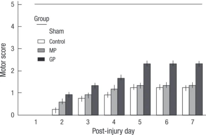
The time course of functional recovery, as measured by the Gale rating score, was recorded (Fig. 1). The results of the “motor func- tion score” evaluation are presented as a mean value with SEM (Table 1). The mean motor function scores at post-injury day 7 were 1.92 ± 0.2, 6 ± 0, 2.0 ± 0.3, and 3.17 ± 0.2 in group I to IV re- spectively. On the statistical analysis, the mean motor function scores of each group had a statistically significant difference (P = 0.01), which was tested for with a one-way ANOVA. The recov- ery of hindlimb function in the GP group was superior to that of the control and the MP groups (P = 0.01).
Inclined plane test
The results of the “inclined plane score” evaluation are present- ed as a mean value with SEM. The mean inclined plane angles were 28.7 ± 1.5°, 46 ± 1.0°, 27.7 ± 1.5°, and 31.3 ± 0.8° in group I
to IV respectively. On the statistical analysis, the mean inclined plane scores of each group had a statistical significance (P = 0.01).
Comparing the GP group with the MP group, there was also sta- tistical significance (P = 0.01).
Body weights changes
Most of the injured rats, except the sham operated rats, suffered from loss of body weight during the observation period. The mean changes for the control group and the MP group are larg- er than that for the GP group (P < 0.05, Table 1).
Effect of pregabalin on the caspase-3, bcl-2 and p38 MAPK expression
The relative optical densities for caspase-3 were 1.6 ± 0.16, 1.33
± 0.02, 1.63±0.2, and 0.97 ± 0.05 in group I-IV, respectively (Fig.
2A). The caspase-3 expression was significantly decreased in GP
group compared to the control and MP groups (f = 3.97, P < 0.05, Bonferroni post hoc analysis, P < 0.05, n = 10). The relative opti- cal densities for bcl-2 are 0.97 ± 0.27, 0.27 ± 0.04, 0.74 ± 0.17, and 0.8 ± 0.26 in group I-IV, respectively (Fig. 2B). However, it revealed no statistical difference between groups (f = 0.52, P = 0.67). The relative optical densities for phosphorylated p38 MAPK are 0.52
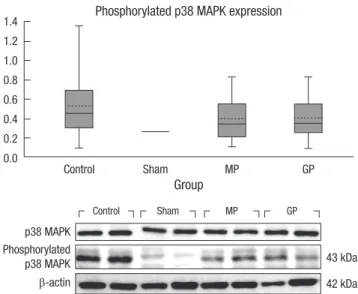
± 0.12, 0.25 ± 0.01, 0.39 ± 0.08, and 0.4 ± 0.08 in group I-IV, respec- tively (Fig. 3). The control group tends to express higher levels phosphorylated p38 MAPK than the other groups. However, there was no statistically significant difference (f = 0.65, P = 0.58).
Activation of the astrocytes and microglia
Except for the sham operated rats, the spinal cord of all the in- jured groups demonstrates the abundant activation of astro- cytes (Fig. 4). The number of GFAP positive cells was 47.3 ± 2.5, 21.5 ± 3.7, 32.5 ± 5.2, and 29.3 ± 2.5 in group I-IV, respectively.
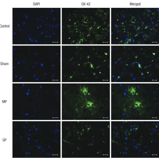
The control group demonstrate a statistically significant increase in the number of the activated astrocytes compared to that of the other groups (P < 0.05). In most specimens, the activated astrocytes were located around the injured site and recruitment was also observed from the adjacent gray and white matter. The mean number of OX-42 positive cells in the × 200 magnified specimen was 21.2 ± 1.2, 5.3 ± 2.4, 18.6 ± 2.3, and 19.6 ± 1.5 in group I-IV, respectively. The injured group demonstrated a sig- nificantly elevated number of activated microglia. However, there was no statistical difference among the injured groups (Fig. 5).
Fig. 2. (A) The effect of intraperitoneal pregabalin (30 mg/kg) on the expression of caspase-3 in contusive spinal cord injury. One-way ANOVA reveals significant differences between the groups for the expression of caspase-3 expression in the spinal cord tissue and the post hoc comparison (Bonferroni) test revealed that pregabalin treatment induced the least expression of caspase-3.This difference showed a statistical significance compared to the control group and the methylprednisolone treated group (P < 0.05, n = 10 for the experimental group, n = 3 for sham group). (B) The effect of intraperitoneal pregabalin (30 mg/kg) on the expression of bcl-2 in contusive spinal cord injury.
One-way ANOVA test and post hoc analysis reveal no significant difference between the groups for the expression of bcl-2 expression in the spinal cord tissue (f = 0.52, P = 0.67, n = 10 for the experimental group, n = 3 for the sham group).
4.0 3.5 3.0 2.5 2.0 1.5 1.0
5 4 3 2 1 0
Caspase-3 expression bcl-2 expression
Group Group
19 kDa 28 kDa
Caspase-3 bcl-2
42 kDa 42 kDa
β-actin β-actin
*
Control Sham MP GP Control Sham MP GP
Control Sham MP GP Control Sham MP GP
A B
Table 1. Behaviour assessments
Group I Group II Group III Group IV Motor Function score 1.92 ± 0.2 6 2.0 ± 0.3 3.17 ± 0.2 Inclined plane test (°) 28.7 ± 1.5 46.0 ± 1.0 27.8 ± 1.5 31.2 ± 0.8 Body weight change -11.5 ± 5.0 37.2±11.0 -62.3 ± 3.7 3.75 ± 6.5
*The post-injury 7 days scores were assessed. (-) means loss of body weight.
Fig. 1. Motor scores for the each group. The rats in all the groups show a sequential recovery of hindlimb function. At the postoperative 7th day, the pregabalin treated group show a more significant recovery compared to that of the other groups.
Horizontal line at motor score means the score in the sham-operated rats.
Motor score
Post-injury day
1 2 3 4 5 6 7
5
4
3
2
1
0
Control Sham Group
MP GP
DISCUSSION
Many diverse approaches for neuroprotection are being attempt- ed as details of the mechanisms of secondary injury after SCI have been disclosed in the last few years. Through our previous study, we were able to verify the possibility of pregabalin as a neuroprotector after SCI (14). In that study, we only presented the anti-apoptotic and anti-inflammatory effects by histopathol- ogy. The present study used not only functional outcomes but also more detailed immunohistochemistry to demonstrate the feasibility of pregabalin on SCI. First, we analyzed the anti-apop- totic and anti-inflammatory effects by measuring the expression of caspase-3, bcl-2 and p38 MAPK. These molecules are known to be key players in the processes of apoptosis and inflammation (4, 15, 16, 21). Following spinal cord injury, neurons and glial cells undergo apoptosis; and the subsequent demyelination by apoptosis of oligodendrocyte is a key feature of secondary spi- nal cord injury. Previous studies have demonstrated that this phenomenon peaks at 48 hr and 7 days after SCI (1, 2, 15). And excitotoxicity is known to be one of the major pathomechanisms to elicit the apoptosis of the neuron and the supporting glial cells in the spinal cord injury. Presynaptic release, disrupted neurons, 1.4
1.2 1.0 0.8 0.6 0.4 0.2 0.0
Phosphorylated p38 MAPK expression
Group
p38 MAPK
43 kDa 42 kDa Phosphorylated
p38 MAPK β-actin
Control Sham MP GP
Control Sham MP GP
Fig. 3. The effect of intraperitoneal injection of pregabalin (30 mg/kg) on the expres- sion of phosphorylated p38 MAPK in contusive spinal cord injury. The values were examined on post-injury day 7. The methylprednisolone and pregabalin treatment groups demonstrate a reduced expression of phosphorylated p38 MAPK as compared to that of the control group. However, ANOVA indicated no statistically significant dif- ference (f = 0.65, P = 0.58, n = 10 for the experimental group, n = 3 for the sham group). The following Bonferroni post hoc analysis also indicated no statistical difference between the groups.
DAPI
Control
Sham
MP
GP
GFAP Merged
Fig. 4. Immunofluorescence detection of the activated astrocyte labeled with anti- body GFAP. Abundant activation of astro- cytes is observed in the most of the spe- cimen of the injured groups. The control group demonstrate a higher number of activated astrocytes than that of the MP and the GP groups. The astrocytes are mostly located around the periphery of the injured site, and the astrocytes also show a tendency of recruitment from the surrounding gray and white matter (mag- nification × 200, scale bar 20 µm).
DAPI
Control
Sham
MP
GP
OX-42 Merged
Fig. 5. Immunofluorescence detection of the activated microglia labeled with anti- body OX-42. The injured group demon- strates a significantly elevated number of activated microglia. However, there was no statistical difference among the injured groups (magnification × 200, scale bar 20 µm).
axons and/or glial cells, and especially astrocytes and oligoden- drocytes, have been suggested as sources of glutamate in the gray matter and white matter. And subsequently these over-ex- pressed excitotoxic neurotransmitters can induce the apoptosis by means of the direct damage of the mitochondria and the in- duction of apoptosis inducing enzymes including caspases. In this study, we observed a statistically significant reduction of cas- pase-3 expression in the pregabalin-treated group. The reduced expression of caspase-3 in the pregabalin group might be relat- ed to the pharmacologic action of this drug. Pregabalin may re- duce extracellular glutamate concentration after SCI and this down-regulating effect of pregabalin was associated with rescu- ing cells from excitotoxicity. When we first designed this experi- ment, we decided to take the sample at the 7th day post-injury.
This was based on the previous references that have demonstrat- ed the apoptosis of neuronal damage after SCI persisted through the first week (2, 15). Although the present study demonstrated the statistical significant reduction of caspase-3 expression, we were not able to observe the significant differences in other pro- teins that are also related to apoptosis pathway. However, if we evaluate more of the acute stage samples (for example 24 hr af- ter injury), we look forward to finding more statistical significant
differences. We also measured phosphorylated p38 MAPK. p38 MAPK has been reported to have inflammation-modulating function at the level of transcription and translation (18, 19, 22).
In the central nervous system, the glia, and mainly microglia, upregulate the expression and activation of p38 MAPK in patho- logic conditions, and the subsequent activation of other cyto- kines such as IL-1, IL-6, TNF-α, and COX-2 (17, 23, 24). In the present investigation, although not statistically significant, the pregabalin treated group showed lower expression of phosphor- ylated p38 MAPK than that of the control group. This finding is further supported by the histologic differences noted with regard to the activation of microglia. During histopathologic evaluation, we focused on the observation of reactive astrocytosis and pre- gabalin effects on it. Astrocytes have been shown to regulate of the extracellular level of glutamate in synapses. This scavenger action on overexpressed glutamate is exerted by excitatory ami- no acid transporters (EAATs) and glutamine synthetase (25, 26).
In response to injury in the central nervous system, astrocytes change their appearance and they undergo a characteristic hy- pertrophy of their processes with the upregulation of interme- diate filaments that are composed of glial fibrillary acid protein, vimentin and nestin. These changes are known as gliosis and
they are a suitable reaction to overcome the pathologic condi- tion (25). Astrocyte proliferation after injury has been known to have both detrimental and beneficial effects (26, 27). Astrocyte scarring has long been considered as a major impediment to the regeneration of damaged axons. Chondroitin sulfate glyco- proteins are well known key molecules in inhibiting the regen- eration of axons (28). However, beneficial effects of astrocytes have recently been established as well. Astrocyte protective ac- tions are exerted by restoring altered homeostasis (the ionic and neurotransmitter balance) and re-establishing the anatomical blood-brain barrier (29). At later stages, astrocytes can aid in re- generation by production of a variety of cytokines such as TGF- β, glial cell line derived neurotrophic factor, β-FGF and VEGF (29, 30). Some of these growth factors can facilitate oligodendro- cyte precursor migration, proliferation and differentiation (30).
Taking these backgrounds of the astrocyte into account, we care- fully hypothesize that pregabalin may reduce excitotoxicity and minimize the need for astrocytic clearance of glutamate during the acute pathologic condition of SCI. Because astrocytes can provide positive effects, however, it remains to be seen if the low- er proliferation of astrocytes can provide long-term benefit.
In summary, we were able to get more promising results that might support the feasibility of pregabalin following SCI. Anti- inflammatory and anti-apoptotic effects of pregabalin were re- peatedly demonstrated by the histopathological and biochemi- cal methods. Further studies are needed to determine the safe- ty, ideal dosages, methods of administration and defining long- term results. We are encouraged, however, that pregabalin might be another neuroprotective agent for the treatment of spinal cord injury.
REFERENCES
1. Kwon BK, Tetzlaff W, Grauer JN, Beiner J, Vaccaro AR. Pathophysiology and pharmacologic treatment of acute spinal cord injury. Spine J 2004;
4: 451-64.
2. Dumont RJ, Okonkwo DO, Verma S, Hurlbert RJ, Boulos PT, Ellegala DB, Dumont AS. Acute spinal cord injury, Part I: pathophysiologic mecha- nisms. Clin Neuropharmacol 2001; 24: 254-64.
3. Fehlings MG, Baptiste DC. Current status of clinical trials for acute spi- nal cord injury. Injury 2005; 36 Suppl 2: B113-22.
4. Rowland JW, Hawryluk GW, Kwon B, Fehlings MG. Current status of acute spinal cord injury pathophysiology and emerging therapies: prom- ise on the horizon. Neurosurg Focus 2008; 25: E2.
5. Zhang Y, Bhavnani BR. Glutamate-induced apoptosis in primary corti- cal neurons is inhibited by equine estrogens via down-regulation of cas- pase-3 and prevention of mitochondrial cytochrome c release. BMC Neu- rosci 2005; 6: 13.
6. Xu GY, Liu S, Hughes MG, McAdoo DJ. Glutamate-induced losses of oli- godendrocytes and neurons and activation of caspase-3 in the rat spinal cord. Neuroscience 2008; 153: 1034-47.
7. Globus MY, Alonso O, Dietrich WD, Busto R, Ginsberg MD. Glutamate
release and free radical production following brain injury: effects of post- traumatic hypothermia. J Neurochem 1995; 65: 1704-11.
8. Park E, Velumian AA, Fehlings MG. The role of excitotoxicity in second- ary mechanisms of spinal cord injury: a review with an emphasis on the implications for white matter degeneration. J Neurotrauma 2004; 21:
754-74.
9. Káradóttir R, Cavelier P, Bergersen LH, Attwell D. NMDA receptors are expressed in oligodendrocytes and activated in ischaemia. Nature 2005;
438: 1162-6.
10. Joshi I, Taylor CP. Pregabalin action at a model synapse: binding to pre- synaptic calcium channel alpha2-delta subunit reduces neurotransmis- sion in mice. Eur J Pharmacol 2006; 553: 82-8.
11. Tassone DM, Boyce E, Guyer J, Nuzum D. Pregabalin: a novel gamma- aminobutyric acid analogue in the treatment of neuropathic pain, par- tial-onset seizures, and anxiety disorders. Clin Ther 2007; 29: 26-48.
12. Fehrenbacher JC, Taylor CP, Vasko MR. Pregabalin and gabapentin re- duce release of substance P and CGRP from rat spinal tissues only after inflammation or activation of protein kinase C. Pain 2003; 105: 133-41.
13. Million M, Wang L, Adelson DW, Roman F, Diop L, Taché Y. Pregabalin decreases visceral pain and prevents spinal neuronal activation in rats.
Gut 2007; 56: 1482-4.
14. Ha KY, Kim YH, Rhyu KW, Kwon SE. Pregabalin as a neuroprotector after spinal cord injury in rats. Eur Spine J 2008; 17: 864-72.
15. Keane RW, Kraydieh S, Lotocki G, Bethea JR, Krajewski S, Reed JC, Di- etrich WD. Apoptotic and anti-apoptotic mechanisms following spinal cord injury. J Neuropathol Exp Neurol 2001; 60: 422-9.
16. Citron BA, Arnold PM, Haynes NG, Ameenuddin S, Farooque M, Santa- cruz K, Festoff BW. Neuroprotective effects of caspase-3 inhibition on functional recovery and tissue sparing after acute spinal cord injury. Spine (Phila Pa 1976) 2008; 33: 2269-77.
17. Horiuchi H, Ogata T, Morino T, Chuai M, Yamamoto H. Continuous in- trathecal infusion of SB203580, a selective inhibitor of p38 mitogen-acti- vated protein kinase, reduces the damage of hind-limb function after tho- racic spinal cord injury in rat. Neurosci Res 2003; 47: 209-17.
18. Guo G, Bhat NR. p38alpha MAP kinase mediates hypoxia-induced mo- tor neuron cell death: a potential target of minocycline’s neuroprotective action. Neurochem Res 2007; 32: 2160-6.
19. Genovese T, Esposito E, Mazzon E, Muià C, Di Paola R, Meli R, Bramanti P, Cuzzocrea S. Evidence for the role of mitogen-activated protein kinase signaling pathways in the development of spinal cord injury. J Pharma- col Exp Ther 2008; 325: 100-14.
20. Gale K, Kerasidis H, Wrathall JR. Spinal cord contusion in the rat; behav- ioral analysis of functional neurologic impairment. Exp Neurol 1985; 88:
123-34.
21. Austin JW, Fehlings MG. Molecular mechanisms of Fas-mediated cell death in oligodendrocytes. J Neurotrauma 2008; 25: 411-26.
22. Obata K, Yamanaka H, Kobayashi K, Dai Y, Mizushima T, Katsura H, Fukuoka T, Tokunaga A, Noguchi K. Role of mitogen-activated protein kinase activation in injured and intact primary afferent neurons for me- chanical and heat hypersensitivity after spinal nerve ligation. J Neurosci 2004; 24: 10211-22.
23. Nakahara S, Yone K, Sakou T, Wada S, Nagamine T, Niiyama T, Ichijo H.
Induction of apoptosis signal regulating kinase 1 (ASK1) after spinal cord injury in rats: possible involvement of ASK1-JNK and -p38 pathways in neuronal apoptosis. J Neuropathol Exp Neurol 1999; 58: 442-50.
24. Kwak EK, Kim JW, Kang KS, Lee YH, Hua QH, Park TI, Park JY, Sohn YK.
The role of inducible nitric oxide synthase following spinal cord injury in rat. J Korean Med Sci 2005; 20: 663-9.
25. Anderson CM, Swanson RA. Astrocyte glutamate transport: review of properties, regulation, and physiological functions. Glia 2000; 32: 1-14.
26. Williams A, Piaton G, Lubetzki C. Astrocytes--friends or foes in multiple sclerosis? Glia 2007; 55: 1300-12.
27. Faulkner JR, Herrmann JE, Woo MJ, Tansey KE, Doan NB, Sofroniew MV. Reactive astrocytes protect tissue and preserve function after spinal cord injury. J Neurosci 2004; 24: 2143-55.
28. Barritt AW, Davies M, Marchand F, Hartley R, Grist J, Yip P, McMahon SB, Bradbury EJ. Chondroitinase ABC promotes sprouting of intact and injured spinal systems after spinal cord injury. J Neurosci 2006; 26: 10856- 67.
29. Buffo A, Rolando C, Ceruti S. Astrocytes in the damaged brain: molecu- lar and cellular insights into their reactive response and healing poten- tial. Biochem Pharmacol 2010; 79: 77-89.
30. Rosenstein JM, Krum JM. New roles for VEGF in nervous tissue-beyond blood vessels. Exp Neurol 2004; 187: 246-53.
AUTHOR SUMMARY
Pregabalin as a Neuroprotector after Spinal Cord Injury in Rats: Biochemical Analysis and Effect on Glial Cells
Kee-Yong Ha, Eugene Carragee, Ivan Cheng, Soon-Eok Kwon, and Young-Hoon Kim
Pregabalin was used as a neuroprotective agent after spinal cord injury (SCI). Here we compared the effects of pregabalin with methylprednisolone. 50 rats were divided into four groups; the control group, the sham operated group, the methylprednisolone treated group, and the pregabalin-treated group. The pregabalin-treated group showed improved mean motor function score and the inclined plane score. Caspase-3 and phosphorylated p38MAPK expression of the pregabalin group were lower than that of the control group. Bcl-2 expression was not different between the control and the treated groups. In histopathological analysis, pregabalin treated group demonstrated less proliferation of microglia and astrocyte. The above data suggest neuroprotective effects of pregabalin.