© 2012 The Korean Academy of Medical Sciences.
This is an Open Access article distributed under the terms of the Creative Commons Attribution Non-Commercial License (http://creativecommons.org/licenses/by-nc/3.0) which permits unrestricted non-commercial use, distribution, and reproduction in any medium, provided the original work is properly cited.
pISSN 1011-8934 eISSN 1598-6357
High Incidence of Rickets in Extremely Low Birth Weight Infants with Severe Parenteral Nutrition-Associated Cholestasis and Bronchopulmonary Dysplasia
Risk factors for rickets of prematurity have not been re-examined since introduction of high mineral formula, particularly in ELBW infants. We analyzed the incidence and the risk factors of rickets in extremely low birth weight (ELBW) infants. As a retrospective case- control study from 2004 to 2008, risk factors were analyzed in 24 patients with rickets versus 31 patients without. The frequency of rickets in ELBW infants was 24/55 (44%).
Infants with rickets were diagnosed at 48.2 ± 16.1 days of age, and improved by 85.3 ± 25.3 days. By radiologic evaluation, 29% were grade 1 rickets, 58% grade 2 and 13% grade 3. In univariate analysis, infants with rickets had significantly higher incidence of patent ductus arteriosus, parenteral nutrition associated cholestasis (PNAC), severe PNAC and moderate/severe bronchopulmonary dysplasia (BPD). In multiple regression analysis, after adjustment for gestation and birth weight, rickets significantly correlated with severe PNAC and with moderate/severe BPD. Serum peak alkaline phosphatase levels were significantly elevated in rickets (P < 0.001). In ELBW infants, the incidence of rickets of prematurity remains high and the incidence of severe PNAC and moderate/severe BPD was significantly increased 18 and 3 times, respectively.
Key Words: Cholestasis; Bronchopulmonary Dysplasia; Risk Factors; Rickets; Infant, Extremely Low Birth Weight
Soon Min Lee, Ran Namgung,
Min Soo Park, Ho Sun Eun, Kook In Park, and Chul Lee
Division of Neonatology, Department of Pediatrics, Severance Children’s Hospital, Yonsei University College of Medicine, Seoul, Korea
Received: 15 June 2012 Accepted: 20 September 2012 Address for Correspondence:
Ran Namgung, MD
Division of Neonatology, Department of Pediatrics, Severance Children’s Hospital, Yonsei University College of Medicine, 50 Yonsei-ro, Seodaemun-gu, Seoul 120-752, Korea Tel: +82.2-2228-2050, Fax: +82.2-313-4614 E-mail: [email protected]
This research was supported by Basic Science Research Program through the National Research Foundation of Korea funded by the Ministry of Education, Science and Technology (No. 2011- 0014207).
http://dx.doi.org/10.3346/jkms.2012.27.12.1552 • J Korean Med Sci 2012; 27: 1552-1555
ORIGINAL ARTICLE
Pediatrics
INTRODUCTION
In the 1980s, the prevalence of rickets in extremely low birth weight (ELBW) infants was around 50% and was 17% for frac- tures (1, 2). After introduction of a high mineral containing diet in the late 1980s, the prevalence decreased to 30% or lower (3, 4). There is little information, however, beyond 1985, concern- ing the incidence of rickets in extreme prematurity and factors that relate to its occurrence.
The pathogenesis of rickets in preterm infants is considered multifactorial. In addition to inadequate mineral intake as a ma- jor contributing factor (5), chronic co-morbidities, like chronic lung disease, short bowel syndrome, and parenteral nutrition associated cholestasis (PNAC) could also be related factors (6).
Medications used in the neonatal intensive care unit (NICU), such as steroids, methylxanthines, and diuretics could theoreti- cally increase the risk of inadequate bone mineralization (7-9).
Other risk factors, such as immobilization from sepsis, cerebral pathology, muscular disorders and paralysis are associated with bone disease of prematurity (10). Since mineral enriched for- mula for the preterms began 30 yr ago, it is the standard prac- tice to provide minerals in an attempt to reach in utero mineral
accretion rates. In theory, under ideal conditions, the advent- age of high mineral intake formulas should be associated with a marked reduction of rickets. However, it is often practically dif- ficult to achieve ideal nutrition support in sick ELBW infants, the current incidence of rickets in ELBW infants is unknown, and the relationship of the severity of overall illness and degree of prematurity in this population is unclear.
The aim of this study was to characterize the risk factors for development of rickets in ELBW infants fed with high mineral containing preterm formula.
MATERIALS AND METHODS
As a retrospective case-control study, medical records of 55 ELBW infants admitted into the Severance Children’s Hospital NICU from 2004 to 2008 that survived until 1 month of age were reviewed. All infants had their serum alkaline phosphatase (ALP), calcium (Ca) and phosphate (P) levels reported weekly and bone screen radiography at 1 month of age. For nutritional care, fortified human milk (Ca 131 mg/100 kcal, P 71 mg/100 kcal) or a mineral fortified preterm formula (Ca 112-141 mg/kcal, P 52-63 mg/kcal) was used. During parenteral nutrition, Ca, P
Lee SM, et al. • High Incidence and Risk Factors of Rickets in ELBW Infants
http://jkms.org 1553
http://dx.doi.org/10.3346/jkms.2012.27.12.1552
and multivitamins with trace minerals were added to parenter- al nutrition, appropriately.
Rickets of prematurity was defined by radiologic screening criteria of Koo et al. (12). Grade 1 was defined as a loss of dense white line, increased lucency of bone shaft, and thinning of cor- tex; Grade 2 showed irregularity and fraying of a metaphysis with splaying and cupping; and Grade 3 presented evidence of fractures.
As co-morbidities, hyaline membrane disease (HMD), pat- ent ductus arteriosus (PDA), necrotizing enterocolitis (NEC;
using the modified Bell’s staging criteria), parenteral nutrition associated cholestasis (PNAC, defined as direct bilirubin [DB]
greater than 2.0 mg/dL), bronchopulmonary dysplasia (BPD;
using NIH classification), retinopathy of prematurity (ROP, in- cluded above stage 2), and intraventricular hemorrhage (IVH, above grade 3) were analyzed. Severe PNAC was defined as ele- vation of DB greater than 4.0 mg/dL for more than 1 month and aspartate aminotransferase (AST) and alanine aminotranster- ase (ALT) greater than 60 IU/L and 35 IU/L, respectively. Mod- erate/severe BPD was defined in infants requiring either 28 days of supplemental oxygen therapy with oxygen, nasal continuous positive airway pressure (CPAP) or positive pressure ventilation at 36 weeks postmenstrual age or discharge, whichever came first, whose gestational age was younger than 32 weeks, or at 56 days postnatal age or discharge, whichever came first, in infants whose gestational age was older than 32 weeks. Statistics were calculated with the Student t-test and chi-square test for means and frequencies. Numeric data are presented as mean and stan- dard deviation because the data were normally distributed. Cal- culations were performed with SPSS software version 17.0 (SPSS, Chicago, IL, USA). P values less than 0.05 were considered sta- tistically significant.
Ethics statement
The present retrospective review of database in this study was
approved by the institutional review board of the Yonsei Univer- sity Health System, Severance Hospital (Approval No., 4-2012- 0157). The informed consent was exempted by the board.
RESULTS
Twenty-four ELBW infants (26.3 ± 1.9 weeks, 792 ± 99 g) were diagnosed as rickets of prematurity, and 31 infants without rick- ets (27.2 ± 2.3 weeks, 871 ± 93 g) were the control group. There was no significant difference of gestation and birth weight be- tween the two groups. Rickets of prematurity was diagnosed in 43.9% of ELBW infants and fracture in 13%. For the rickets group, 7 infants (29%) showed G1, 14 infants (58%) G2 and 3 infants (13%) G3. Rickets of prematurity was diagnosed at 48.2 ± 16.1 days of life and improved by 85.3 ± 25.3 days of life.
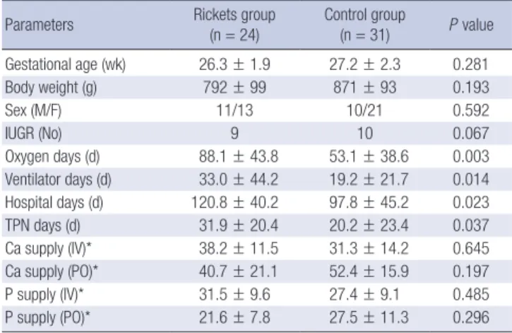
In patient characteristics such as mean gestational ages, birth weight, gender, and frequency of intrauterine growth retarda- tion (IUGR), there was no difference between the rickets and control groups. The cumulative amounts (either enteral or par- enteral) of Ca and P administration during hospitalization were not different between the groups. For the rickets group in com- parison with the control group, mean days on oxygen and ven- tilator use were significantly longer (P < 0.05). The duration of hospitalization and parenteral nutrition were significantly lon- ger in the rickets group versus the control group (Table 1).
In univariate analysis, ELBW infants with rickets had a signif- icantly higher occurrence of PDA (P = 0.025), PNAC (P = 0.041), severe PNAC (P = 0.013), BPD (P = 0.019), and moderate/se- vere BPD (P = 0.012) (Table 2). Common neonatal illnesses such as HMD, air leak, NEC, ROP, IVH and periventricular leucoma- lacia (PVL) were not different between the groups.
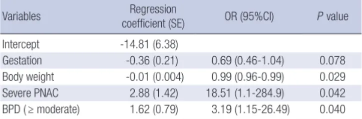
In multiple regression analysis after adjustment of gestation and birth weight, rickets in ELBW infants significantly correlat-
Table 1. Patient characteristics of ELBW infants between the rickets and control group
Parameters Rickets group
(n = 24)
Control group
(n = 31) P value Gestational age (wk) 26.3 ± 1.9 27.2 ± 2.3 0.281
Body weight (g) 792 ± 99 871 ± 93 0.193
Sex (M/F) 11/13 10/21 0.592
IUGR (No) 9 10 0.067
Oxygen days (d) 88.1 ± 43.8 53.1 ± 38.6 0.003 Ventilator days (d) 33.0 ± 44.2 19.2 ± 21.7 0.014 Hospital days (d) 120.8 ± 40.2 97.8 ± 45.2 0.023
TPN days (d) 31.9 ± 20.4 20.2 ± 23.4 0.037
Ca supply (IV)* 38.2 ± 11.5 31.3 ± 14.2 0.645 Ca supply (PO)* 40.7 ± 21.1 52.4 ± 15.9 0.197
P supply (IV)* 31.5 ± 9.6 27.4 ± 9.1 0.485
P supply (PO)* 21.6 ± 7.8 27.5 ± 11.3 0.296
*mg/kg/d. IUGR, intrauterine growth retardation; TPN, total parenteral nutrition; Ca, calcium; P, phosphorus; IV, intravenous; PO, per os.
Table 2. Comparison of co-morbidities between the rickets and control group Co-morbidities Rickets group
(n = 24)
Control group
(n = 31) P value
HMD 100% 100% 1
PDA 67% 29% 0.025
Air leak 4% 6% 0.495
PNAC 54% 23% 0.041
Severe PNAC* 33% 6% 0.013
BPD 89% 71% 0.019
Moderate/severe BPD 54% 26% 0.012
NEC 13% 3% 0.324
ROP 5% 3% 0.095
IVH 21% 19% 0.328
PVL 29% 10% 0.054
Death 8% 3% 0.245
*DB ≥ 4.0 mg/dL for move than 1 month, and AST ≥ 60 IU/L, ALT 35 IU/L.HMD, hya- line membrane disease; PDA, patent ductus arteriosus; PNAC, parenteral nutrition associated cholestasis; BPD, bronchopulmonary dysplasia; NEC, necrotizing entero- colitis; ROP, retinopathy of prematurity; IVH, intraventricular hemorrhage; PVL, peri- ventricular leukomalacia.
Lee SM, et al. • High Incidence and Risk Factors of Rickets in ELBW Infants
1554 http://jkms.org http://dx.doi.org/10.3346/jkms.2012.27.12.1552 ed with severe PNAC (OR 18.5; 95% CI, 1.1-285; P = 0.042) and
moderate/severe BPD (OR 3.2; 95% CI, 1.2-26.5; P = 0.04) (Ta- ble 3).
The mean level of peak ALP was significantly higher in the rickets group than the control group (952.2 ± 413.8 vs 524.7 ± 158.2 IU/L), while the mean level of the lowest Ca and P levels were not different between groups (Table 4).
DISCUSSION
In the 1980s, fortified mineral formulas were introduced to re- duce the rates of rickets and fractures in very low birth weight (VLBW < 1,500 g) infants (11). Reports at that time indicated that up to 30% of VLBWs had rickets with fractures (12).
With advances in preterm nutrition, especially with the intro- duction of mineral fortified formulas, theoretically the incidence of rickets of prematurity should have dropped (3, 4). However, for increasing the survival of ELBW infants, significant co-mor- bid conditions could affect actual mineral intake which might mitigate the effect of high mineral fortification (13).
This is the first study to focus on the incidence of rickets in ELBW infants after 2 decades of fortified formula use in Korea.
It is also the first to characterize and compare risk factors and neonatal morbidities for rickets of prematurity in ELBW infants.
We found that the incidence of radiologic rickets in ELBW in- fants was extremely high at 44% (24/55) due to increased surviv- al, and the incidence of significantly increased 18 times and 3 times for severe PNAC and moderate to severe BPD, respective- ly. Early recognition and treatment of active rickets may prevent subsequent bone damage and fractures.
Factors commonly thought to relate to rickets of prematurity include mismatch of postnatal intake of minerals compared to intrauterine mineral transfer, low bone loading effects in the
NICU, intrauterine growth restriction, chorioamnionitis, steroids, methylxanthines and diuretic usage and necrotizing enteroco- litis (7-10, 14-16). Inadequate mineral nutrient (calcium and phosphorous) intake appears to play a major role in causing rickets of prematurity (10, 17). Prolonged parenteral nutrition (PN) and delayed enteral nutrition complicate delivery of ade- quate mineral intake and would be particularly acute in ELBW infants (10). It is standard practice in NICU to provide high min- eral preterm formula and human milk fortifier as an attempt to reach in utero mineral accretion rate, including the use of mini- mal early enteral feeding, which appears to improve bone mass at term (3, 17-19). However, in ELBW infants, co-morbid illness- es often preclude full enteral tolerance and the actual delivery of mineral nutrients (13). Further, when PN is needed, the pro- vision of minerals in PN can be limited by solubility issues com- pounded by fluid restriction in sick infants (3).
Specific chronic co-morbidities, particularly chronic lung disease and short bowel syndrome which is generally associat- ed with prolonged parenteral nutrition and PNAC, are poten- tially serious risk factors for poor bone mineralization (6, 20, 21).
Thus our findings of markedly higher risk of rickets in ELBW in- fants with PNAC (18 times) and BPD (3 times) support these associations.
The limitations of this study are a small sample size of a single institution and a retrospective design. In conclusion, there is a high incidence (44%) of rickets in ELBW infants, which shows an 18-fold increase in severe PNAC and a 3-fold increase in moderate to severe BPD. We suggest that in ELBW infants with severe PNAC or moderate/severe BPD, aggressive prevention or treatment for rickets and fractures can be instituted earlier in the course of management.
REFERENCES
1. Lyon AJ, McIntosh N, Wheeler K, Williams JE. Radiological rickets in ex- tremely low birthweight infants. Pediatr Radiol 1987; 17: 56-8.
2. McIntosh N, Livesey A, Brooke OG. Plasma 25-hydroxyvitamin D and rickets in infants of extremely low birthweight. Arch Dis Child 1982; 57:
848-50.
3. Greer FR. Osteopenia of prematurity. Annu Rev Nutr 1994; 14: 169-85.
4. MacDonald MG, Seshia MM, Mullett MD. Avery’s neonatology: patho- physiology & management of the newborn. 6th ed. Philadelphia: Lip- pincott Williams & Wilkins, 2005.
5. Weber G, Guarneri MP, Corbella E, Gallia P, Chiumello G. Osteopenia in premature children: an emerging problem. Minerva Pediatr 1989; 41:
347-52.
6. Ferrone M, Geraci M. A review of the relationship between parenteral nutrition and metabolic bone disease. Nutr Clin Pract 2007; 22: 329-39.
7. Venkataraman PS, Han BK, Tsang RC, Daugherty CC. Secondary hyper- parathyroidism and bone disease in infants receiving long-term furose- mide therapy. Am J Dis Child 1983; 137: 1157-61.
8. Weiler HA, Wang Z, Atkinson SA. Dexamethasone treatment impairs Table 3. Multiple regression analysis predicting rickets of prematurity
Variables Regression
coefficient (SE) OR (95%CI) P value
Intercept -14.81 (6.38)
Gestation -0.36 (0.21) 0.69 (0.46-1.04) 0.078
Body weight -0.01 (0.004) 0.99 (0.96-0.99) 0.029 Severe PNAC 2.88 (1.42) 18.51 (1.1-284.9) 0.042 BPD ( ≥ moderate) 1.62 (0.79) 3.19 (1.15-26.49) 0.040 SE, standard errors; BPD, bronchopulmonary dysplasia; PNAC, parenteral nutrition associated cholestasis.
Table 4. Comparison of biochemical factors between rickets and control group
Items Rickets group
(n = 24) Control group
(n = 31) P value Peak ALP (IU/L) 952.2 ± 413.8 524.7 ± 158.2 < 0.001
Lowest Ca (mg/dL) 7.86 ± 0.77 8.01 ± 0.79 0.078
Lowest P (mg/dL) 2.56 ± 0.64 2.92 ± 0.96 0.118
ALP, alkaline phosphatase; Ca, calcium; P, phosphorus.
Lee SM, et al. • High Incidence and Risk Factors of Rickets in ELBW Infants
http://jkms.org 1555
http://dx.doi.org/10.3346/jkms.2012.27.12.1552
calcium regulation and reduces bone mineralization in infant pigs. Am J Clin Nutr 1995; 61: 805-11.
9. Zanardo V, Dani C, Trevisanuto D, Meneghetti S, Guglielmi A, Zacchel- lo G, Cantarutti F. Methylxanthines increase renal calcium excretion in preterm infants. Biol Neonate 1995; 68: 169-74.
10. Harrison CM, Johnson K, McKechnie E. Osteopenia of prematurity: a national survey and review of practice. Acta Paediatr 2008; 97: 407-13.
11. American Academy of Pediatrics Committee on Nutrition. American Academy of Pediatrics Committee on Nutrition: nutritional needs of low-birth-weight infants. Pediatrics 1985; 75: 976-86.
12. Koo WW, Sherman R, Succop P, Krug-Wispe S, Tsang RC, Steichen JJ, Crawford AH, Oestreich AE. Fractures and rickets in very low birth weight infants: conservative management and outcome. J Pediatr Orthop 1989;
9: 326-30.
13. Rusk C. Rickets screening in the preterm infant. Neonatal Netw 1998; 17:
55-7.
14. Cakir M, Mungan I, Karahan C, Can G, Okten A. Necrotizing enterocoli- tis increases the bone resorption in premature infants. Early Hum Dev
2006; 82: 405-9.
15. Holland PC, Wilkinson AR, Diez J, Lindsell DR. Prenatal deficiency of phosphate, phosphate supplementation, and rickets in very-low-birth- weight infants. Lancet 1990; 335: 697-701.
16. Ryan S, Congdon PJ, James J, Truscott J, Horsman A. Mineral accretion in the human fetus. Arch Dis Child 1988; 63: 799-808.
17. Horsman A, Ryan SW, Congdon PJ, Truscott JG, James JR. Osteopenia in extremely low birthweight infants. Arch Dis Child 1989; 64: 485-8.
18. Rigo J, Pieltain C, Salle B, Senterre J. Enteral calcium, phosphate and vi- tamin D requirements and bone mineralization in preterm infants. Acta Paediatr 2007; 96: 969-74.
19. Steichen JJ, Gratton TL, Tsang RC. Osteopenia of prematurity: the cause and possible treatment. J Pediatr 1980; 96: 528-34.
20. Miller ME. The bone disease of preterm birth: a biomechanical perspec- tive. Pediatr Res 2003; 53: 10-5.
21. Vestergaard P. Bone loss associated with gastrointestinal disease: preva- lence and pathogenesis. Eur J Gastroenterol Hepatol 2003; 15: 851-6.