Inhalation Therapy of Calcitonin Relieves Osteoarthritis of the Knee
This study was conducted to determine if nasal salmon calcitonin has additional beneficial effects on clinical symptoms, serum NO, IL-1β, matrix metalloproteinase 3, urinary C-terminal telopeptide type II collagen (CTX-II) levels and MRI findings in knee
osteoarthritis (OA) when used concomitantly with exercise therapy. Fifty female patients with knee OA were randomized into two groups. The first group (n = 30) received 200 IU/
day nasal salmon calcitonin and a home exercise program; the second group (n = 20) received a home exercise program for 6 months. Compared with baseline, while significant improvements were observed in visual analogue scale (VAS), WOMAC pain, physical function scores, 20-m walking time (P < 0.001) and WOMAC stiffness score (P = 0.041) in the first group, walking and resting VAS, and WOMAC physical function scores were improved (P = 0.029) in the second group after treatment. Significantly increased levels of serum NO and urinary CTX-II (P < 0.001) and significant improvements in the area of medial femoral condyle (P < 0.05) were noted only in the first group. There were
significant differences in VAS activation values (P = 0.032) and NO levels (P < 0.001) in the favor of the first group. In conclusion, nasal salmon calcitonin may have possible
chondroprotective effects besides its known effects on symptoms in patients with knee OA.
Key Words: Nasal Salmon Calcitonin; Knee Osteoarthritis; Exercise Onur Armagan1, Dilek Kaya Serin2,
Cüneyt Calisir3, Ali Dokumacioglu4, Merih Ozgen1, Setenay Oner5, and Ozkan Alatas4
1Department of Physical Therapy and Rehabilitation, Eskisehir Osmangazi University Faculty of Medicine;
2Department of Physical Therapy and Rehabilitation, Eskisehir State Hospital; Departments of 3Radiology,
4Biochemistry, and 5Biostatistics, Eskisehir Osmangazi University Faculty of Medicine, Eskisehir, Turkey
Received: 8 February 2012 Accepted: 27 August 2012 Address for Correspondence:
Onur Armagan, MD
Department of Physical Therapy and Rehabilitation, Eskisehir Osmangazi University Faculty of Medicine, Meselik 26480 Eskisehir, Turkey
Tel: +902222392979/2457, Fax: +902222393774 E-mail: [email protected]
http://dx.doi.org/10.3346/jkms.2012.27.11.1405 • J Korean Med Sci 2012; 27: 1405-1410 Musculoskeletal Disorders
INTRODUCTION
Osteoarthritis (OA) is a chronic degenerative disease character- ized by the loss of cartilage in the joints covered by synovia. Ma- trix homeostasis of the cartilage is maintained by the balance of synthesis and degeneration of the matrix, which is produced by chondrocytes. Both synthesis and degeneration are under the control of messenger proteins, growth factors and cytokines.
While growth factors induce the production of connective tissue in general, cytokines lead to the matrix destruction (1). Major cytokines triggering the cartilage destruction are Interleukin 1β (IL-1β) and tumor necrosis factor alpha (TNF-α), both of which are secreted from synovial macrophages. Interleukin 1β de- creases the prostaglandin synthesis, impairs repair of the carti- lage matrix damage and facilitates the secretion of the matrix metalloproteinase (MMPs). In particular, MMP-3 has a major role in OA (2). On the other hand, several cytokines, including NO, are secreted from chondrocytes with the stimulation of IL- 1β . Moreover, increased expression of NO and thus increased ratio of nitrite/nitrate were found in OA (1).
Collagen type II (CTX-II) is the main structural component of the cartilage tissue. It was shown that urinary excretion of the degradation products of CTX-II is increased in patients with OA
and it correlates with the progression of articular damage (3) . Experimental and clinical observations suggest that structural integrity of the articular cartilage depends on normal subchon- dral bone turnover and healthy function of chondrocytes, as well as normal biomechanical stresses (4). Degenerative process in OA is not limited to the cartilage. Given that there is a strong re- lationship between subchondral bone and articular cartilage, an optimal therapeutic agent should regulate the metabolic activi- ty of both the bone and the cartilage (4). There are some studies carried out and still ongoing on preventing the OA-associated articular damage and developing an effective pharmacological agent for the treatment of OA. Antiresorptive agents have been shown to act both on bone and cartilage and they were proposed to be considered as OA-modifying agents (5).
Recent in vivo and in vitro experiments indicated that calci- tonin, an antiresorptive agent, acts both on cartilage and sub- chondral bone, decreases the severity of the cartilage lesions in OA, and involves in altered biochemical content and supramo- lecular organization of the cartilage matrix (6-9). Moreover, cal- citonin showed an analgesic effect on bone pain associated with osteoporotic vertebral fractures, Paget’s disease or other mus- culoskeletal disorders (10).
Exercise is a widely used non-pharmacological treatment
modality for OA and it was found effective in a vast number of studies (11, 12). However, given that calcitonin has analgesic and beneficial effects on cartilage and subchondral bone, we hypothesized that it may be a potential therapeutic option for the treatment of OA. Therefore, we aimed to determine if nasal salmon calcitonin treatment has additional beneficial effects on clinical symptoms, cartilage degeneration-associated bio- chemical parameters, and magnetic resonance image (MRI) findings in patients with knee OA when used concomitantly with the exercise therapy.
MATERIALS AND METHODS
Female patients with the diagnosis of primary knee OA accord- ing to the ACR criteria were included to the study. All patients were postmenopausal. Informed consent was obtained from all patients. Kellgren Lawrence radiological stage II or III patients with ACR criteria-based bilateral OA and knee pain were in- cluded to the study. Patients with a previous lower extremity surgery, intraarticular injections or physical therapy for knee within the last year, any previous calcitonin treatment, allergy to calcitonin, peripheral or central neurological disorder, or se- vere cardiac disease and incorporated patients were excluded from the study.
After a detailed anamnesis and physical examination, com- plete blood count (CBC), erythrocyte sedimentation rate (ESR), C-reactive protein (CRP) and rheumatoid factor (RF) levels were measured and the tests of liver and kidney functions were per- formed in all patients included to the study. In addition, conven- tional standing anteroposterior (AP) and lateral knee radiogra- phies were taken.
The present study was designed as a prospective, random- ized, single-blind and controlled clinical study. At the begin- ning of the study, the first and second groups consisted of 32 and 23 patients, respectively. However, 2 patients from the first group and 3 from the second group were lost at follow-up, leav- ing 30 and 20 patients respectively in each group.
Treatment protocol
The first group of patients (n = 30) received 200 IU/day nasal salmon calcitonin (Miacalcic nasal 200, Novartis, Basel, Swit- zerland) by inhalation and a home exercise program, and the second group (n = 20) received a home exercise program for 6 months. Home exercise program was given in the form of an illustrated brochure. Quadriceps isometric and isotonic strength- ening, joint range of motion (ROM), and hamstring stretching exercises were planned as once daily, with 2 sets of 20 repeti- tions and 2 min rests between the sets. Proper practical instruc- tions were given to all patients. During the 6 months follow-up, compliance of the patients to the treatment program was as- sessed by weekly phone calls.
Evaluations
All patients were evaluated before and after the treatment pro- tocol by a blinded investigator with respect to the following pa- rameters. Visual Analogue Scale (VAS), Western Ontario and McMaster Universities Osteoarthritis Index (WOMAC) and 20-m walking time were used for the clinical assessment. The pain at rest, walking and activation were assessed from 0 (no pain) to 10 (worst possible pain) on VAS scale. 20-m walking time was assessed with having all patients walking on the same straight ground, and using the same standard words and the same chro- nometer. The WOMAC consists of 24 items divided into 3 sub- scales: pain, stiffness and physical function. Global score has a range of 0 (no symptom) to 96 (worst symptoms), with standard- izing the score to have a range of 0 to 100.
Biochemical evaluations
From all patients a 6-7 mL of blood sample was taken twice, be- fore and after the treatment protocol. After centrifugation of the blood, serum samples were collected and stored at -80°. Nitric oxide, IL-1β and MMP-3 levels were determined in serum sam- ples.
Nitric oxide levels were measured manually by Griess Reac- tion Assay (Shimadzu UV-1201, Kyoto, Japan). In vivo stabile end-products of the NO are nitrite and nitrate, the levels of which are used as an index of NO production. The levels of these end- products based on cadmium reaction were determined spec- trophotometrically at a wavelength of 545 nm and NO levels were calculated in µM/L. Interleukin 1β levels were measured using a chemiluminescence assay (IMMULITE, Siemens Health- care Diagnostics GmbH, Erlangen, Germany). Serum MMP-3 levels were analyzed using a quantitative sandwich ELISA ac- cording to the manufacturer’s instructions (R&D Systems Inc, Minneapolis, USA). Absorbency of the samples read at 450 nm;
MMP-3 levels were automatically calculated by the device and expressed as ng/mL. CTX-II levels in the 24 hr urine samples were measured with using a commercially available ELISA kit (Immundiagnostik AG, Bensheim, Germany); the results divid- ed by the serum creatinine concentration and were expressed as ng/mM.
Radiological evaluations
Magnetic resonance images (MRIs) of the knees with more sig- nificant symptoms were obtained from all patients, with em- ploying a cartilage sequence. All the analyses were performed on a 1.5 Tesla MRI unit (VB33D Vision Plus; Siemens, Erlangen, Germany) using an extremity coil for signal reception and de- livery. FS 3D FLASH MRI parameters were defined as TR/TE:
60/11 and FA: 40°; and gradient-echo images were obtained in axial, sagittal and coronal planes. Wide slice thickness was 65 mm in axial, 70 mm in sagittal and 80 mm in coronal planes.
Slice thickness was 2.5 mm for axial, sagittal and coronal planes.
An imaging area of 25 cm, a spatial resolution of 1.2 × 1 mm and a matrix of 256 × 192 were used to minimize the artifacts from the contralateral knee. Sequence imaging time was 6.28 min and fat suppression prolonged the total scan time about 3 min.
MRI scans were read by a blinded musculoskeletal radiolo- gist. In all patients, 4 articular surfaces were identified on knee:
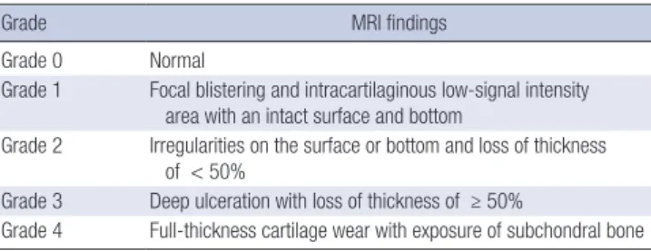
medial and lateral femoral condyles (MFC and LFC), and me- dial and lateral tibial plateaus (MTP and LTP). Each single plane was evaluated and graded for the presence and appearance of articular cartilage defects using a standard arthroscopic grading scheme adapted to MR imaging (SPGR). In the case of the mul- tiple cartilage defects with different grades on any articular sur- face, the highest grade was considered as the grade of the carti- lage defect (Table 1) (13).
Statistical analysis
Statistical procedures were performed using PASW 18.0 and SigmaStat 3.5 software. Normally distributed continuous quan- titative data were expressed as n, mean and standard deviation;
Kolmogorov-Smirnov test were used to for normality test and not normally distributed ones as n, median (25%-75% quartile);
and qualitative data as n. Repeated and independent measure- ments with not normally distributed variables were analyzed by using Wilcoxon, Mann-Whitney U test and those with normally distributed variables were analyzed by using t test. Chi-square analyses were used for independent categorical variables and Marginal Homogeneity test for dependent categorical variables.
Ethics statement
The study was conducted in full compliance with the amended Declaration of Helsinki, following approval from the institution-
al review board of Eskisehir Osmangazi University (31-07-2009/
332). Informed consent was obtained from all patients.
RESULTS
Two patients from the first group and three from the second group were lost at follow-up and data of these patients were not included to the analysis. Demographical characteristics of the patients did not differ significantly between the two groups (P = 0.074 between P = 0.807) (Table 2).
Before the treatment, there were no differences between the groups in clinical parameters, biochemical analysis and MRI findings. At the post treatment evaluation, significant improve- ments were noted in walking, activity and resting VAS scores, WOMAC pain and physical function scores, and 20-m walking time (P < 0.001) and in WOMAC stiffness score (P = 0.041) of the patients in the first group. On the other hand, walking and resting VAS scores, and WOMAC physical function score (P = 0.029) were significantly improved compared to the pretreat-
Table 1. Grading of the cartilage defects by SPRG MRI
Grade MRI findings
Grade 0 Normal
Grade 1 Focal blistering and intracartilaginous low-signal intensity area with an intact surface and bottom
Grade 2 Irregularities on the surface or bottom and loss of thickness of < 50%
Grade 3 Deep ulceration with loss of thickness of ≥ 50%
Grade 4 Full-thickness cartilage wear with exposure of subchondral bone
Table 2. Comparison of demographic characteristic of patients group Parameters Group I (n = 30)
(mean ± SD) Group II (n = 20)
(mean ± SD) P value
Age (yr) 63.30 ± 6.45 59.85 ± 6.67 0.074
Height (cm) 156.77 ± 5.90 159.60 ± 5.81 0.101
Weight (kg) 75.80 ± 7.49 79.15 ± 8.89 0.157
BMI (kg/m2) 30.85 ± 3.66 31.14 ± 4.26 0.799 Disease duration (yr) 4.00 ± 2.12 3.5 ± 1.64 0.807 KGL scale
Stage 2
Stage 3 20
9 12
8 0.517
0.109
Table 3. Inter- and intra-group comparisons of the clinical evaluation parameters Parameters Group 1 (n = 30)
Median (25%-75%) Group 2 (n = 20)
Median (25%-75%) P value VAS rest
Pre-treatment Post-treatment P value
2 (1-5) 0 (0-1.25)
< 0.001
2 (1-3) 1 (0-2) 0.032
0.125 0.201 VAS activation
Pre-treatment Post-treatment P value
6 (5-7.25) 4 (2-5.25)
< 0.001
7 (6-8) 5 (4.25-8)
0.089
0.063
VAS walking Pre-treatment Post-treatment P value
8 (6.75-9) 5 (3-7)
< 0.001
8 (6.25-9) 6 (4-8)
0.021
0.520
WOMAC pain Pre-treatment Post-treatment P value
10.5 (8-11.25) 5 (2-7.25)
< 0.001
11 (9-13) 6.5 (5-10.75)
0.065
0.730
WOMAC stiffness Pre-treatment Post-treatment P value
3 (1-4) 1.5 (0-3) 0.041
3 (0-4) 2.5 (0-4) 0.352
0.098
20 m. walking time (m/sn) Pre-treatment Post-treatment P value
19.50 (18-22) 18 (17-20)
< 0.001
18.50 (16-20) 18 (17-20)
0.635 Group 1 (n = 30)
Mean ± SD
Group 2 (n = 20)
Mean ± SD P value VAS activation
Post-treatment 4.13 ± 2.36 5.70 ± 2.66 0.032 VAS walking
Post-treatment 5.20 ± 2.62 5.80 ± 2.61 0.251 WOMAC pain
Post-treatment 5.30 ± 3.88 7.70 ± 4.75 0.105 WOMAC physical function
Pre-treatment Post-treatment P value
40.57 ± 16.72 20.70 ± 17.27
< 0.001
39.85 ± 13.90 31.75 ± 21.68
0.029
0.763 0.075
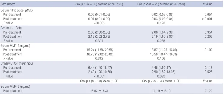
ment status in patients of the second group (Table 3). Signifi- cant increases in serum NO and urinary CTX-II levels (P < 0.001) and significant improvements in MFC area (P = 0.012) were found only in the first group of patients (Tables 4, 5). Compari- son of the groups revealed significant differences in VAS activity scores (P = 0.032) and serum NO levels (P < 0.001) in the favor of the first group (Tables 3, 4).
DISCUSSION
The aim of the OA treatment was to decrease the pain severity and functional limitation, as well as to slow the progression of the cartilage damage. The present study was conducted to deter- mine if nasal salmon calcitonin treatment has additional bene- ficial effects when used concomitantly with the exercise thera- py. In patients included to the present study, although exercise
therapy had beneficial effects on the symptoms of knee OA, con- comitant use of the nasal salmon calcitonin resulted in a better improvement. With improvements observed in the biochemi- cal parameters related to chondroprotection and cartilage de- generation, nasal salmon calcitonin was considered to be effec- tive in the treatment of knee OA.
Osteoarthritis results in pain, decreased movement, articular instability and muscle atrophy, all of them impairing the quality of life. As a result of pain, patients usually tend to limit their phys- ical activities (14). On the other hand, calcitonin is suggested to have analgesic effects. Several experimental and clinical studies established the analgesic effects of calcitonin via central seroto- nergic system and increased serum beta endorphin levels (15- 17). Therefore, it can be proposed that beneficial effects of cal- citonin on clinical symptoms of the patients with OA may be related to its analgesic properties.
Salmon calcitonin has been found effective in the treatment of OA patients in a limited number of clinical studies (9, 18).
Manicourt et al. (9) evaluated the effectiveness of 0.5 or 1.0 mg oral calcitonin preparation on 41 patients with knee OA in a ran- domized, double-blinded, placebo-controlled study. Authors found significant improvements in pain and functional ability in the two groups of patients receiving salmon calcitonin. Simi- larly, 200 IU nasal calcitonin was also reported to have benefi- cial effects on pain scores of the patients with knee osteoarthri- tis (18). Our patients treated with calcitonin also showed signif- icant improvements in pain severity, all VAS scores assessing the functional status, WOMAC scores, and 20-m walking time.
Calcitonin has potent antiresorptive effects by directly acting on osteoclasts. Calcitonin receptors have been also identified on chondrocytes (7, 19-21). In vitro and in vivo studies demon- strated that calcitonin increases the synthesis of proteoglycans and glycosaminoglycans, increases the production of collagen Table 4. Inter- and intra-group comparisons of the biochemical parameters
Parameters Group 1 (n = 30) Median (25%-75%) Group 2 (n = 20) Median (25%-75%) P value
Serum nitric oxide (µM/L) Pre-treatment Post-treatment P value
0.02 (0.01-0.02) 0.01 (0.01-0.02)
< 0.001
0.02 (0.02-0.05) 0.03 (0.02-0.04)
0.123
0.654
< 0.001 Serum I.L-1 Beta
Pre-treatment Post-treatment P value
2.36 (2.00-2.85) 2.16 (2.02-2.72)
0.301
2.06 (1.84-2.39) 2.19 (1.60-3.00)
0.235
0.354 0.205 Serum MMP-3 (ng/mL)
Pre-treatment Post-treatment P value
15.24 (11.56-20.58) 16.75 (12.82-20.82)
0.312
13.87 (11.25-16.46) 13.58 (10.47-16.83)
0.106
0.102
Urinary CTX-II (ng/mmoL) Pre-treatment Post-treatment P value
6.44 (1.40-18.47) 2.40 (1.20-10.50)
< 0.001
4.46 (1.50-17) 2.90 (1.52-18.05)
0.093
0.116 0.526
Group 1 (n = 30) Mean ± SD Group 2 (n = 20) Mean ± SD P value
Serum MMP-3 (ng/mL)
Post-treatment 16.82 ± 5.31 14.19 ± 5.10 0.120
Table 5. Inter- and intra-group comparisons of the knee MRI findings MRI findings Group 1 (n = 30)
Median (25%-75%)
Group 2 (n = 20)
Median (25%-75%) P value MFC
Pre-treatment Post-treatment P value
3 (2-3) 2 (2-3) 0.012
3 (2-3) 2 (2-3) 0.429
0.128 0.135 LFC
Pre-treatment Post-treatment P value
2.5 (2-3) 2 (1-3) 0.219
2 (2-2.75) 2 (1-3)
0.325
0.119 0.200 MTP
Pre-treatment Post-treatment P value
3 (2-3) 2 (2-3) 0.425
2 (1.25-3) 2 (1-3)
0.351
0.235 0.141 LTP
Pre-treatment Post-treatment P value
3 (2-3) 2 (1-3) 0.321
2 (2-2.75) 2 (1.25-3)
0.251
0.116 0.201
II, stimulates growth of the cartilage, prevents cartilage erosions, and decreases the proteolytic and metalloproteinase activities (7, 21-23). In addition to the beneficial effects on cartilage, cal- citonin also decreases subchondral bone resorption and increas- es the trabecular thickness (26). Thus, calcitonin is thought to have a chondroprotective effect.
In the present study, serum NO, IL-1β, and MMP-3 levels and urinary CTX-II levels were measured and the changes in the thickness of cartilage were evaluated on MRI scans in order to assess the chondroprotective effects of calcitonin. Collagen type II is the main component of the cartilage. Several studies demon- strated increased urinary excretion of CTX-II degradation prod- ucts in patients with OA, as well as correlation of these products with the severity of OA (25, 26). In a placebo-controlled study, salmon calcitonin treatment significantly decreased 24-hr uri- nary CTX-II excretion dose-dependently after 3 months of treat- ment (27). Another placebo-controlled study comparing the ef- fects of 0.5 and 1.0 mg oral salmon calcitonin in patients with knee OA reported significantly decreased CTX-II levels in pa- tients receiving 1 mg oral calcitonin (9). In a similar designed placebo-controlled study, patients received twice daily 0.6 or 0.8 mg oral calcitonin for 14 days and authors reported decreased urinary excretion of CTX-II in patients receiving 0.8 mg oral cal- citonin (28). Consistent with other studies, our results also con- firmed that calcitonin treatment decreases urinary excretion of CTX-II.
Nasal calcitonin administered by inhalation at a dose of 200 IU did not alter serum IL-1β and MMP-3 levels. While, there are no studies evaluating the effects of calcitonin on IL-1β levels.
Manicourt et al. (9) reported that 1 mg oral calcitonin treatment resulted in decreased MMP-3 levels. This discrepancy from our results may be due to using a different dose and preparation of calcitonin in that study.
On the other hand, 200 IU nasal calcitonin treatment decreased serum NO levels in patients with OA. Nitric oxide is one of the major catabolic factors synthesized in chondrocytes. NO was shown to inhibit collagen and proteoglycan synthesis, to increase the activation of MMPs, and to increase apoptosis and sensitiv- ity to oxidants such as hydrogen peroxide. Because NO increases the bone resorption and contributes to the alterations in sub- chondral bone, it is deemed to have a role in the pathogenesis of OA (29). Literature is lacking about the effects of calcitonin treatment on serum NO levels. The results of the present study suggest that 200 IU nasal calcitonin might have beneficial effects of on subchondral bone.
The effects of nasal CT on OA is evaluated with MRI scans and some beneficial effects on cartilage defects were observed in the MFC area. Although a linear relationship was reported between urinary excretion of CTX-II and MRI findings of the cartilage defect in patients with OA (30), there are no studies evaluating the effects of calcitonin on MRI findings of OA. The
beneficial effects of calcitonin on MRI findings of OA in the present study was thought to be related to the fact that calcito- nin decreases the degradation of calcitonin, a major compo- nent of the cartilage.
Although, it is well known that calcitonin is beneficial on symptoms of OA, to our knowledge, this is the first study evalu- ating the effects of calcitonin on cartilage degradation-related mediators as well as on MRI findings when used concomitantly with the exercise therapy. Limitations of the present study are small sample size and a short follow-up time. Despite consider- able advancements in the treatment of OA, disease-modifying and chondroprotective treatment modalities are still under in- vestigation. Many newer potential agents are being tested in preclinical trials and the results of the clinical studies are yet insufficient to draw a clear conclusion. Although nearly 10 yr passed since the disease-modifying drugs were first introduced, neither of them was approved by FDA for the treatment of OA.
The aim of the present study was to determine if calcitonin, which is recently shown to play a role in supramolecular orga- nization of the cartilage matrix, has a disease-modifying effect in OA. Because the use of nasal calcitonin by inhalation con- comitantly with the exercise therapy shows beneficial effects on symptoms of the disease and MRI findings of the cartilage thick- ness and levels of cartilage degradation-related mediators in patients with OA, it may have the potential to be a disease-mod- ifying agent.
REFERENCES
1. Sellam J, Berenbaum F. The role of synovitis in pathophysiology and clinical symptoms of osteoarthritis. Nat Rev Rheumatol 2010; 6: 625-35.
2. Fernandes JC, Martel-Pelletier J, Pelletier JP. The role of cytokines in os- teoarthritis pathophysiology. Biorheology 2002; 39: 237-46.
3. Meulenbelt I, Kloppenburg M, Kroon HM, Houwing-Duistermaat JJ, Garnero P, Hellio Le Graverand MP, Degroot J, Slagboom PE. Urinary CTX-II levels are associated with radiographic subtypes of osteoarthritis in hip, knee, hand, and facet joints in subject with familial osteoarthritis at multiple sites: the GARP study. Ann Rheum Dis 2006; 65: 360-5.
4. Hayami T, Pickarski M, Wesolowski GA, McLane J, Bone A, Destefano J.
The role of subchondral bone remodeling in osteoarthritis: reduction of cartilage degeneration and prevention of osteophyte formation by alen- dronate in the rat anterior cruciate ligament transection model. Arthritis Rheum 2004; 50:1193-206.
5. Karsdal MA, Tanko LB, Riis BJ, Sondergard BC, Henriksen K, Altman RD, Qvist P, Christiansen C. Calcitonin is involved in cartilage homeo- stasis: is calcitonin a treatment for OA? Osteoarthritis Cartilage 2006;
14: 617-24.
6. Manicourt DH, Altman RD, Williams JM, Devogelaer JP, Druetz-Van Egeren A, Lenz ME, Pietryla D, Thonar EJ. Treatment with calcitonin suppresses the responses of bone, cartilage, and synovium in the early stages of canine experimental osteoarthritis and significantly reduces the severity of the cartilage lesions. Arthritis Rheum 1999; 42: 1159-67.
7. Sondergaard BC, Wulf H, Henriksen K, Schaller S, Oestergaard S, Qvist
P, Tankó LB, Bagger YZ, Christiansen C, Karsdal MA. Calcitonin directly attenuates collagen type II degradation by inhibition of matrix metallo- proteinase expression and activity in articular chondrocytes. Osteoar- thritis Cartilage 2006; 14: 759-68.
8. Sondergaard BC, Oestergaard S, Christiansen C, Tankó LB, Karsdal MA.
The effect of oral calcitonin on cartilage turnover and surface erosion in an ovariectomized rat model. Arthritis Rheum 2007; 56: 2674-8.
9. Manicourt DH, Azria M, Mindeholm L, Thonar EJ, Devogelaer JP. Oral salmon calcitonin reduces Lequesne’s algofunctional index scores and decreases urinary and serum levels of biomarkers of joint metabolism in knee osteoarthritis. Arthritis Rheum 2006; 54: 3205-11.
10. Lyritis GP, Paspati I, Karachalios T, Ioakimidis D, Skarantavos G, Lyritis PG. Pain Relief From Nasal Salmon Calcitonin I.n Osteoporotic Vertebral Crush Fractures. Acta Orthop Scand Suppl 1997; 275: 112-4.
11. Petrella RJ, Bartha C. Home based exercise therapy for older patients with knee osteoarthritis: a randomized clinical trial. J Rheumatol 2000; 27:
2215-21.
12. Evcik D, Sonel B. Effectiveness of a home-based exercise therapy and walking program on osteoarthritis of the knee. Rheumatol Int 2002; 22:
103-6.
13. Bredella MA, Tirman PF, Peterfy CG, Zarlingo M, Feller JF, Bost FW, Belzer JP, Wischer TK, Genant HK. Accuracy of T2-weighted fast spin- echo MR imaging with fat saturation in detecting cartilage defects in the knee: comparison with arthroscopy in 130 patients. AJR Am J Roentgen- ol 1999; 172: 1073-80.
14. van Baar ME, Dekker J, Lemmens JA, Oostendorp RA, Bijlsma JW. Pain and disability in patients with osteoarthritis of hip or knee: the relation- ship with articular, kinesiological and psychological characteristics. J Rheumatol 1998; 25: 125-33.
15. Ofluoglu D, Akyuz G, Unay O, Kayhan O. The effect of calcitonin on be- ta-endorphin levels in postmenopausal osteoporotic patients with back pain. Clin Rheumatol 2007; 26: 44-9.
16. Takayama B, Kikuchi S, Konno S, Sekiguchi M. An immunohistochemi- cal study of the antinociceptive effect of calcitonin in ovariectomized rats. BMC Musculoskelet Disord 2008; 9: 164.
17. Ito A, Kumamoto E, Takeda M, Shibata K, Sagai H, Yoshimura M. Mech- anisms for ovariectomy-induced hyperalgesia and its relief by calcitonin:
participation of 5-HT1A-like receptor on C-afferent terminals in substan- tia gelatinosa of the rat spinal cord. J Neurosci 2000; 20: 6302-8.
18. Badurski J, Jeziernicka E, Naruszewicz K, Racewicz A. Comparative analysis of three treatment regimens for treating gonarthritis with cal- citonin, naproxen and flavonoids based on EULAR criteria and visual analogue scale (VAS). Pol Tyg Lek 1995; 50: 37-40.
19. Suzuki H, Nakamura I, Takahashi N, Ikuhara T, Matsuzaki K, Isogai Y, Hori M, Suda T. Calcitonin-induced changes in the cytoskeleton are me- diated by a signal pathway associated with protein kinase A in osteo- clasts. Endocrinology 1996; 137: 4685-90.
20. Shyu JF, Shih C, Tseng CY, Lin CH, Sun DT, Liu HT, Tsung HC, Chen TH, Lu RB. Calcitonin induces podosome disassembly and detachment of os- teoclasts by modulating Pyk2 and Src activities. Bone 2007; 40: 1329-42.
21. Franchimont P, Bassleer C, Henrotin Y, Gysen P, Bassleer R. Effects of human and salmon calcitonin on human articular chondrocytes culti- vated in clusters. J Clin Endocrinol Metab 1989; 69: 259-66.
22. Sondergaard BC, Madsen SH, Segovia-Silvestre T, Paulsen SJ, Christian- sen T, Pedersen C, Bay-Jensen AC, Karsdal MA. Investigation of the di- rect effects of salmon calcitonin on human osteoarthritic chondrocytes.
BMC Musculoskelet Disord 2010; 11: 62.
23. Di Nino DL, Linsenmayer TF. Positive regulation of endochondral carti- lage growth by perichondrial and periosteal calcitonin. Endocrinology 2003; 144: 1979-83.
24. Karsdal MA, Leeming DJ, Dam EB, Henriksen K, Alexandersen P, Pas- toureau P. Should subchondral bone turnover be targeted when treating osteoarthritis? Osteoarthritis Cartilage 2008; 16: 638-46.
25. Reijman M, Hazes JM, Bierma-Zeinstra SM, Koes BW, Christgau S, Chris- tiansen C, Uitterlinden AG, Pols HA. A new marker for osteoarthritis:
cross-sectional and longitudinal approach. Arthritis Rheum 2004; 50:
2471-8.
26. Meulenbelt I, Kloppenburg M, Kroon HM, Houwing-Duistermaat JJ, Garnero P, Hellio-Le Graverand MP, DeGroot J, Slagboom PE. Clusters of biochemical markers are associated with radiographic subtypes of os- teoarthritis (OA) in subject with familial OA at multiple sites. The GARP study. Osteoarthritis Cartilage 2007; 15: 379-85.
27. Bagger YZ, Tankó LB, Alexandersen P, Karsdal MA, Olson M, Minde- holm L, Azria M, Christiansen C. Oral salmon calcitonin induced suppres- sion of urinary collogen type II degradation in postmenapausal women:
A new potential treatment of osteoarthritis. Bone 2005; 37: 425-30.
28. Karsdal MA, Byrjalsen I, Henriksen K, Riis BJ, Lau EM, Arnold M, Chris- tiansen C. The effect of oral salmon calcitonin delivered with 5-CNAC on bone and cartilage degradation in osteoarthritic patients: a 14-day ran- domized study. Osteoarthritis Cartilage 2010; 18: 150-9.
29. Lotz M. The role of nitric oxide in articular cartilage damage. Rheum Dis Clin North Am 1999; 25: 269-82.
30. Dam EB, Byrjalsen I, Karsdal MA, Qvist P, Christiansen C. Increased urinary excretion of C-telopeptides of type II collagen (CTX-II) predicts cartilage loss over 21 months by MRI. Osteoarthritis Cartilage 2009; 17:
384-9.