pISSN: 0378-6471⋅eISSN: 2092-9374
http://dx.doi.org/10.3341/jkos.2014.55.6.795
Original Article
눈물샘 상피종양의 임상적 분석
Clinical Analysis of Epithelial Tumors of the Lacrimal Gland
원재연⋅정수경⋅백지선⋅양석우
Jae Yon Won, MD, Su Kyung Jung, MD, Ji Sun Paik, MD, Suk Woo Yang, MD, PhD
가톨릭대학교 의과대학 안과 및 시과학교실
Department of Ophthalmology and Visual Science, The Catholic University of Korea College of Medicine, Seoul, Korea
Purpose: To analyze the clinical and radiologic findings and to evaluate the treatment and prognosis of epithelial tumor patients.
Methods: We retrospectively reviewed clinical and radiologic findings of 36 patients who had been histopathologically diagnosed with epithelial tumors of the lacrimal gland after biopsy and surgery at Seoul St. Mary’s Hospital from May 2005 to October 2012.
Results: Among the patients with epithelial tumors of the lacrimal gland based on histopathological findings, there were 21 cases of pleomorphic adenoma, seven cases of dacryops, four cases of adenoid cystic carcinoma, two cases of adenocarcinoma, one case of benign oncocytoma, and one case of mucoepidermoid carcinoma. The characteristic clinical finding of epithelial tumors of the lacrimal gland was proptosis (52.7%). In contrast with benign epithelial tumors of the lacrimal gland, the characteristic clin- ical findings of malignant epithelial tumors of the lacrimal gland were limitation of motion (57.1%), diplopia (57.1%), ocular pain (42.9%), and decreased visual acuity (42.9%). The mean duration of the symptoms of malignant epithelial tumors (5.0 ± 4.2 months) was shorter than that of benign epithelial tumors (11.2 ± 11.1 months) (t-test, p = 0.034). In radiologic CT and MRI find- ings, there was minimal bony destruction in two cases of pleomorphic adenoma and calcification in one case of pleomorphic adenoma. Malignant epithelial tumors of the lacrimal gland, in contrast to benign tumors, showed characteristic bony destruction (57.1%), poorly marginated tumor outline (42.9%) and calcification (14.3%). The 57.1% of patients in this study with malignant tumors were treated with chemotherapy and radiotherapy after surgical treatment, and there was one case (14.3%) of re- currence after treatment.
Conclusions: Careful analysis of clinical and radiologic findings can lead to early diagnosis of malignant tumors.
J Korean Ophthalmol Soc 2014;55(6):795-800
Key Words: Clinical findings, Epithelial tumors of the lacrimal gland, Radiologic findings
■Received: 2013. 9. 13. ■ Revised: 2014. 1. 6.
■Accepted: 2014. 5. 1.
■Address reprint requests to Suk Woo Yang, MD, PhD Department of Ophthalmology, Seoul St. Mary’s Hospital,
#222 Banpo-daero, Seocho-gu, Seoul 137-701, Korea Tel: 82-2-2258-1188, Fax: 82-2-599-7405
E-mail: [email protected]
* This study was presented as a narration at the 110th Annual Meeting of the Korean Ophthalmological Society 2013.
ⓒ2014 The Korean Ophthalmological Society
This is an Open Access article distributed under the terms of the Creative Commons Attribution Non-Commercial License (http://creativecommons.org/licenses/by-nc/3.0/) which permits unrestricted non-commercial use, distribution, and reproduction in any medium, provided the original work is properly cited.
눈물샘종양은 안와의 공간을 침범하는 질환 중 6-12%를 차지하며 그중 22-28%는 눈물샘상피종양이 차지한다.1-3 과
거 눈물샘상피종양에 대한 국내 연구가 있었으나, 그 수가 적고 오래 전에 시행된 연구이므로 최근의 눈물샘상피종양 에 대한 새로운 고찰이 필요하다.4 이에 저자들은 눈물샘상 피종양으로 확진된 환자들을 대상으로 눈물샘상피종양의 임상양상과 방사선 소견 및 치료 결과와 예후에 대하여 알 아보고자 하였다.
대상과 방법
2005년 5월부터 2012년 10월까지 본원에서 생검 및 종양 제거술을 시행하여 병리조직학적으로 눈물샘상피종양으로
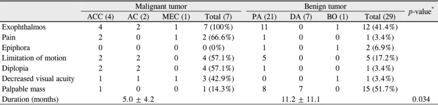
Table 1. Symptoms, signs and duration of symptom of epithelial tumors in the lacrimal gland
Malignant tumor Benign tumor p-value*
ACC (4) AC (2) MEC (1) Total (7) PA (21) DA (7) BO (1) Total (29)
Exophthalmos 4 2 1 7 (100%) 11 0 1 12 (41.4%)
Pain 2 0 1 2 (66.6%) 1 0 0 1 (3.4%)
Epiphora 0 0 0 0 (0%) 1 0 1 2 (6.9%)
Limitation of motion 2 2 0 4 (57.1%) 5 0 0 5 (17.2%)
Diplopia 2 2 0 4 (57.1%) 1 0 0 1 (3.4%)
Decreased visual acuity 1 1 1 3 (42.9%) 0 0 1 1 (3.4%)
Palpable mass 1 0 0 1 (14.3%) 8 7 0 15 (51.7%)
Duration (months) 5.0 ± 4.2 11.2 ± 11.1 0.034
Values are presented as mean ± SD.
ACC = adenoid cystic carcinoma; AC = adenocarcinoma; MEC = mucoepidermoid carcinoma; PA = pleomorphic adenoma; DA = dacryops; BO = benign oncocytoma.
*t-test.
Table 2. Computed tomography and magnetic resonance imaging findings of epithelial tumors in the lacrimal gland
Malignant tumor Benign tumor
ACC (4) AC (2) MEC (1) Total (7) PA (21) DA (7) BO (1) Total (29)
Bone destruction 3 1 0 4 (57.1%) 2 0 0 2 (6.9%)
Calcification 1 0 0 1 (14.3%) 1 0 0 1 (3.4%)
Outline
Well marginated 2 0 1 3 (42.9%) 19 7 1 27 (93.1%)
Poorly marginated 1 2 0 3 (42.9%) 2 0 0 2 (6.9%)
ACC = adenoid cystic carcinoma; AC = adenocarcinoma; MEC = mucoepidermoid carcinoma; PA = pleomorphic adenoma; DA = dacryops; BO = benign oncocytoma.
확진된 36명 환자를 대상으로 하였다. 남자 14명, 여자 22 명이었고 평균 나이는 48.7 ± 14.2세였다. 종양의 종류, 성 별, 연령 분포, 임상증상, 증상발현 기간, 수술 전에 시행한 전산화 단층촬영과 자기 공명영상의 방사선 소견, 치료방 법, 재발, 예후 및 경과 등을 분석하여 조기진단에 도움이 되는 악성종양과 양성 종양의 임상적 특징과 방사선 소견 의 차이점에 대해서 알아보았다.
결 과
총 36명의 환자 중, 다형성 선종 21예(58.3%), 눈물샘낭 7예(19.4%), 선낭암종 4예(11.1%), 선암종 2예(5.5%), 양성 호산성 과립세포종 1예(2.7%), 점액표피양 암종 1예(2.7%) 였다. 양성종양에서는 여자가 더 많았지만 악성종양에서는 남자와 여자의 비는 4:3, 평균 연령은 악성종양이 51.0 ± 9.4세, 양성종양이 48.2 ± 15.2세였고 통계학적으로 유의한 차이를 보이지 않았다(Mann-Whitney test, p=0.64).
악성눈물샘상피종양의 임상증상으로 안구돌출이 100%, 안구운동장애 및 복시가 57.1%, 시력저하는 42.9%, 안구통 증은 42.9%, 종괴감이 14.3%에서 나타났다. 양성눈물샘상 피종양의 임상증상은 종괴감이 51.7%, 안구 돌출이 41.4%, 안구운동장애가 17.2%, 유루는 6.9%, 복시, 시력저하, 안구
통증이 각각 3.4%에서 나타났다(Table 1). 평균증상 발현기 간도 악성눈물샘상피종양(5.0 ± 4.2개월)은 양성눈물샘상피 종양(11.2 ± 11.1개월)과 비교하여 짧았다(t-test, p=0.034).
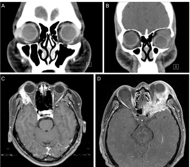
악성종양의 전산화 단층촬영 및 자기공명영상 소견에서 골 파괴가 57.1% (4/7안), 종양의 불확실한 경계면이 42.9% (3/7 안), 석회화가 14.3% (1/7안)에서 나타났다. 양성종양의 경우 다형성 선종에서만 골파괴 소견이 9.5% (2/21안), 종양의 불확실한 경계면이 9.5% (2/21안), 석회화가 4.8% (1/21안) 에서 보였으나 다른 양성종양에서는 종양의 확실한 경계면 이 93.1% (29/31안)에서 관찰되었다(Table 2) (Fig. 1).
7예의 악성종양 환자 중 1예에서 안와내용제거술(orbital exenteration) 후 방사선치료, 3예에서 종양제거, 1예에서 종 양제거술 후 동시항암방사선 치료 및 사이버나이프 치료, 1 예에서 종양제거술 후 동시항암방사선 치료, 1예에서 종양 제거술 후 항암치료를 받았다. 안와내용제거술을 받은 1예 의 경우 술 전에 앞안와절개술과 생검술 후 안와내용제거 술을 시행하였고 그 후에 방사선치료를 시행하였다. 종양 제거술을 시행한 총 6예의 환자 중 1예에서 종양제거술 후 동시항암방사선치료를 받은 후 재발소견 보여 사이버나이 프 치료를 시행하였다. 7예의 악성종양 환자 중 2예에서 다 른 부위로 전이된 소견 보였는데 2예 모두 폐전이 소견을 보여 추적관찰 중이다.
Figure 1. (A) Pleomorphic adenoma. CT shows a well-marginated round mass with isodensity in the Rt lacrimal
gland. There is no bone destruction and caldification. (B) Pleomorphic adenoma. CT shows a well-marginated round mass with isodensity in the left lacrimal gland. Subtle bony erosive changes is seen on the wall of the left lacrimal fossa. (C) Adenoid cystic carcinoma. T1W MR imaging shows a ill-defined mass and bony destruction at the right orbital wall. (D) Adenocarcinoma. T1W MR imaging shows a heterogeneously enhancing mass and bony destruction at the left orbital wall. CT = computed tomography; Rt = right; T1W MR = T1 weighted magnetic resonance.Table 3. Treatments of epithelial tumors in the lacrimal gland
Malignant tumor Benign tumor
ACC (4) AC (2) MEC (1) PA (21) DA (7) BO (1)
Exenteration + RTx 1 0 0 0 0 0
Removal + CCRTx + cyberknife 1 0 0 0 0 0
Removal + CCRTx 0 1 0 0 0 0
Removal + CTx 0 1 0 0 0 0
Removal 2 0 1 21 7 1
ACC = adenoid cystic carcinoma; AC = adenocarcinoma; MEC = mucoepidermoid carcinoma; PA = pleomorphic adenoma; DA = dacryops; BO = benign oncocytoma; RTx = radiotherapy; CCRTx = concurrent chemoradiotherapy; CTx = chemotherapy.
양성종양의 경우는 모두 술전 절제생검을 시행하지 않았 고, 29예 모두 앞외측안와절개술(antero-lateral orbitotomy) 및 종양제거술을 시행하였다(Table 3).
현재 36명의 환자 중 27명은 외래경과 관찰 중이고, 양성
종양의 평균 외래 경과 관찰 기간은 39.69 ± 24.97개월, 악 성종양의 평균 외래 경과 관찰 기간은 66.71 ± 35.30개월이 며 현재까지 사망한 경우는 없었으며 안와에 재발 소견 보 이는 환자도 관찰되지 않고 있다.
A B
C D
고 찰
눈물샘상피종양의 약 50%가 다형성 선종이고 나머지 50%
는 악성종양으로 알려졌다.5 과거 국내에서 시행된 연구에 서도 악성 종양은 52.6%로 비슷하였으나 본 연구에서는 악 성종양이 19.4%로 다른 연구에 비해 적었다. 하지만 다형성 선종은 58.3%를 보여 다른 연구 결과와 거의 일치하였다.
눈물샘상피종양은 주로 중년에서 발생하며 그중에 다형 성 선종은 80세까지 모든 연령에서 발생할 수 있으나 주로 평균 40대에서 발생하며선낭암종은 40대에서 주로 발생하 지만 10대에서도 많이 발생한다고 하였다.6 그 외의 종양에 서 다형성 선종유래암종, 선암종 그리고 점막표피암은 대 략 평균 50-52세에서 호발한다고 하였다.3 악성종양의 경우 는 평균 51세에서 발생하여 다른 연구결과와 분포가 유사 하였다. 악성종양의 성비의 경우 보통은 성비의 발생차이 가 없는 반면 점막표피암에서는 3:2로 여자에서 호발하고 선암종의 경우 여자보다 남자에서 더 잘 호발한다고 하였 다.7 과거 국내 연구에서 악성종양은 여자에서 더 많이 호 발하였으나 본 연구에서는 전체 악성 종양의 발생에 대한 성비는 4:3이었으나 증례수가 적어 성별에 따른 차이는 확 인하기 어려웠다.
양성종양에서는 통증이 흔하지 않아 통증 발생 시 악성 종양을 의심해볼 수 있으며 이런 통증은 초기 말초 신경과 외안근에 종양이 침범해서 발생하며 선낭암종과 선암종에 서 통증은 상대적으로 흔하지만 다형성 선종유래암종과 점 막표피암에서는 거의 나타나지 않는다고 하였고7-9 과거 국 내 연구에서도 안구통증은 악성 눈물샘상피종양에서만 특 징적으로 나타났다. 다른 증상으로 유루증과 시력 변화는 선낭암종에서 발생적이 있다는 보고가 있다.10 본 연구에서 도 다형성선종 1예에서 제외하고 안구통증은 양성종양에서 발생하지 않았으나 악성 종양의 경우 42.9%에서 안구통증 이 발생하였으며 안구통증은 악성종양과 통계학적으로 관 련이 있다(Chi-square test, p=0.047).
평균 증상 발현기간을 살펴보면 다형성선종은 대략 2년, 서서히 자라는 종양의 경우는 20년까지 지속되었다고 보고 된 바 있다.8 이와는 달리 악성눈물샘종양의 경우 전형적으 로 평균 증상 발현기간이 짧았는데 선낭암종의 경우 대략 6개월, 다른 악성눈물샘종양의 경우는 1년보다 짧았었 다.8-11 본 연구에서도 평균증상 발현 기간에서 악성눈물샘 상피종양(5.0 ± 4.2개월)은 양성눈물샘상피종양(11.2 ± 11.1 개월)과 비교하여 짧았다. 따라서 눈물샘상피종양의 악성 종양과 양성종양을 조기에 감별하기 위해서는 증상 및 발 현 기간이 중요하며 초기에 통증 유무에 대한 자세한 병력 정취가 진단에 악성종양을 감별하는 데 도움을 줄 수 있음
을 알 수 있다.
이런 임상 증상 외에도 눈물샘상피종양의 수술 전 진단에 있어서 방사선 소견은 중요하다. 전산화단층촬영(CT)에서 다형성선종은 구형 또는 타원형의 경계가 명확한 단단한 형 태이며 때때로 석회화와 골 재형성 과정이 나타나며 이와는 달리 선낭암종의 경우 전형적으로 경계가 불명확하며 주변 조직으로 침윤소견 보이며 때때로 골파괴 소견이 보인다.12 CT에서 모든 악성눈물샘상피종양의 전형적인 방사선학적 소견은 불명확한 경계, 골미란 그리고 석회화이고, 이는 자 기공명영상(MRI)에서 동일한 소견을 보인다.7,12,13 본 연구 에서는 CT 및 MRI에서 악성종양은 골파괴가 57.1% (4/7안), 석회화가 14.3% (1/7안), 종양의 불확실한 경계면이 42.9%
(3/7안)에서 나타났다. 이는 과거 국내 연구와 유사한 성향 을 보였다. 과거 국내 연구에서 양성종양의 경우에 골파괴, 석회화, 불확실한 경계면이 보이지 않았으며 본 연구에서 는 다형성 선종에서만 골파괴 소견이 9.5% (2/21안), 석회 화가 4.8% (1/21안), 종양의 불확실한 경계면이 9.5% (2/21 안)에서 보였으며 다른 양성종양에서는 93.1% (27/29안)에 서 종양의 경계면이 명확했고 골파괴 소견은 관찰되지 않 았다.
이러한 임상양상과 방사선 소견은 진단에 중요한 역할을 하지만 눈물샘상피종양의 최종 진단은 조직검사로만 이루 어진다. 하지만 양성눈물샘상피종양의 대부분을 차지하는 다형성 선종의 경우 불충분하게 절제할 때 재발되거나 악 성으로 변형될 수 있다고 알려졌다.14 이러한 이유로 과거 눈물샘종양에서 조직검사를 하지 않고 초기에 수술적 절제 를 권장하였지만15 최근 Lai et al16과의 연구에 의하면 다형 성선종 환자에서 생검술 후 생검길을 제거할 경우 재발률 이 낮았다.16,17 또한 눈물샘상피종양은 쉽게 방사선 소견을 양성종양으로 잘못 해석할 수 있어 현재는 눈물샘상피종양 의 절개 생검술도 선택할 수 있는 방법의 하나로 인식되고 있다. 그러나 여전히 미용적인 문제나 재건의 문제로 종양 의 초기 완전절제술이 선호되고 있다. 본 연구에서도 21명 의 다형성선종환자에서 모두 생검술 없이 앞외측안와절개 술 및 종양제거술을 받았으며 아직까지 재발되거나 악성으 로 전환된 예는 없었다.
Shields and Shields17는 눈물샘종양 중 악성종양에서 크 기가 작고 국한되어 있는 경우만 “en bloc” 종양절제술로 종양을 완전히 제거할 수 있으며 그 외의 경우는 종양절제 술 후 방사선 치료나 항암치료를 해야 한다고 하였다.또한 안와내용제거술(orbital exenteration)은 악성종양이 확장되 거나 종양을 싸고 있는 막을 넘어서 주변조직에 침윤될 때 시행한다고 하였지만 심각한 미용적인 문제와 생존율에 큰 도움이 되지 않는다는 보고가 있다.18 본 연구에서 7예의 악
성종양 환자 중 1예에서 안와내용제거술(orbital exentera- tion) 후 방사선치료, 3예에서 종양제거, 1예에서 종양제거 술 후 동시항암방사선 치료 및 사이버나이프 치료, 1예에서 종양제거술 후 동시항암방사선 치료, 1예에서 종양제거술 후 항암치료를 시행하였다. 이는 과거 국내 연구에서 악성 종양 환자에서 외과적 절제술과 방사선 치료만을 시행했던 것과 비교해 치료 방법이 조금 더 다양하게 이루어진 양상 을 보인다. 현재 7예의 악성종양의 환자들은 모두 생존해 있으며 안와에 재발소견 보이지 않고 추적관찰 중이다.
눈물샘상피양성종양은 일반적으로 예후가 좋다. 만약 초 기 수술에 종양을 완전히 절제했다면 추가 치료 없이 환자 는 퇴원할 수 있으나 만약 종양이 부서지면 재발과 악성변 화로 장기적인 외래 경과 관찰이 필요하다.19 이와는 달리 악성종양의 경우 임상양상이 공격적이며 그중 다형성 선종 유래암종의 경우 진단 후 수년 내에 사망한다고 알려졌고,20 선낭암은 75%까지 재발하고 진단 후 10년 생존율이 20-30%이며, 선암은 평균 생존기간이 1.5년이라 하였다.3 본 연구에서는 악성종양의 평균 외래 경과 관찰 기간은 66.7 ± 35.3개월이며 이전 연구 결과와는 달리 현재까지 조 사한 바로는 악성종양 환자들 중에 재발되거나 사망한 환 자는 보고되지 않고 있다.
결론적으로 서울성모병원에서 악성눈물샘상피 종양으로 진단된 환자에서 특징적인 임상증상과 방사선학적 소견상 골파괴, 불확실한 경계면 등이 보일 경우 조직병리학적으 로 악성이었다. 눈물샘상피종양이 의심되는 환자에서 임상 양상과 방사선학적 소견이 종양의 악성 유무를 예상하는 데 있어 도움이 되며, 이에 좀 더 주의를 기울여 빠른 진단 및 치료가 가능할 것으로 생각한다.
REFERENCES
1) Shields JA, Shields CL, Scartozzi R. Survey of 1264 patients with orbital tumors and simulating lesions: The 2002 Montgomery Lecture, part 1. Ophthalmology 2004;111:997-1008.
2) Kennedy RE. An evaluation of 820 orbital cases. Trans Am Ophthalmol Soc 1984;82:134-57.
3) Font RL, Croxatto JO, Rao NA. Tumours of the eye and ocular adnexa. AFIP Atlas of Tumor Pathology. Series 4 Vol. 5.
Washington DC: American Registry of Pathology, 2006;111-25.
4) Jung JG, Jang JW, Lee SY. The clinical review of epithelial tumors in the lacrimal gland. J Korean Opthalmol Soc 1999;40:3258-65.
5) Shields CL, Shields JA, Eagle RC, Rathmell JP. Clinicopathologic review of 142 cases of lacrimal gland lesions. Ophthalmology 1989;96:431-5.
6) Tellado MV, Mclean IW, Specht C, Varga J. Adenoid cystic carci- nomas of the lacrimal gland in childhood and adolescence.
Ophthalmology 1997;104:1622-5.
7) Eviatar JA, Hornblass A. Mucoepidermoid carcinoma of the lac- rimal gland: 25 cases and a review and update of the literature.
Ophthal Plast Reconstr Surg 1993;9:170-81.
8) Rose GE, Wright JE. Pleomorphic adenoma of the lacrimal gland.
Br J Ophthalmol 1992;76:395-400.
9) Heaps RS, Miller NR, Albert DM, et al. Primary adenocarcinoma of the lacrimal gland. A retrospective study. Ophthalmology 1993;100:1856-60.
10) Lee DA, Campbell RJ, Waller RR, Ilstrup DM. A clinicopathologic study of primary adenoid cystic carcinoma of the lacrimal gland.
Ophthalmology 1985;92:128-34.
11) Zeng J, Shi JT, Li B, et al. Epithelial tumours of the lacrimal gland in the Chinese: a clinicopathologic study of 298 patients. Graefes Arch Clin Exp Ophthalmol 2010;248:1345-9.
12) Vaidhyanath R, Kirke R, Brown L, Sampath R. Lacrimal fossa le- sions: pictorial review of CT and MRI features. Orbit 2008;27:
410-8.
13) Henderson JW, Farrow GM. Primary malignant mixed tumors of the lacrimal gland report of 10 cases. Ophthalmology 1980;87:
466-75.
14) Potter PD, Shield JA, Shield CL. MRI of the Eye and Orbit, 1st ed.
Philadelphia: J. B. Lippincott, 1995;227-35.
15) Hornblass A, Jakobiec FA, Bosniak S, Flanagan J. The diagnosis and management of epithelial tumors of the lacrimal sac.
Ophthalmology 1980;87:476-90.
16) Lai T, Prabhakaran VC, Malhotra R, Selva D. Pleomorphic ad- enoma of the lacrimal gland: is there a role for biopsy? Eye (Lond) 2009;23:2-6.
17) Shields JA, Shields CL. Eyelid, conjunctival, and orbital tumors:
An atlas and textbook, 2nd ed. Philadelphia: Lippincott Williams
& Wilkins, 2008;700-14.
18) Polito E, Leccisotti A. Epithelial malignancies of the lacrimal gland: survival rates after extensive and conservative therapy.
Annals of Ophthalmology 1993;25:422-6.
19) Currie ZI, Rose GE. Long-term risk of recurrence after intact ex- cision of pleomorphic adenomas of the lacrimal gland. Arch Ophthalmol 2007;125:1643-6.
20) Font RL, Smith SL, Bryan RG. Malignant epithelial tumours of the lacrimal gland: a clinicopathologic study of 21 cases. Arch Ophthalmol 1998;116:613-6.
= 국문초록 =
눈물샘 상피종양의 임상적 분석
목적: 눈물샘 상피종양 환자의 임상 양상과 방사선 소견을 분석하고 치료 및 예후에 대하여 알아보고자 한다.
대상과 방법: 2005년 5월부터 2012년 10월까지 본원에서 조직생검이나 외과적 절제 후 병리조직학적으로 눈물샘 상피종양으로 확진 받은 36명 환자의 의무기록을 후향적으로 고찰하였다.
결과: 총 36명의 눈물샘 상피종양 중 병리조직학적으로 다형성 선종 21예, 눈물샘낭 7예, 선낭암종 4예, 선암종 2예, 양성 호산성 과립 세포종 1예, 점액표피양 암종이 1예였다. 가장 흔한 증상은 안구돌출(52.7%)이었고 악성눈물샘상피종양은 양성눈물샘상피종양에 비해 안구운동 장애 및 복시(57.1%), 안구 통증(42.9%), 시력 저하(42.9%)가 특징적인 증상이었다. 평균증상 발현기간도 악성눈물샘상피종양 (5.0 ± 4.2개월)은 양성눈물샘상피종양(11.2 ± 11.1개월)에 비하여 짧았다(t-test, p=0.034). 전산화단층촬영과 자기공명영상에서 다형 성선종 2예(9.5%)에서 경한 골파괴 소견과 1예(3.4%)에서 석회화가 관찰된 것을 제외하고는, 악성 눈물샘 상피종양은 양성눈물샘상피 종양과 비교하여 특징적인 골파괴 소견(57.1%), 종양의 경계가 불확실한 경우(42.9%) 그리고 석회화(14.3%)가 관찰되었다. 악성눈물샘 상피종양은 외과적 절제술 후 방사선 치료 및 항암치료를 57.1%에서 받았으며 그중 1예(14.3%)에서 치료 후 재발소견을 보였다.
결론: 눈물샘상피종양 환자의 임상증상 및 방사선 소견을 주의 깊게 분석하면 악성종양을 조기 진단하는 데 도움을 줄 수 있을 것으로 생각한다.
<대한안과학회지 2014;55(6):795-800>