J Korean Ophthalmol Soc 2015;56(4):509-514 ISSN 0378-6471 (Print)⋅ISSN 2092-9374 (Online)
http://dx.doi.org/10.3341/jkos.2015.56.4.509
Original Article
전안부 빛간섭 단층촬영을 이용해 측정한 결막이완증 수술 전후의 결막 및 눈물띠 면적 변화
Changes in Area of Conjunctiva and Tear Meniscus Measured Using Anterior Segment Optical Coherence Tomography after Conjunctivochalasis Surgery
김종수⋅배정범⋅서장원⋅박우찬
Jong Soo Kim, MD, Jeong Bum Bae, MD, Jang Won Seo, MD, PhD, Woo Chan Park, MD, PhD
동아대학교 의과대학 안과학교실
Department of Ophthalmology, Dong-A University College of Medicine, Busan, Korea
Purpose: To evaluate cross-sectional areas of conjunctiva and tear meniscus of conjunctivochalasis using Fourier-Domain RTVue-100 optical coherence tomography (OCT) before and after conjunctivochalasis surgery.
Methods: Thirty-one patients (33 eyes) with symptomatic conjunctivochalasis were recruited for this study between June 2013 and April 2014. All patients underwent crescent-shaped conjunctiva resection and amniotic membrane transplantation. Anterior segment OCT (AS-OCT) imaging was performed and tear break-up time was evaluated prior to and 3 months after the con- junctivochalasis surgery. Cross-sectional areas of conjunctiva and tear meniscus of conjunctivochalasis at 7 locations (1 center, 3 nasal and 3 temporal areas) were measured in all patients.
Results: The mean age of patients was 66.3 ± 10.8 years. Cross-sectional areas of conjunctivochalasis at all locations sig- nificantly decreased from 0.487 ± 0.42 mm2 to 0.007 ± 0.011 mm2 (p < 0.001), whereas no significant changes in cross-sectional areas of tear meniscus at all 7 locations were observed after the surgery. Mean tear break-up time significantly increased from 2.26 ± 0.69 sec to 3.81 ± 1.22 sec following the surgery.
Conclusions: Using AS-OCT, in this study we showed that areas of conjunctiva decreased and areas of tear meniscus were un- changed after conjunctivochalasis surgery.
J Korean Ophthalmol Soc 2015;56(4):509-514
Key Words: Amniotic membrane transplantation, Anterior segment optical coherence tomography, Conjunctivochalasis, Tear meniscus
■Received: 2014. 6. 13. ■ Revised: 2014. 11. 23.
■Accepted: 2015. 3. 23.
■Address reprint requests to Woo Chan Park, MD, PhD Department of Ophthalmology, Dong-A University Hospital,
#26 Daesingongwon-ro, Seo-gu, Busan 602-715, Korea Tel: 82-51-240-5227, Fax: 82-51-254-1987
E-mail: [email protected]
* This study was presented as a narration at the 111th Annual Meeting of the Korean Ophthalmological Society 2014.
* This article has been supported by the Research Fund of the Dong‐A University.
ⓒ2015 The Korean Ophthalmological Society
This is an Open Access article distributed under the terms of the Creative Commons Attribution Non-Commercial License (http://creativecommons.org/licenses/by-nc/3.0/) which permits unrestricted non-commercial use, distribution, and reproduction in any medium, provided the original work is properly cited.
결막이완증은 변성에 의해 늘어난 결막이 아래눈꺼풀과 안구 사이에 결막주름을 형성한 것으로, 과도한 주름은 눈 물막의 안정성을 방해해 이물감 및 건성안 관련 증상을 일 으키는 것으로 알려졌다.1-3 증상이 있을 경우 먼저 약물 치 료를 시행하며, 증상 호전이 없을 경우 수술적 치료를 고려 한다.4,5
수술적 치료로는 다양한 방법들이 소개되었는데, 이완된 결막의 단순 절제술,6 결막 절제술 후 양막이식술을 하는 방법들이 있으며,7-11 두극 지짐기로 소작하거나 고주파 전 기수술기를 사용하는 방법,12,13 아르곤레이저를 이용한 결
Figure 1. The seven arrows indicate the locations and direc-
tions scanned by RTvue.Figure 2. Measurement of cross-sectional conjunctivochalasis area using RTVue-100. (A) Photograph of lower eye-
lid and cornea where the vertical scans were taken. Arrow means the location and direction scanned by RTvue. (B) The obtained scan image. (C) After the image was digitally magnified, tissue boundaries among the lower lid, pro- lapsed redundant conjunctiva, and bulbar conjunctiva/inferior part of the cornea were discriminated from each based on the different levels the brightness between tissues. (D) Cross-sectional area of the outlined conjunctivochalasis was measured as 0.221 mm2 using the instrument's software.막이완증 수술법 등도 소개되었다.14
빛간섭 단층촬영은 1990년대 처음 소개된 이후 비침습적 인 방법으로 조직의 단면을 관찰할 수 있는 수단으로 널리 이용되었으며, 특히 황반 부종, 연령관련 황반변성 등 유리 체 망막 영역 및 녹내장 영역에서 질병의 진단과 경과관찰 에 이용되었다.15-20 그 중 전안부 빛간섭 단층촬영은 안구 전방 구조물, 각결막, 전방각 및 눈물띠 등에 대한 높은 해 상도의 영상을 제공할 수 있으며, 최근에는 이를 이용하여
결막이완증의 진단 및 정도의 평가에 사용되고 있다.21-28 본 연구는 약물치료에 증상 호전이 없는 결막이완증 환 자들을 대상으로 결막 절제술 및 양막이식술을 시행하고, 수술 전후의 결막 및 눈물띠 면적 변화를 전안부 빛간섭 단 층촬영을 통해 알아보고자 하였다.
대상과 방법
2013년 6월부터 2014년 4월까지 본원에서 결막이완증을 진단받고 약물 치료에 호전이 없어 수술을 시행한 31명 33 안을 대상으로 하였다. 안검 내반 및 외반, 첩모난생 등의 눈꺼풀 질환이나 누점 또는 누소관 협착, 비루관 폐쇄 등의 눈물길 질환이 있는 환자, 결막이나 눈꺼풀 수술 과거력이 있는 환자, 그리고 콘택트 렌즈 사용자는 대상에서 제외하 였다.
모든 환자에서 한 명의 검사자가 세극등 현미경 검사를 통해 결막이완증의 진단하였으며, 수술 전과 수술 후 3개월 째에 눈물막 파괴 시간을 측정하였다. 눈물막 파괴 검사는 Fluorescein strip (Haag-Streit AG, Koniz, Switzerland)을 하 측 결막낭에 접촉시킨 후 환자로 하여금 수초간 몇 번 눈을
A B
C D
Figure 3. Measurement of cross-sectional tear meniscus using RTVue-100. (A) Photograph of lower eyelid and cor-
nea where the vertical scans were taken. Arrow means the location and direction scanned by RTvue. (B) After the im- age was digitally magnified. (C) Triangular-shaped tear meniscus is seen between the lower cornea and lower lid. (D) Cross-sectional area of the outlined tear meniscus was measure as 0.060 mm2 using the instrument's software.Figure 4. Measurement of cross-sectional tear meniscus using
RTVue-100. In case of many tear meniscus caused by tears dispersed among the conjunctival folds, each tear meniscus area was measured separately and then the total meniscus area was determined by summation of each tear meniscus area.감았다 뜨게 한 후 세극등 현미경의 코발트 블루 필터를 통 해 마지막 깜박임 후 각막 눈물막의 첫 번째 건조점이 발생 한 시간을 3회 측정 후 평균을 초 단위로 기록하였다.
수술 전과 수술 후 3개월에서 6개월 사이에 Corneal ante- rior modulator (CAM)를 장착한 전안부 빛간섭 단층촬영 (RTVue-100, Optovue Inc., Fremont, California, USA)을 시 행하였다. 윤부의 6시를 기준으로 중심부 한 곳, 그리고 3 시와 9시의 윤부를 기준으로 비측 3곳, 이측 3곳까지 포함 해 총 7곳에서 촬영(Fig. 1) 후 RTVue-100 내의 소프트웨어 를 이용하여 이완된 결막 및 눈물띠의 경계를 따라 수동으 로 선을 그어 그 면적을 측정하였다(Fig. 2, 3). 한 단면에서 눈물띠가 여러 개가 있는 경우에도 같은 방법으로 수동으 로 선을 그어 각각의 면적을 따로 측정한 다음, 모두 더해 눈물띠 면적을 구하였다(Fig. 4). 전안부 빛간섭 단층촬영에 대한 측정은 두 명의 연구자에 의해 이루어졌으며, 모든 결 과는 두 측정치의 평균으로 나타내었다.
수술은 모든 환자에서 한 명의 술자가 결막이완증 절제 술 및 봉합사를 이용한 양막 이식술을 시행하였다. 구후마 취와 Nadbath-Rehman-Ellis 마취 후 개검기로 눈꺼풀을 벌 리고 6-0 Black silk 봉합사로 3시와 9시 결막에 견인 봉합 을 하여 안구가 위를 향하도록 견인하였다. Gentin violet으 로 검열반이 포함되도록 절제할 범위를 표시한 후 Westcott scissor로 절제하였다. 준비된 양막(AmniSite-Cornea®, Bioland
A B
C D
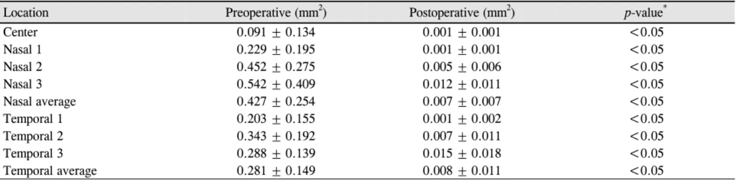
Table 1. Cross-sectional conjunctivochalasis area at baseline and following the surgery
Location Preoperative (mm2) Postoperative (mm2) p-value*
Center 0.091 ± 0.134 0.001 ± 0.001 <0.05
Nasal 1 0.229 ± 0.195 0.001 ± 0.001 <0.05
Nasal 2 0.452 ± 0.275 0.005 ± 0.006 <0.05
Nasal 3 0.542 ± 0.409 0.012 ± 0.011 <0.05
Nasal average 0.427 ± 0.254 0.007 ± 0.007 <0.05
Temporal 1 0.203 ± 0.155 0.001 ± 0.002 <0.05
Temporal 2 0.343 ± 0.192 0.007 ± 0.011 <0.05
Temporal 3 0.288 ± 0.139 0.015 ± 0.018 <0.05
Temporal average 0.281 ± 0.149 0.008 ± 0.011 <0.05
Values are presented as mean ± SD.
*Paired t-test.
Table 2. Cross-sectional tear meniscus area at baseline and following the surgery
Location Preoperative (mm2) Postoperative (mm2) p-value*
Center 0.03 ± 0.02 0.01 ± 0.00 0.073
Nasal 1 0.02 ± 0.02 0.04 ± 0.04 0.415
Nasal 2 0.02 ± 0.02 0.02 ± 0.02 0.789
Nasal 3 0.02 ± 0.02 0.03 ± 0.04 0.545
Nasal average 0.02 ± 0.02 0.03 ± 0.04 0.513
Temporal 1 0.04 ± 0.03 0.02 ± 0.01 0.377
Temporal 2 0.02 ± 0.03 0.01 ± 0.01 0.689
Temporal 3 0.02 ± 0.03 0.07 ± 0.05 0.211
Temporal average 0.03 ± 0.03 0.04 ± 0.04 0.104
Values are presented as mean ± SD.
*Paired t-test.
Figure 5. Schematic design for conjunctivochalasis surgery.
After resection of the conjunctivochalasis area (inside of dot- ted line) including the pingueculae (arrows), amniotic mem- brane transplantations was done in the exposed scleral area.
Ltd., Cheonan, Korea)의 상피면에 25% 알코올을 묻힌 후
#15 Parker blade를 이용하여 상피를 제거한 다음, 노출된 공막의 크기대로 디자인하여 상피 기저막이 위를 향하도록 덮어준 후 10-0 nylon으로 상공막에 고정하고, 결막의 가장 자리 부분과 봉합하였다(Fig. 5).
수술 후 치료용 콘택트렌즈를 착용시키고, 양막 위로 상 피가 완전히 재생될 때까지 압박안대를 시행하였다. 압박 안대를 시행한 평균기간은 7일이었다. 그 후로는 0.3% gati- floxacin (Gatiflo®, Handok Pharmaceutical Co., Ltd., Seoul,
Korea)을 하루 4번, Fortified 1% Methylprednisolone (Solu- Medrol®, Pfizer Inc., New York, NY, USA)을 하루 3번, 무 보존제 인공누액을 2시간마다 점안하였고, Fortified 1%
Methylprednisolone는 매 방문 시마다 점안 횟수를 줄이도 록 하였다. 봉합사는 수술 후 2주경에 제거하였다. 추적관 찰은 술 후 첫 1주 동안은 매일, 그리고 술 후 2주, 3주, 6주, 12주째 시행하였으며, 이후부터는 2개월마다 시행하 였다.
자료의 분석은 SPSS 18.0에서 Paired t-test를 이용하여 수술 전과 후의 결막 단면적, 눈물띠 단면적, 눈물막 파괴 시간을 비교하였으며, p값이 0.05 미만인 경우를 유의하다 고 판정하였다.
결 과
총 대상 환자는 31명 33안(남자 10명 11안, 여자 21명 22 안)이었고, 평균 연령은 65.8 ± 9.8세(범위, 49-85세)였다.
눈물막 파괴 시간은 수술 전 2.26 ± 0.69초에서 수술 후 3개 월째에 33.81 ± 1.22초로 증가하였으며, 통계적으로 유의하였 다(p<0.05). 각 구역별 결막 단면적은 중심부 0.09 ± 0.13 mm2 에서 0.001 ± 0.001 mm2로(p<0.05), 비측 평균 0.43 ± 0.25 mm2
에서 0.007 ± 0.007 mm2로(p<0.05), 이측 평균 0.28 ± 0.15 mm2 에서 0.008 ± 0.011 mm2로(p<0.05) 모두 감소하였다(Table 1).
반면 눈물띠 면적은 중심부 0.03 ± 0.02 mm2에서 0.01 ± 0.00 mm2로(p=0.073), 비측 평균 0.02 ± 0.02 mm2에서 0.03
± 0.04 mm2로(p=0.513), 이측 평균 0.03 ± 0.03 mm2에서 0.04 ± 0.04 mm2로(p=0.104) 술 전과 비교하여 술 후에 통 계적인 차이가 없었다(p>0.05) (Table 2).
고 찰
결막이완증의 발생원인은 현재까지 명확하게 밝혀지지 않았으나, 노인성 변화와 결막탄력 섬유의 변성 혹은 감소 로 인해 결막 주름이 발생한다는 보고와 결막과 눈꺼풀의 기계적인 자극으로 림프관 유출에 장애를 일으켜 결막림프 관 확장증이 발생하며, 그 결과 결막이완증이 발생한다고 보고들이 있다.3,6
결막이완증은 대부분 무증상으로 치료가 필요 없는 경우 가 많으나, 건조감, 이물감, 눈물흘림, 결막하 출혈 등과 같 은 증상이 있을 경우 인공눈물, 윤활제, 스테로이드, 항히스 타민 점안액 등을 사용해볼 수 있으며, 약물치료에 효과가 없을 경우 수술적 치료가 고려된다.
본 연구에서는 이완된 결막을 절제한 후 절제된 결막 부 위를 양막을 이용하여 덮은 뒤 봉합사로 고정하여 이식하 는 방법을 사용하였는데, 이 방법은 이완된 결막 제거와 함 께 시행한 결손 부위의 양막 이식이 항염증 효과와 빠른 상 피재생, 정상적인 결막 상피 표현형으로의 회복 등 많은 장 점이 있다.7,8
이미 많은 연구에서 전안부 빛간섭 단층촬영이 눈물띠 측정에 대한 높은 정확도와 좋은 재현성을 갖고 있다고 보 고되었다.23,29 이에 저자들은 이전의 다른 연구에서 정상인 과 결막이완증 환자의 결막 면적과 눈물띠 면적을 측정하 여 비교하였고, 결막이완증 정도를 객관적으로 평가할 수 있다는 가능성을 확인한 바가 있다. 따라서, 본 연구에서는 전안부 빛간섭 단층촬영을 이용해 결막이완증 수술 전과 비교하여 수술 후에 결막의 단면적과 눈물띠의 단면적과 같은 아래 결막의 측정치들에 어떠한 변화가 일어나는지 접근해보고자 하였다.
결막이완증 수술 전과 비교해 수술 후에 눈물막 파괴시 간은 증가하였고, 평균 결막의 단면적은 모든 방향에서 의 미 있는 감소를 보였으나, 눈물띠의 단면적은 수술 전과 후 에서 통계적으로 차이가 없는 것으로 나타났다. 이것은 결 막이완증이 있는 환자에서도 눈물막의 총량은 같으나, 결 막 주름들 때문에 눈물 분포에 문제를 일으켜 불편감을 유 발하는 것으로 보이며, 결막이완증 수술 후에는 분산되어
있던 눈물이 합쳐지면서 BUT가 증가해 증상이 호전되는 것으로 판단해 볼 수 있다. 즉, 결막이완증 환자는 눈물분 비가 적은 것이 아니라, 같은 양이라도 눈물이 분산되어 있 어 그 기능이 떨어지는 것으로 보인다는 것을 객관적으로 확인하였다. 다만, 결막의 이완이 심해 누점을 막아 오히려 눈물의 양이 많아지는 형태의 결막이완증은 이에 해당하지 않는다.
본 연구의 한계점으로 결막과 눈물띠 단면적의 측정에 있어 환자의 눈 깜박임이나, 촬영 시간 등에 의해 결과가 영향을 받을 수 있는 가능성을 배제할 수 없으며, 7군데의 다른 방향에서 촬영을 하였으나 그 측정치가 전체 결막이 나 눈물띠의 양을 정확히 반영하지는 못한다는 점이다. 또 한 환자가 주관적으로 느끼는 증상을 객관화시키지 못한다 는 것에 대하여 향후 연구에서 반영시켜야 할 부분으로 생 각한다.
결론적으로, 본 연구에서 전안부 빛간섭 단층촬영을 통 해 결막이완증 환자에서 결막과 눈물띠의 양을 보다 객관 적으로 정량화시켜보고자 하였으며, 결막이완증 수술 후 결막의 양이 효과적으로 감소한 반면 눈물띠의 면적은 변 화가 없는 것을 알 수 있었다. 결막절제술 및 양막이식술은 이러한 결막의 이완을 효과적으로 제거하여 증상을 해결할 수 있는 좋은 수술 방법으로 생각하며, 전안부 빛간섭 단층 촬영은 이러한 결과를 객관적으로 확인할 수 있는 하나의 도구로써 향후 결막이완증 환자의 진료 및 치료 결과 판단 에 도움을 줄 수 있을 것으로 생각한다.
REFERENCES
1) Meller D, Tseng SC. Conjunctivochalasis: literature review and possible pathophysiology. Surv Ophthalmol 1998;43:225-32.
2) Wang Y, Dogru M, Matsumoto Y, et al. The impact of nasal con- junctivochalasis on tear functions and ocular surface findings. Am J Ophthalmol 2007;144:930-7.
3) Watanabe A, Yokoi N, Kinoshita S, et al. Clinicopathologic study of conjunctivochalasis. Cornea 2004;23:294-8.
4) Jordan DR, Pelletier CR. Conjunctivochalasis. Can J Ophthalmol 1996;31:192-3.
5) Krachmer JH, Mannis MJ, Holland EJ. Cornea, 3rd ed. St. Louis:
Mosby, 2011; v. 1:635-9.
6) Hughes WL. Conjunctivochalasis. Am J Ophthalmol 1942;25:
48-51.
7) Tseng SC, Prabhasawat P, Lee SH. Amniotic membrane trans- plantation for conjunctival surface reconstruction. Am J Ophthalmol 1997;124:765-74.
8) Meller D, Maskin SL, Pires RT, Tseng SC. Amniotic membrane transplantation for symptomatic conjunctivochalasis refractory to medical treatments. Cornea 2000;19:796-803.
9) Kheirkhah A, Casas V, Blanco G, et al. Amniotic membrane trans- plantation with fibrin glue for conjunctivochalasis. Am J
= 국문초록 =
전안부 빛간섭 단층촬영을 이용해 측정한 결막이완증 수술 전후의 결막 및 눈물띠 면적 변화
목적: Fourier-Domain RTVue-100 전안부 빛간섭 단층촬영을 이용해 결막이완증 수술 전과 후의 결막 면적 및 눈물띠 면적을 측정하 여 그 결과를 비교, 분석하고자 하였다.
대상과 방법: 2013년 6월부터 2014년 4월까지 결막이완증 환자 31명 33안을 대상으로 결막이완증 절제술 및 양막이식술을 시행하였 다. 수술 전과 수술 후 3개월 경에 RTVue-100 전안부 빛간섭 단층촬영을 시행하였고, 세극등 현미경 검사를 통해 눈물막 파괴시간을 측정하였다. 전안부 빛간섭 단층촬영은 중심부(1곳), 비측(3곳), 이측(3곳)에서 시행하여 결막과 눈물띠의 단면적을 측정하였다.
결과: 총 대상자의 평균 나이는 65.8 ± 9.8세였다. 평균 결막의 단면적은 수술 전 0.487 ± 0.42 mm2에서 수술 후 0.007 ± 0.011 mm2 (p<0.001)로 감소하였으며, 모든 방향에서 수술 후에 의미 있는 감소를 보였다(p<0.05). 수술 전과 후의 눈물띠의 단면적은 통계적으로 차이가 없었으며, 눈물막 파괴 시간은 수술 전 2.26 ± 0.69초에서 수술 후 3개월째에 3.81 ± 1.22초로 증가하였다(p<0.05).
결론: 결막이완증에서 전안부 빛간섭 단층촬영을 이용하여 술 전과 술 후 비교 시 결막의 면적은 효과적으로 감소하였으나 눈물면적 에는 유의한 차이가 없었다.
<대한안과학회지 2015;56(4):509-514>
Ophthalmol 2007;144:311-3.
10) Lim HJ, Lee JK, Park DJ. Conjunctivochalasis surgery: amniotic membrane transplantation with fibrin glue. J Korean Ophthalmol Soc 2008;49:195-204.
11) Nam K, Jo YJ, Lee SB. The efficacy of fibrin glue in surgical treat- ment of conjunctivochalasis with epiphora. J Korean Ophthalmol Soc 2010;51:498-503.
12) Oh SJ, Byon DS. Treatment of conjunctivochalasis using bipolar cautery. J Korean Ophthalmol Soc 1999;40:707-11.
13) Youm DJ, Kim JM, Choi CY. Simple surgical approach with high-frequency radio-wave electrosurgery for conjunctivochalasis.
Ophthalmology 2010;117:2129-33.
14) Shin KH, Hwang JH, Kwon JW. New approach for con- junctivochalasis with argon laser photocoagulation. Can J Ophthalmol 2012;47:380-2.
15) Huang D, Swanson EA, Lin CP, et al. Optical coherence tomography.
Science 1991;254:1178-81.
16) Blumenthal EZ, Williams JM, Weinreb RN, et al. Reproducibility of nerve fiber layer thickness measurements by use of optical co- herence tomography. Ophthalmology 2000;107:2278-82.
17) Chen TC, Cense B, Pierce MC, et al. Spectral domain optical co- herence tomography: ultra-high speed, ultra-high resolution oph- thalmic imaging. Arch Ophthalmol 2005;123:1715-20.
18) González-García AO, Vizzeri G, Bowd C, et al. Reproducibility of RTVue retinal nerve fiber layer thickness and optic disc measure- ments and agreement with Stratus optical coherence tomography measurements. Am J Ophthalmol 2009;147:1067-74, 1074.e1.
19) Menke MN, Dabov S, Knecht P, Sturm V. Reproducibility of reti- nal thickness measurements in patients with age-related macular degeneration using 3D Fourier-domain optical coherence tomog- raphy (OCT) (Topcon 3D-OCT 1000). Acta Ophthalmol 2011;89:
346-51.
20) Jittpoonkuson T, Garcia PM, Rosen RB. Correlation between fluo-
rescein angiography and spectral-domain optical coherence tomog- raphy in the diagnosis of cystoid macular edema. Br J Ophthalmol 2010;94:1197-200.
21) Sakata LM, Lavanya R, Friedman DS, et al. Comparison of gonio- scopy and anterior segment ocular coherence tomography in de- tecting angle closure in different quadrants of the anterior chamber angle. Ophthalmology 2008;115:769-74.
22) Doors M, Tahzib NG, Eggink FA, et al. Use of anterior segment op- tical coherence tomography to study corneal changes after collagen cross-linking. Am J Ophthalmol 2009;148:844-51.e2.
23) Wang J, Aquavella J, Palakuru J, Chung S. Repeated measurements of dynamic tear distribution on the ocular surface after instillation of artificial tears. Invest Ophthalmol Vis Sci 2006;47:3325-9.
24) Chen Q, Wang J, Shen M, et al. Lower volumes of tear menisci in contact lens wearers with dry eye symptoms. Invest Ophthalmol Vis Sci 2009;50:3159-63.
25) Singh M, Aung T, Aquino MC, Chew PT. Utility of bleb imaging with anterior segment optical coherence tomography in clinical de- cision-making after trabeculectomy. J Glaucoma 2009;18:492-5.
26) Ciancaglini M, Carpineto P, Agnifili L, et al. Filtering bleb func- tionality: a clinical, anterior segment optical coherence tomog- raphy and in vivo confocal microscopy study. J Glaucoma 2008;
17:308-17.
27) Gumus K, Crockett CH, Pflugfelder SC. Anterior segment optical coherence tomography: a diagnostic instrument for conjuncti- vochalasis. Am J Ophthalmol 2010;150:798-806.
28) Gumus K, Pflugfelder SC. Increasing prevalence and severity of conjunctivochalasis with aging detected by anterior segment opti- cal coherence tomography. Am J Ophthalmol 2013;155:238-42.e2.
29) Zhou S, Li Y, Lu AT, et al. Reproducibility of tear meniscus meas- urement by Fourier-domain optical coherence tomography: a pilot study. Ophthalmic Surg Lasers Imaging 2009;40:442-7.