pISSN: 0378-6471 eISSN: 2092-9374 DOI : 10.3341/jkos.2010.51.2.203
= 증례보고 =
거대세포바이러스 망막염의 임상양상 및 예후에 대한 고찰
권영교1⋅채주병2⋅함돈일1
성균관대학교 의과대학 삼성서울병원 안과학교실1, 울산대학교 의과대학 서울아산병원 안과학교실2
목적: 거대세포바이러스 망막염은 후천성 면역 결핍증, 세포독성 화학요법 등으로 면역기능이 저하된 환자들에게 나타난다. 저자들은 거대세포바이러스 망막염의 임상 양상 및 예후에 대하여 알아보고자 하였다.
대상과 방법: 거대세포바이러스 망막염으로 진단 받은 21명 32안을 대상으로 임상 양상 및 시력예후를 후향적으로 분석하였다.
결과: 평균연령은 24.4±19.8세이며, 백혈병 환자가 9명, 장기이식으로 인한 면역억제 환자가 3명, 후천성 면역 결핍증 환자가 2명, 기타 7명이었고, 양측성이 11명이었다. 경과관찰기간은 평균 31.3개월이었고, 진단시의 평균 교정시력은 0.70±0.31이었으며, 최종 평 균교정시력은 0.77±0.20이었다. 시력저하의 주요원인은 황반부의 망막염 침범과 위축성 변화였다. 항바이러스제 치료로 망막염은 성공적으로 치료되었으나, 백내장이 10안, 낭포황반부종 4안, 망막박리 2안, 그리고 면역회복포도막염이 2안에서 발생하였다.
결론: 거대세포바이러스 망막염은 항바이러스제로 적절히 치료하면 시력예후는 비교적 양호하지만, 치료 후 망막박리, 낭포황반부종 이나 면역회복포도막염과 같은 합병증이 발생할 수 있다.
<대한안과학회지 2010;51(2):203-209>
■ 접 수 일: 2009년 6월 1일 ■ 심사통과일: 2009년 11월 24일
■ 책 임 저 자: 함 돈 일
서울시 강남구 일원동 50 성균관대학교 삼성서울병원 안과 Tel: 02-3410-3567, Fax: 02-3410-0084 E-mail: [email protected]
* 본 논문의 요지는 2008년 대한안과학회 제100회 추계학술대회에서 포스터로 발표되었음.
거대세포바이러스는 DNA 바이러스로 헤르페스바이러스 군에 속하며, 일반인의 약 50%에서 항체가 발견이 되지만 주로 세포매개 면역기능이 저하되어 있는 환자들에서 여러 신체기관에 감염을 일으키게 된다. 특히 거대세포바이러스 망막염은 후천성면역결핍증에서 발생하는 가장 흔한 안과 적 기회감염으로 임파종, 백혈병, 장기이식 환자 등의 면역 이 저하된 환자에서도 발생할 수 있는 것으로 알려져 있다.
거대세포바이러스 망막염은 보통 주변부 망막에서 괴사성 망막염의 형태로 발생한 후 중심 방향으로 진행하며, 황반 부위와 시신경 주변까지 침범하면 심각한 시력손상을 일으 킬 수 있다. 따라서 치료의 목표는 망막괴사가 더 진행하는 것을 방지하면서 염증을 없애는 것이다.1,2
현재 국내에서는 거대세포바이러스 망막염에 대하여 단 편적인 증례 보고3,4가 있을 뿐 많은 환자들의 임상양상에 대한 분석은 없는 실정이다. 따라서 저자들은 비록 한 병원 에서 짧은 시기에 조사하였다는 제한점이 있지만 최근에 경험한 이 병의 임상양상, 경과 및 예후에 대해 알아보고자 하였다.
대상과 방법
2002년부터 2007년까지 삼성서울병원 안과 포도막염 클리닉을 방문하여 거대세포바이러스 망막염을 진단받은 21명 32안을 대상으로 하여 후향적으로 의무기록을 조사 하였다. 모든 검사는 한 명의 망막 전문의에 의하여 시행되 었고, 거대세포바이러스 망막염은 산동 후 시행한 안저검사 에서 특징적인 괴사성 망막염이 있을 때 진단하였다.5시력 에 영향을 줄 수 있는 다른 기저 질환이 있는 환자들은 연 구에서 제외되었다.
거대세포바이러스 망막염의 발병 시기는 장기이식이나 골수이식, 면역억제치료, 그리고 후천성면역결핍증후군이 나 백혈구 증식성 질환의 진단 이후부터 거대세포바이러스 망막염을 진단받았을 때까지의 기간으로 정의하였다.
성별, 거대세포바이러스 망막염을 진단받을 때의 나이와 혈청학적 거대세포바이러스 PCR 결과, 원인이 되는 전신질 환의 종류 및 치료약물을 조사하였다. 모든 환자들은 외래 방문 때마다 세극등검사, 안저검사, 현성굴절검사, 스넬렌 차트를 이용한 시력검사를 받았다.
거대세포바이러스 망막염으로 진단되면 즉시 항바이러 스제 정맥주사 치료를 시작하였고, 새로운 병변의 발생여 부, 기존 병변의 악화 또는 호전 여부, 그리고 합병증 발생 여부를 알기 위하여 안과검사를 반복하였다. 면역회복포도 막염(IRU; immune recovery uveitis)은 면역기능이 회복 된 환자에서 안구내 염증이 다시 발생하였을 때 임상적으
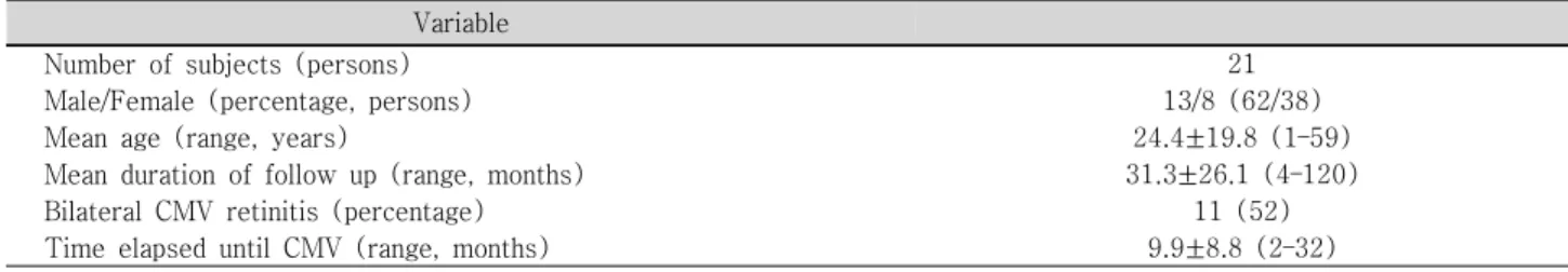
Table 1. Subject characteristics Variable
Number of subjects (persons) 21
Male/Female (percentage, persons) 13/8 (62/38)
Mean age (range, years) 24.4±19.8 (1–59)
Mean duration of follow up (range, months) 31.3±26.1 (4–120)
Bilateral CMV retinitis (percentage) 11 (52)
Time elapsed until CMV (range, months) 9.9±8.8 (2–32)
Table 2. Symptoms of CMV retinitis
Symptoms Eyes (percentage)
Decreased visual acuity 8 (35%)
Floater 6 (26%)
Photopsia 2 (9%)
No symptom 7 (30%)
Total 23 (100%)
*Infants who could not complain about their symptom were excluded.
Figure 1. Predisposing conditions of subjects.
*Thrombotic thrombocytopenic purpura, Wiskott-Aldrich syndrome, congenital immune deficiency syndrome.
로 진단하였다.5
통계 분석은 SPSS (version 15.0) 통계프로그램을 이용 하여 분석하였다. 거대세포바이러스 망막염의 양안에서 발 생여부와 나이, 시력과의 관계를 알기 위하여 Mann-Whitnry U 검정을 이용하였으며 p값이 0.05 미만일 경우를 통계적 으로 유의하다고 하였다.
결 과
조사기간 동안 거대세포바이러스 망막염을 진단받아 최 종 분석에 포함된 환자는 모두 21명 32안이었다. 남자는 13명, 여자는 8명이었고 평균 연령은 24.4±19.8 (1~59) 세 이었다. 11명(52%)에서 양안에 발생하였으며, 통계적 으로 유의하게 양안에서 발생한 군이 단안에서 발생한 군
보다 더 낮은 연령임을 확인할 수 있었다(p<0.01). 평균 추적관찰기간은 31.3±26.1(4~120)개월 이었고, 심한 면 역 저하 발생시기로 추정된 때부터 거대세포바이러스 망막 염의 발병시기까지의 기간은 9.9±8.8 (2~32)개월 이었다 (Table 1). 거대세포바이러스 망막염이 처음 진단되었을 때 호소하는 증상은 시력저하가 8안(35%)으로 가장 흔하 였으며, 그 다음으로 무증상이 7안(30%), 비문증이 6안 (26%), 광시증이 2안(9%)이었다(Table 2).
원인이 되는 질환은 백혈병이 9명(42%)으로 가장 많았 으며, 그 외에 장기이식으로 인한 면역억제가 3명(14%), 후천성면역결핍증이 2명(9%), 재생불량성빈혈이 2명(9%), 항암제에 의한 경우가 2명(9%) 이었고, 그밖에 혈전성 혈 소판감소 자반증, Wiskott-Aldrich 증후군, 선천성 면역결 핍 증후군이 각각 1명이었다(Fig. 1).
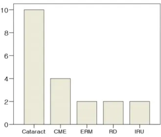
거대세포바이러스 망막염의 치료로서 15명에서 ganciclovir (Fig. 2), 2명에서는 foscarnet을 정맥주사 하였으며 4명은 처음에 ganciclovir를 정맥주사 하다가 foscarnet으로 바꾸 어 정맥주사 하였다(Table 3). 거대세포바이러스 망막염의 진단시의 평균 교정시력은 0.17±0.21이었으며, 최종 평균 교정시력은 0.16±0.19이었다(Fig. 3). 거대세포바이러스 망막염의 합병증으로 백내장은 10안(31.3%), 낭포황반부 종은 4안(12.5%), 망막박리는 2안(6.3%), 망막앞막은 2안 (6.3%), 그리고 면역회복포도막염이 2안(6.3%)에서 발생 하였다(Fig. 4).
망막염이 양안에서 발생한 경우와 단안에서 발생한 경우 망막박리의 발생 빈도를 비교하면 양안인 경우 9.1% (1/11 안), 단안인 경우 10% (1/10안)로 두군 사이에 망막박리의 빈도가 유의한 차이를 보이지 않았다(p=0.738, Fisher’s exact test). 또한 양안에서 망막염이 발생한 경우에 초진 과 최종시력(LogMAR)의 차이가 -0.31±0.18로 단안인 경우(0.07±0.13)와 비교하여 유의한 차이를 보이지 않았 다(p=0.129, Mann Whitney U test).
고 찰
거대세포바이러스는 항체가 미국 성인의 50~85%에서
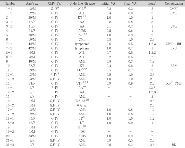
Table 3. Clinical characteristics of CMV retinitis patients
Number Age/Sex CMV Tx* Underline disease Initial VA† Final VA† Zone‡ Complication
1-1 12/M G IV§ ALL§§ 0.2 0.2 2 CME**
1-2 12/M G IV ALL 0.4 0.8 2 CME
2 50/M G IV KT∏∏ 1.0 1.0 1
3-1 16/F G IV AA 0.8 0.8 3
3-2 16/F G IV AA 0.1 0.7 1 CME
4 34/F G IV AIDS 0.5 0.8 1
5 38/M G IV CML## 1.0 0.9 2
6 10/M G IV CML 0.5 1.0 2,3
7-1 42/M G IV lymphoma 0.6 0.5 1,2,3 ERM††, IRU
7-2 42/M G IV lymphoma 1.0 0.7 1 IRU
8-1 4/M G IV ALL 0.7 0.5 1,2,3
8-2 4/M G IV ALL 0.2 0.6 2
9 40/M G IV AML 0.8 0.7 1,2
10 58/F G IV KT 0.4 0.6 2 ERM
11 59/M G IV PC### 0.5 0.7 2
12-1 13/M F IV∏ AML 0.9 1.0 2,3
12-2 13/M G,F IV AML 1.0 1.0 2,3
13 31/F G IV TTP∏∏∏ 0.9 0.9 2,3 RD‡‡, CME
14-1 2/F F IV AA*** - - 1,2,3,
14-2 2/F F IV AA - - 1,2,3
15 2/F F IV AML - - 2
16-1 1/M G,F IV WA sd.§§§ - - 2,3
16-2 1/M G,F IV WA sd. - - 2,3
17-1 12/M G,F IV AML 1.0 0.8 1,2
17-2 12/M G,F IV AML 1.0 0.8 1,2
18-1 45/F G IV LT††† 1.0 1.0 1,2
18-2 45/F G IV LT 1.0 0.9 1
19-1 1/M G IV IDS‡‡‡ - - -
19-2 1/M G IV IDS - - -
20 33/M G IV AIDS 1.0 0.9 2
21-1 9/F G,F IV AML 0.9 0.5 2,3
21-2 9/F G,F IV AML 0.6 0.2 2,3 RD
*Cytomegalovirus treatment regimen; †Visual acuity; ‡Zone 1=the area within 1500 μm of the optic nerve or within 3000 μm of the fovea, Zone 2=from zone 1 to the equator, Zone 3=anterior to the equator; §Intravenous ganciclovir; ∏Intravenous foscarnet; **Cystoids macular edema; ††Epiretinal membrane; ‡‡Retinal detachment; §§Acute lymphoblastic leukemia; ∏∏Kidney transplantation; ##Chronic myeloid leukemia; ***Aplastic anemia; †††Liver transplantation; ‡‡‡Immune deficiency syndrome; §§§Wiskott- Aldrich syndrome; ∏∏∏Thrombotic thrombocytopenic purpura; ###Prostate cancer.
발견이 되고 있으며, 사회경제적 수준이 낮거나 개발도상국 의 거주자, 남자 동성연애자 등에서는 약 90%까지 발견될 만큼 도처에 편재되어 있다.6-8 거대세포바이러스 감염은 평생에 걸쳐 지속되지만 건강한 사람들에서는 증상을 일으 키지 않고 잠복하여 있으나 면역이 저하되어 있는 환자에 서 활성화되어 폐, 위장관계, 중추신경계와 망막 등에 감염 증상을 보이게 된다. 후천성 면역결핍증후군의 환자들에서 는 평생 25~40%가 발생하지만 장기나 골수이식 후 면역 억제된 환자들에서 거대세포바이러스 망막염은 매우 적은 것으로 알려져 있다.9-13 그러나 후천성면역결핍중후군 환 자에서 고활성항레트로바이러스요법(Highly Active Anti- retroviral Therapy, HAART)을 사용한 이후 거대세포바이 러스 망막염의 유병률은 저하되었으며, 실제 7~15%정도
라고 한다.14-16본 연구에서는 전체 발생 중 백혈병 환자가 43%로 가장 많았으며 후천성 면역결핍증후군은 9%였다.
이는 한국에서 후천성 면역결핍증후군의 유병률이 전체인 구 중 0.003~0.1%로 전 세계의 1.0%에 비교하여 매우 적 기 때문으로 생각된다.
초반에 나타나는 증상은 망막에 발생한 병변의 위치에 영향을 받게 되는데 거대세포바이러스 망막염은 발병 시 보통 망막 주변부에서 작은 병변으로 나타나기 때문에 초 반에는 증상이 미비한 것으로 알려져 있으며, 시력저하 (67%), 비문증(49%), 광시증(49%) 등이 나타날 수 있다.
즉, 후천성 면역결핍증후군을 포함한 면역이 저하된 환자들 에서 시력저하나 광시증, 비문증이 나타나는 경우 거대세포 바이러스 망막염의 가능성을 고려해봐야 한다.17
A B
Figure 2.(A) Color fundus photograph of a transplant patient with cytomegalovirus retinitis manifested as a creamy- colored area with overlying retinal hemorrhages. The lesions are located in the temporal part of the macula and do not involve the fovea. (B) Color fundus photographs after repeated intravitreal ganciclovir injections for 20 days.
Figure 3.Change of visual outcome through follow-up period Figure 4. Complications of CMV retinitis
성인 거대세포바이러스 망막염의 경우 진단 당시 양안에 서 병변이 발견되는 경우가 약 30% 정도로 보고되고 있지 만,18-20소아의 경우 Baumal et al21의 연구결과에 의하면 거대세포바이러스망막염이 발생한 8명의 환아 모두에서 양 안에 이미 병변이 발생하여 성인과는 다른 양상을 보이고 있다. 이에 대하여 Kim and Kim4은 소아의 경우 발병 초기 에 시력저하에 대한 증상호소가 없어 진단이 늦어졌을 가 능성에 대하여 말한 바 있다. 본 연구에서도 병변이 양안에 발생한 경우는 11명(52%)이었으며, 이중 소아환자의 경우 75%가 양안에 발생하였다. 따라서 전체 환자 중 소아환자 가 차지하는 비중이 57%로 많아 양안에 발생한 경우가 다 른 연구들보다 양안에 더 많이 발생하였음을 확인할 수 있 었다. Yoon et al16은 0.4 이하의 중등도 시력저하는 52%에 서 발생하였으며 망막박리와 황반부 침범에 중등도 시력저
하 원인의 80%를 차지하였다고 하였다. 본 연구에서는 중 등도 시력저하는 2안(6%)에서 발생하였으며 그 원인은 망 막박리와 황반부 침범이었다. 또한 양안에서 망막염이 발생 한 경우 망막박리의 발생률과 시력저하는 단안에서 발생한 경우와 차이가 없었다. 이는 백혈병이 전체 기저질환의 48%
이며 이로 인하여 평균 나이는 24.4세였으며 특히 3명은 망 막염이 양안에서 발생하였지만 3세 이하로 시력측정을 할 수 없었던 점을 고려해야 할 것이다.
거대세포바이러스 망막염의 치료를 위하여 ganciclovir를 사용할 수 있는데 바이러스를 사멸시키기 보다는 증식을 저지하기 때문에 충분히 사용하지 않고 중단할 경우 망막 염이 다시 재발할 수 있다.22 경구복용을 하면 위장관계에 서 흡수가 10% 미만으로 생체이용률이 낮기 때문에 보통 정맥주사로 투여하게 되며, 보통 하루에 5~7.5 mg/kg을
2~3주 사용한 후 유지용량으로 하루에 5 mg/kg으로 사용 하게 된다. 이번 연구에서도 ganciclovir를 사용한 모든 환 자에서 정맥주사를 하였다. Foscarnet은 정맥주사를 하면 ganciclovir와 시력을 보존하는데 효과가 거의 비슷한 것으 로 보고되고 있다.23또한 후천성 면역결핍증후군 환자에서 는 foscarnet으로 치료한 군에서 ganciclovir로 치료한 군보 다 사망률이 낮은 것으로 보고되었다.24그러나 ganciclovir 와 비교할 때 주사하는 시간이 더 오래 걸리며 신장독성, 전해질 이상과 같은 이상반응이 잘 발생하여 사용이 제한 되고 있다.25
인간 면역결핍바이러스에 감염되지 않은 성인 면역저하 환자에서 발생한 거대세포바이러스 망막염은 후천성 면역 결핍증후군 환자에서 발생한 망막염과 임상양상 및 예후가 비슷하다.26 전층 망막 괴사가 발생하는 경우 합병증으로 열공성 망막박리가 발생할 수 있고, 거대세포바이러스 망막 염을 진단받은 후 1년 안에 33%에서 발생한다.27근래에는 후천성 면역결핍증후군의 치료에서 고활성항레트로바이러 스요법을 사용함으로써 거대세포바이러스 망막염의 발생률 과 망막박리의 발생률이 60%정도까지 줄었다고 보고되었 다.28 망막박리는 망막염 병변이 크거나 앞에 위치해 있는 경우 발생 위험도가 증가하고29 ganciclovir를 안내주사로 치료할 경우 정맥주사만으로 치료한 경우보다 발생이 감소 된다고 보고되었다.30그 밖에 합병증으로 망막하출혈31및, 시신경32, 망막 그리고 홍채33의 신생혈관이 발생할 수 있다.
이전에 발표된 거대세포바이러스 망막염에 대한 연구는 후천성 면역결핍증후군 환자에서 발생한 것만을 제한적으 로 보고하였다. 그러나 국내의 상황을 고려해볼 때 후천성 면역결핍증후군의 유병률은 매우 적으며 임상에서는 장기 이식이나 혈액종양 등에 의한 항암치료 후에 발생하는 경 우가 더 많은데 저자들도 기저질환에서 백혈병이 43%로 가장 많은 비율을 차지함을 확인할 수 있었다. 또한 백혈병 을 포함한 혈액질환이 비교적 어린 나이에서 호발하며 유 아에서는 증상의 호소를 할 수 없음을 고려할 때 정기적인 안과검사의 중요성을 확인할 수 있었다. 하지만 이번 연구 에서는 환자의 수가 적어 의미 있는 분석을 시행할 수 없었 으며 추후 많은 대상으로 시행한 연구가 필요할 것이다.
결론적으로 한국에서 발생한 거대세포바이러스 망막염 의 원인질환으로는 백혈병이 가장 흔하였고, 발병 나이가 낮았으며, 양안에 발생하는 경우가 많았음을 확인할 수 있 었다. 면역이 저하되어 있는 환자들의 경우 시력저하, 광시 증, 비문증 등의 증상이 거대세포바이러스 망막염과 관련될 수 있으므로 주의 깊게 관찰할 필요가 있다. 또한 항바이러 스제 치료 후 시력예후는 비교적 양호하였지만 백내장이나 황반부종, 망막박리, 망막전막 등의 합병증이 발생할 수 있
으므로 정기적인 경과 관찰이 필요할 것이다.
참고문헌
1) Dujić M, Jevtović Dj, Salemović D, et al. The prognosis of CMV retinitis among patients with AIDS in Serbia. Biomed Pharmaco- ther 2008;62:443-7.
2) Gallant JE, Moore RD, Richman DD, et al. Incidence and natural history of cytomegalovirus disease in patients with advanced human immunodeficiency virus disease treated with zidovudine.
The Zidovudine Epidemiology Study Group. J Infect Dis 1992;
166:1223-7.
3) Kim NR, Moon YS, Chin HS, Yoon JH. A case of valganciclovir treatment for cytomegalovirus retinitis. J Korean Ophthalmol Soc 2008;49:531-8.
4) Kim YH, Kim SK. Cytomegalovirus retinitis in a child with acute lymphoblastic leukemia. J Korean Ophthalmol Soc 2006;47:1009-15.
5) Hughes MD, Johnson VA, Hirsch MS, et al. Monitoring plasma HIV-1 RNA levels in addition to CD4+ lymphocyte count im- proves assessment of antiretroviral therapeutic response. ACTG 241 Protocol Virology Substudy Team. Ann Intern Med 1997;
126:929-38.
6) Marshall GS, Rabalais GP, Stewart JA, Dobbins JG. Cytomeg- alovirus seroprevalence in women bearing children in Jefferson County, Kentucky. Am J Med Sci 1993;305:292-6.
7) Pass RF. Epidemiology and transmission of cytomegalovirus. J Infect Dis 1985;152:243-8.
8) Sohn YM, Oh MK, Balcarek KB, et al. Cytomegalovirus infection in sexually active adolescents. J Infect Dis 1991;163:460-3.
9) Ciardella AP, Barile G, Langton K, Chang S. Cytomegalovirus retinitis and FK 506. Am J Ophthalmol 2003;136:386-9.
10) Crippa F, Corey L, Chuang EL, et al. Virological, clinical, and op- hthalmologic features of cytomegalovirus retinitis after hematop- oietic stem cell transplantation. Clin Infect Dis 2001;32:214-9.
11) Montaner JS, Le T, Hogg R, et al. The changing spectrum of AIDS index diseases in Canada. AIDS 1994;8:693-6.
12) Munoz A, Schrager LK, Bacellar H, et al. Trends in the incidence of outcomes defining acquired immunodeficiency syndrome (AIDS) in the Multicenter AIDS Cohort Study: 1985-1991. Am J Epidemiol 1993;137:423-38.
13) Pertel P, Hirschtick R, Phair J, et al. Risk of developing cytomega- lovirus retinitis in persons infected with the human immunodef- iciency virus. J Acquir Immune Defic Syndr 1992;5:1069-74.
14) Holtzer CD, Jacobson MA, Hadley WK, et al. Decline in the rate of specific opportunistic infections at San Francisco General Hospital, 1994-1997. AIDS 1998;12:1931-3.
15) Jacobson MA, Stanley H, Holtzer C, et al. Natural history and outcome of new AIDS-related cytomegalovirus retinitis diagn- osed in the era of highly active antiretroviral therapy. Clin Infect Dis 2000;30:231-3.
16) Yoon CK, Woo SJ, Yu HG. Visual outcome of cytomegalovirus retinitis in korean patients with acquired immune deficiency syndrome. J Korean Ophthalmol Soc 2009;50:92-8.
17) Hodge WG, Boivin JF, Shapiro SH, et al. Clinical risk factors for cytomegalovirus retinitis in patients with AIDS. Ophthalmology 2004;111:1326-33.
18) Gross JG, Bozzette SA, Mathews WC, et al. Longitudinal study of cytomegalovirus retinitis in acquired immune deficiency synd- rome. Ophthalmology 1990;97:681-6.
19) Roarty JD, Fisher EJ, Nussbaum JJ. Long-term visual morbidity of cytomegalovirus retinitis in patients with acquired immune deficiency syndrome. Ophthalmology 1993;100:1685-8.
20) Winston DJ, Ho WG, Champlin RE. Cytomegalovirus infections after allogeneic bone marrow transplantation. Rev Infect Dis 1990;12:S776-92.
21) Baumal CR, Levin AV, Kavalec CC, et al. Screening for cytomeg- alovirus retinitis in children. Arch Pediatr Adolesc Med 1996;150:
1186-92.
22) Dunn JP, Jabs DA. Cytomegalovirus retinitis in AIDS: natural hi- story, diagnosis, and treatment. AIDS Clin Rev 1995-1996:99-129.
23) Foscarnet-Ganciclovir Cytomegalovirus Retinitis Trial. 4. Visual outcomes. Studies of Ocular Complications of AIDS Research Group in collaboration with the AIDS Clinical Trials Group.
Ophthalmology 1994;101:1250-61.
24) Mortality in patients with the acquired immunodeficiency syn- drome treated with either foscarnet or ganciclovir for cytomeg- alovirus retinitis. Studies of Ocular Complications of AIDS Research Group, in collaboration with the AIDS Clinical Trials Group. N Engl J Med 1992;326:213-20.
25) Morbidity and toxic effects associated with ganciclovir or fos- carnet therapy in a randomized cytomegalovirus retinitis trial.
Studies of ocular complications of AIDS Research Group, in collaboration with the AIDS Clinical Trials Group. Arch Intern
Med 1995;155:65-74.
26) Kuo IC, Kempen JH, Dunn JP, et al. Clinical characteristics and outcomes of cytomegalovirus retinitis in persons without human immunodeficiency virus infection. Am J Ophthalmol 2004;138:
338-46.
27) Rhegmatogenous retinal detachment in patients with cytome- galovirus retinitis: the Foscarnet-Ganciclovir Cytomegalovirus Retinitis Trial. The Studies of Ocular Complications of AIDS (SOCA) Research Group in Collaboration with the AIDS Clinical Trials Group (ACTG). Am J Ophthalmol 1997;124:61-70.
28) Kempen JH, Jabs DA, Dunn JP, et al. Retinal detachment risk in cytomegalovirus retinitis related to the acquired immunodefici- ency syndrome. Arch Ophthalmol 2001;119:33-40.
29) Biswas J, Choudhry S, Priya K, Gopal L. Detection of cytome- galovirus from vitreous humor in a patient with progressive outer retinal necrosis. Indian J Ophthalmol 2002;50:319-21.
30) Young S, McCluskey P, Minassian DC, et al. Retinal detachment in cytomegalovirus retinitis: intravenous versus intravitreal ther- apy. Clin Experiment Ophthalmol 2003;31:96-102.
31) Freidlin J, Sharma MC, Goldstein DA. Subretinal hemorrhage in cytomegalovirus retinitis. Ophthalmic Surg Lasers Imaging 2005;
36:73-5.
32) Sanislo SR, Lowder CY, Kaiser PK. Optic nerve head neovascu- larization in a patient with inactive cytomegalovirus retinitis and immune recovery. Am J Ophthalmol 1998;126:318-20.
33) Bogie GJ, Nanda SK. Neovascularization associated with cytome- galovirus retinitis. Retina 2001;21:85-7.
=ABSTRACT=
Cinical Manifestations and Prognosis of Cytomegalovirus Retinitis
Young Kyo Kwun, MD1, Ju Byung Chae, MD2, Don Il Ham, MD1
Department of Ophthalmology, Samsung Medical Center, Sungkyunkwan University School of Medicine1, Seoul, Korea Department of Ophthalmology, University of Ulsan, College of Medicine, Asan Medical Center2, Seoul, Korea
Purpose: Cytomegalovirus (CMV) retinitis is common in patients with immunodeficient conditions caused by acquired immunodeficiency syndrome (AIDS), cytotoxic chemotherapy and immunosuppresive treatment. The purpose of this study was to assess the clinical manifestations and prognosis of CMV retinitis cases.
Methords: Thirty-one eyes of 21 patients who were diagnosed with CMV retinitis were retrospectively reviewed. The clinical manifestations and prognosis of all patients were analyzed.
Results: The average age of patients was 24.4±19.8 years. Eight patients were female and 13 patients were male. The predis- posing conditions of patients were leukemia (nine patients), immunosuppressed conditions due to organ transplantation (three patients), AIDS (two patients) and other (seven patients). Eleven patients exhibited bilateral disease. The mean follow-up period was 31.3 months, and there were no differences between mean initial visual acuity (0.70±0.31) and mean visual acuity (0.77±0.20) at final visit. The major causes of visual loss were retinitis and atrophic changes involving the macula. Although retinitis was successfully treated with anti-viral agents in all cases, cataract (10 eyes, 31.3%), cystoid macular edema (four eyes, 12.5%), retinal detachment (two eyes, 6.3%), epiretinal membrane (two eyes, 6.3%) and immune recovery uveitis (two eyes, 6.3%) developed after the initial treatment.
Conclusions: Although the visual prognosis of CMV retinitis was relatively good after administration of appropriate antiviral therapy, clinicians should remain alert for the development of late complications, including retinal detachment, cystoid macular edema and immune recovery uveitis.
J Korean Ophthalmol Soc 2010;51(2):203-209
Key Words: Cinical manifestations, Cytomegalovirus retinitis, Prognosis
Address reprint requests to Don Il Ham, MD
Department of Ophthalmology, Samsung Medical Center, Sungkyunkwan University School of Medicine
#50 Ilwon-dong, Gangnam-gu, Seoul 135-710, Korea.
Tel: 82-2-3410-3567, Fax: 82-3410-0084, E-mail: [email protected]