1
Department of Periodontology, Research Institute for Periodontal Regeneration, College of Dentistry, Yonsei University
Abstract
Implantation with various guided bone regeneration techniques
Young-Taek Kim
1, Ji-Youn Hong
1, Sung-Tae Kim
1, Chang-Sung Kim
1, Kyoo-Sung Cho
1, Chong-Kwan Kim
1, Seong-Ho Choi
1김영택, 홍지연, 김성태, 김창성, 조규성, 김종관, 최성호
1
연세대학교 치과대학병원 치주과
임프란트
술식은 이제 전문의에 의해 행해지는 술식이 아니라 일반적인 치과 술식으로 행해 지고 있다. 현재 전악 무치악 뿐만 아니라 부 분 무치악과 단일치의 수복에 있어서도 널리 사용되는 술식이며, 95%가 넘는 성공율이 보고되고 있다
1).
치조골의 상황이 항상 임프란트 식립을 위하여 적절 한 상태이지는 않다. 수직적 혹은 수평적 골소실은 치주 질환으로 인한 치아의 발치시 필연적으로 나타나며, 다 른 원인으로 발치한다 하더라도 장기간 방치할 시에 치 조골의 퇴축으로 인한 수직, 수평 골소실이 나타난다.
흡수 혹은 파괴된 치조골을 회복하기 위하여 골 이식 (Bone graft)이나 골유도 재생술(Guided bone regeneration)을 시행하게 된다. 골 이식에는 자가골, 동종골, 이종골, 합성골 등의 다양한 골재료가 이용되 며, 결손부위에 적용하여 적절한 골 형태를 형성하도록 한다. 자가골의 경우, 골형성(osteogenesis), 골유도 (osteoinduction), 골전도성(osteoconduction)의 성질 을 모두 가지고 있어 골이식의 gold standard로 여겨
지나
2, 3), 이차 수술 부위를 새로 형성해야 한다는 점과
골양이 제한된다는 점, 그리고 빠른 흡수가 일어난다는 단점이 있다. 자가골을 제외한 동종골, 이종골, 합성골 등은 골전도성의 성질만을 가지고 있으나, 추가적인 수 술 부위를 형성할 필요가 없으며, 자가골에 비해 흡수가 늦음으로 인해 골이 형성될 동안 골의 모양을 유지해 줄 수 있다는 장점이 있다
4, 5).
골유도 재생술은 조직유도 재생술(Guided tissue regeneration)의 원리를 이용하여 개발되어 졌으며, 마 찬가지로 차폐막을 적용함으로써 주변 조직을 배제하고 혈병을 안정화하여 재생을 유도하는 방법이다
6, 7). 티타
늄-강화 고어텍스 차폐막의 경우, 형성해야 하는 골의 모양을 미리 형성하고 지지해주는 역할을 함으로써 골 유도 재생술의 성공율을 더욱더 높일 수 있다. 또한, 골 유도 재생술은 골이식과 같이 시행되어질 수 있다.
Buser 등은 systemic review를 통해 골유도 재생술을 동반한 임프란트 매식술이 장기적인 성공률에 있어서 골유도 재생술을 동반하지 않은 임프란트 매식술에 비 해서 유의성있게 떨어지지 않는다고 발표한 바 있다
8).
골유도 재생술을 위해 비흡수성 차폐막을 사용할 경 우, 차폐막의 조기노출이 빈번하게 일어날 수 있다.
Simion등은 차폐막의 조기노출로 인해 세균이 막의 내 부까지 침투할 수 있음을 조직학적인 관찰을 통해 확인 하였으며, 골형성은 41%까지 떨어질 수 있다고 발표한
바 있다
9, 10). 노출된 차단막은 항상 바로 제거해야 하는
것은 아니며, 적절한 처치를 통해서 차단막을 유지 혹은 제거하여 골재생을 최대한 형성할 수 있다
11).
이에 본 증례보고에서는 다양한 골결손부에 있어서 비흡수성 차단막과 골이식을 동반한 임프란트 매식술의 증례에서 차폐막의 조기노출을 적절히 치료하여 성공적 인 골형성을 보인 치험례를 보고하고자 한다.
증례 1
2007년 2월 24세 여자 환자가 하악의 보철적 수복을 주소로 내원하였다. 환자의 주소 부위인 35, 36, 37, 46, 47번은 발치한 지 4년이 지난 무치악 상태였으며, 환자는 이에 대한 임프란트 치료를 원하였다. 특별한 전 신질환은 없었으며, 대합되는 상악치아의 정출을 교정 하는 치료를 교정과(연세대학교)에서 받고 있었다. 하악
Ⅱ
I
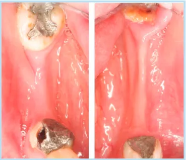
양측의 무치악 부위는 흡수가 빠르게 진행되어 모두 얇 고 낮은 흡수 양상을 보였다(Fig. 1,2).
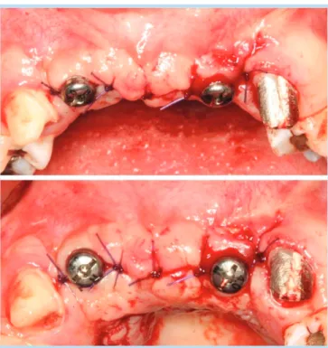
이에 치조골 확장술(ridge expansion)을 동반한 골 유도 재생술(guided bone regeneration, GBR)과 함 께 35,36,46,47번 부위의 임프란트를 계획하였다. 1차 적으로 임프란트를 식립할 충분한 공간을 가진 46번,
47번 부위에는 각각 RP, WP Bra ˚nemark Mk III 10mm 임프란트(Nobel Biocare, Yorba Linda, California)를 식립하였으며, 동시에 healing abutment를 연결하여 1-stage로 진행하였다(Fig. 3).
2달이 지나고 #46,47번의 임프란트 보철물이 진행되었 으며, 35, 36번 부위의 임프란트 식립을 위해 판막을 거 상하였다. 예상대로 치조골은 2mm 정도의 얇은 상태였 으며, 수직적 높이는 임프란트를 식립하기에 충분한 길 이가 계측되었다. 가이드 드릴링 후, 15번 블레이드와 스프레더 1, 2번을 이용하여, 치조골 확장술을 시행한 후, 35번과 36번에 각각 RP,WP Bra ˚nemark Mk III 10mm 임프란트를 식립하였다(Fig. 4). 치조골 확장술 과 협측 치조골편의 green-stick fracture로 인해 비 흡수성막을 이용한 골유도 재생술을 시행하기로 하고, 수술 부위 후방의 retromolar pad부위에서 직경 6.2mm의 trephine bur를 이용하여 자가골을 채취하 였다(Fig. 5). 채취한 자가골을 bone crusher를 이용하 여 분쇄한 후, 분말 형태로 치조골을 확장한 수술 부위 와 협측 치조골에 적용하였다. 분말 형태로 적용하였으 므로, 연조직에 의해 둘러 싸여지게 되거나 형태가 흩어 Fig. 1. Preoperative view.
Young-Taek Kim et al: Implantation with various guided bone regeneration techniques. Implantology 2008
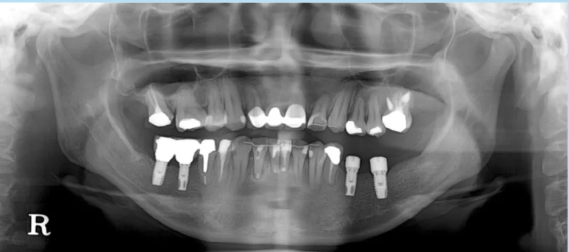
Fig. 2. Preoperative panoramic radiograph.
Young-Taek Kim et al: Implantation with various guided bone regeneration techniques. Implantology 2008
Fig. 4. Ridge expansion and installation of implant fixtures.
Young-Taek Kim et al: Implantation with various guided bone regeneration techniques. Implantology 2008
Fig. 5. Autogenous bone graft with trephine bur.
Young-Taek Kim et al: Implantation with various guided bone regeneration techniques. Implantology 2008
Fig. 6. Adaptation of particulated autogenous bone and application of e-PTFE membrane.
Young-Taek Kim et al: Implantation with various guided bone regeneration techniques. Implantology 2008
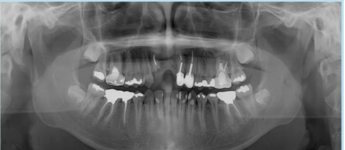
Fig. 3. Preoperative panoramic radiograph.
Young-Taek Kim et al: Implantation with various guided bone regeneration techniques. Implantology 2008
Fig. 7. Postoperative panoramic radiograph.
Young-Taek Kim et al: Implantation with various guided bone regeneration techniques. Implantology 2008
Fig. 9. PPostoperative panoramic radiograph.
Young-Taek Kim et al: Implantation with various guided bone regeneration techniques. Implantology 2008
Fig. 8. Re-entry for membrane removal and healing abutment connection.
Young-Taek Kim et al: Implantation with various guided bone regeneration
techniques. Implantology 2008
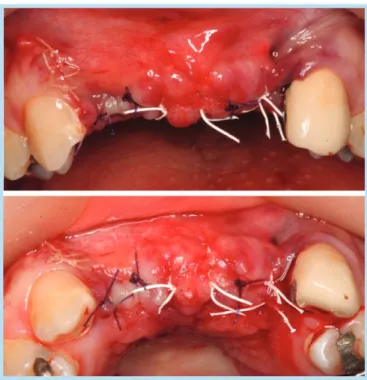
지는 것을 방지하기 위하여 cover screw를 연결한 후 에 TR6Y e-PTFE Gore-Tex
Ⓡmembrane(W.L.
Gore & Associates, Inc., Flagstaff, Arizona)을 적 용하고 Frios
Ⓡpin(Dentsply, Tulsa, Oklahamo)으로 고정하였다(Fig. 6). 봉합은 Gore-Tex
Ⓡsilk를 이용하 였다(Fig. 7).
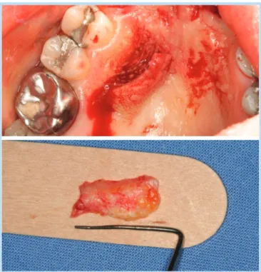
2개월이 지난 후, 35번 부위 crest상방에 fistula가 형성되어 클로르헥시딘으로 이용하여 드레싱을 하고 항 생제 투여를 하였으나, 나아지지 않아 이른 2차수술을 계획하였다. 비흡수성막을 이용하여 유도한만큼의 골이 형성되지는 않았으나, 치조골이 확장되어 자가골이 이 식된 부위와 green-stick fracture이 되었던 협측 치 조골편 모두 완전히 골화되었음을 확인할 수 있다(Fig.
8). healing abutment를 연결하고 봉합하였다(Fig.
9).
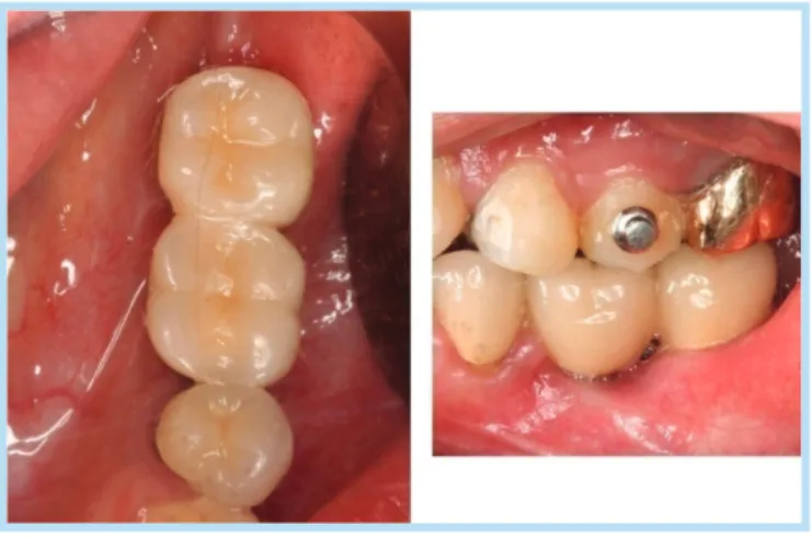
6개월이 지나고 보철물은 모두 완성되었으며, 부착치 은의 부족으로 인해 연조직의 외상을 보였으나, 그 외 특별한 불편함을 호소하지는 않았고, 기능적으로 심미 적으로 만족스러운 결과를 얻을 수 있었다(Fig. 10, 11).
증례 2