REVIEW ARTICLE
비침습적 간섬유화 검사의 종류와 의의
신정우, 박능화
울산대학교 의과대학 울산대학교병원 내과학교실
Clinical Application of Non-invasive Diagnostic Tests for Liver Fibrosis
Jung Woo Shin and Neung Hwa Park
Department of Internal Medicine, Ulsan University Hospital, University of Ulsan College of Medicine, Ulsan, Korea
The diagnostic assessment of liver fibrosis is an important step in the management of patients with chronic liver diseases.
Liver biopsy is considered the gold standard to assess necroinflammation and fibrosis. However, recent technical advances have introduced numerous serum biomarkers and imaging tools using elastography as noninvasive alternatives to biopsy. Serum markers can be direct or indirect markers of the fibrosis process. The elastography-based studies include transient elastography, acoustic radiation force imaging, supersonic shear wave imaging and magnetic resonance elastography. As accumulation of clinical data shows that noninvasive tests provide prognostic information of clinical relevance, non-invasive diagnostic tools have been incorporated into clinical guidelines and practice. Here, the authors review noninvasive tests for the diagnosis of liver fibrosis. (Korean J Gastroenterol 2016;68:4-9)
Key Words: Diagnosis; Fibrosis; Liver
CC This is an open access article distributed under the terms of the Creative Commons Attribution Non-Commercial License (http://creativecommons.org/licenses/
by-nc/4.0) which permits unrestricted non-commercial use, distribution, and reproduction in any medium, provided the original work is properly cited.
Copyright © 2016. Korean Society of Gastroenterology.
교신저자: 박능화, 44033, 울산시 동구 방어진순환도로 877, 울산대학교병원 내과
Correspondence to: Neung Hwa Park, Department of Internal Medicine, Ulsan University Hospital, 877 Bangeojinsunhwan-doro, Dong-gu, Ulsan 44033, Korea.
Tel: +82-52-250-7400, Fax: +82-52-251-8235, E-mail: [email protected] Financial support: None. Conflict of interest: None.
서 론
감염, 비알코올 지방간, 알코올, 유전성 대사질환, 담즙정체 성 질환, 면역질환, 약물 등에 의한 간세포의 손상은 간성상 세포 활성화, 여러 사이토카인 분비 촉진, 콜라겐 침착 증가 등을 유발하고 간섬유화를 일으키며 결국 간경변증으로 진행 하게 된다. 현재까지 간섬유화 유무, 진행 정도를 파악하기 위한 최적 표준 검사는 간생검법이다. 그러나 간생검법은 침 습적 검사로 검사와 관련된 합병증 발생률이 비록 낮기는 하 지만 최악의 경우 사망까지 발생할 수 있어 임상적으로 쉽게 검사할 수 없는 문제점이 있다. 또 생검으로 얻은 조직표본의 크기가 작아서 표집 오차가 발생할 수 있으며 병리학적 등급 결정도 병리학자 간에 결과의 차이를 보일 수 있고1,2 치료 경 과를 확인하기 위해 반복하여 검사하기 어렵다.
간생검을 대체하기 위한 목적으로 비침습적 간섬유화 검사
에 대한 소규모 연구들이 진행되었으며, 만성 간염 치료제의 비약적인 발전으로 인해 간섬유화 정도를 진단하거나 치료 중 간섬유화의 회복을 추적하고자 하는 임상적 필요성이 대두되 었다. 경구용 B형 간염 치료제의 장기간 사용으로 혈액 내 바이러스 음전 및 생화학적 정상화를 유지시킬 뿐 아니라, 간 내 염증을 줄여 간섬유화와 간경변도 회복될 수 있음이 밝혀 졌다.3,4 따라서 만성 B형 간염 치료의 장기 치료 목표인 간섬 유화 억제 및 회복 여부를 확인하기 위한 검사법이 필요하게 되었다. 새로운 경구용 C형 간염 치료제는 높은 치료 효과로 과거 인터페론 치료 대상이 되지 못한 비대상성 간경변증 환 자까지 치료 대상을 넓혔다. 그러나 고가의 약제 비용으로 인 해 치료 시작을 결정하지 못할 때, 간섬유화 정도가 치료 시기 를 결정하는 중요 요인이 될 수 있다. 유럽 간학회 가이드라인 에 따르면 METAVIR F2 이상의 만성 C형 간염은 치료의 적 극적 대상이며, 간경변으로 진단된 경우 치료 기간 및 치료
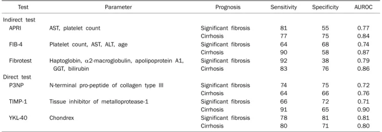
Table 1. Diagnostic Accuracy of Serum Makers
Test Parameter Prognosis Sensitivity Specificity AUROC
Indirect test
APRI AST, platelet count Significant fibrosis
Cirrhosis
81 77
55 75
0.77 0.84 FIB-4 Platelet count, AST, ALT, age Significant fibrosis
Cirrhosis
64 90
68 58
0.74 0.87 Fibrotest Haptoglobin, 2-macroglobulin, apolipoprotein A1,
GGT, bilirubin
Significant fibrosis Cirrhosis
92 83
38 76
0.79 0.86 Direct test
P3NP N-terminal pro-peptide of collagen type III Significant fibrosis Cirrhosis
74 64
75 66
0.72 0.76 TIMP-1 Tissue inhibitor of metalloprotease-1 Significant fibrosis
Cirrhosis
66 91
72 65
0.71 0.90
YKL-40 Chondrex Significant fibrosis
Cirrhosis
78 80
81 71
0.81 0.80 AUROC, area under the receiver operating characteristics curve; APRI, AST to platelet ratio index.
효과에 차이가 있고 완치 후에도 지속적인 간암 스크리닝의 대상이 되므로 간경변 여부를 정확하게 진단하는 것이 중요하 게 되었다.5 최근 증가하고 있는 비알코올 지방간질환 환자에 서 불필요한 간생검을 줄이기 위해 비침습적 간섬유화 검사가 도움이 될 수도 있을 것이다.
비침습적 간섬유화 검사는 혈청에서 섬유화와 관련된 특정 물질을 검사하거나, 물리학적 특성인 탄성력을 영상학적 검사 법을 이용해 측정하는 방법으로 나눌 수 있다. 이 종설에서는 임상에서 흔히 사용하고 있거나 가이드 라인에 소개된 비침습 적 간섬유화 검사법들을 중심으로 각 검사의 장단점, 간섬유 화 진단의 정확성 등을 소개하고자 한다.
본 론
1. 혈청표지자
많은 혈청 표지자들이 간섬유화 진단을 위해 연구되었고 개발되었다(Table 1). 혈청 표지자 검사들은 검사법이 간편하 여 임상에서 쉽게 사용할 수 있으며, 검사실 간 검사 결과 오 차가 적고 재현성이 높다. 반면에 간섬유화 시작과 간경변을 진단할 수 있으나 간섬유화 단계를 구분하기가 어려우며, 간 섬유화에 특이적인 혈청 표지자가 없으므로 간 이외 다른 장 기나 질환에 의해 결과값의 간섭이 나타난다. 혈청 표지자들 은 간경변이 시작되면서 나타나는 간기능 감소를 반영하는 간 접 표지자들과, 세포외 기질의 회전율과 간섬유화와 직접 연 관된 물질을 측정하는 직접 표지자들로 나눌 수 있다.
1) AST/ALT ratio
AST, ALT는 간세포 내에 존재하는 효소로 간세포 괴사의 정도를 반영하는 검사로 널리 사용되고 있다. 대부분의 만성 간염에서 AST/ALT ratio는 1 이하를 나타내나 1 이상을 보 이는 경우 간경변을 시사하는 소견으로 간주될 수 있다.
2) AST to platelet ratio index (APRI)
혈청 AST값과 혈소판 결과를 이용하여 심한 간섬유화와 간경변을 진단할 수 있는 간단한 방법으로, 계산방법은 다음 과 같다: APRI=AST (IU/L)/혈소판(109/L). C형 간염 환자들 을 대상으로 한 메타분석 결과에 따르면 APRI score 1.0 이상 인 경우 간경변으로 예측할 수 있으며(민감도 76%, 특이도 72%), 0.7 이상인 경우 METAVIR score F2 간섬유화로 예측 가능하다(민감도 77%, 특이도 72%).6 B형 간염 환자들을 대 상으로 한 연구도 유사한 결과를 보여주고 있다.7 APRI 검사 는 임상에서 쉽게 검사할 수 있는 혈소판, AST를 이용한다는 장점이 있으나, 다른 혈청 표지자에 비해 진단 및 예후 예측도 가 낮다.
3) Fibrosis-4 (FIB-4)
FIB-4 지표는 혈청 AST, ALT, 나이, 혈소판을 이용하여 계산한다. 초기에 만성 C형 간염 환자들을 대상으로 한 연구 에서 FIB-4 값이 3.25 이상인 경우 간경변 진단 정확도의 area under the receiver operating characteristics curve (AUROC)가 0.91이었으며, 비알코올 지방간 환자들을 대상으 로 한 연구에서는 다른 혈청 표지자 검사법보다 높은 진단 정확도를 나타내었다.8,9
4) Fibrotest
Fibrotest는 2개의 임상지표(나이, 성별)와 5개의 생화학적 표지자(alpha-2-macroglobulin, apolipoprotein A1, hapto- globin, total bilirubin, gamma-glutamyl transpeptidase) 를 조합하여 계산한다. 계산된 값은 0에서 1까지로 이 값들은 METAVIR 섬유화 단계와 상관관계를 갖는다. 메타분석 결과 에 의하면 진행된 간경변 진단의 AUROC가 0.84였으며, 알코 올, 만성 B형 간염, 만성 C형 간염, 비알코올성 지방간질환 등의 원인 질환과 무관하게 비슷한 진단 정확도를 보였다.10 Fibrotest와 transient elastograrphy를 동시 검사하여 섬유
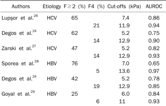
Table 2. Diagnostic Performance of Transient Elastography in Patients with Hepatitis B and C
Authors Etiology F≥2 (%) F4 (%) Cut-offs (kPa) AUROC
Lupşor et al.26 HCV 65 7.4 0.86
21 11.9 0.94
Degos et al.24 HCV 62 5.2 0.75
14 12.9 0.90
Zarski et al.27 HCV 47 5.2 0.82
14 12.9 0.93
Sporea et al.28 HBV 76 7.0 0.65
5 13.6 0.97
Degos et al.24 HBV 42 5.2 0.78
19 12.9 0.85
Goyal et al.29 HBV 25 6.0 0.84
6 11 0.93
AUROC, area under the receiver operating characteristics curve.
화 진단 정확도를 높이기 위한 연구 및 치료 후 섬유화 진행을 추적하기 위한 연구 등도 진행되었다.11,12
5) Non alcoholic fatty liver disease (NAFLD) fibrosis score (NFS)
NFS는 당뇨병, 나이, 성병, AST, ALT, 혈소판, 체질량지 수, 혈청 알부민으로 구성되어 있으며, 13개의 임상연구를 분 석한 메타분석에서 F3 이상의 간섬유화 진단에서 AUROC값 은 0.85를 보였다.13
6) Hyaluronic acid (HA)
HA는 세포외액기질에서 발견된 N-아세틸글루코사민과 글 루쿠론산으로 이루어진 고분자 화합물로 간염 및 간섬유화 진 행 정도에 비례하여 혈중 농도가 증가한다. 만성 C형 간염에 서 간경변 진단의 AUROC값은 0.78이었으나, F2, F3 섬유화 진단의 정확도는 만족스럽지 못했다.14 Hepascore는 HA와 빌 리루빈, gamma-glutamyl transpeptidase, 알파2-마크로글 로불린, 나이, 성별을 조합한 지표로, 간경변 진단에서 AUROC값은 0.89였다.15
7) N-terminal pro-peptide of collagen type III (P3NP) P3NP는 콜라겐 대사를 반영하는 물질로 간섬유화의 지표 로 연구되었다. 초기 연구들은 원발성 담즙성 간경변증에서 진행되었으며 간섬유화 및 담즙 정체 정도와 상관관계가 높다 고 발표되었다.16 비알코올 간질환에서는 단순 지방증과 비알 코올 지방간염을 구별할 수 있는 생체 표지자로 연구되었으 며, 바이러스 간염환자들을 대상으로 한 연구에서는 혈소판과 조합하여 APRI, FIB-4, enhanced liver fibrosis (ELF) 점수 보다 간경변 진단의 정확도가 높았다.17,18
8) Tissue inhibitor of metalloprotease-1 (TIMP-1) 간섬유화는 콜라겐 합성 증가와 콜라겐 제거의 불균형 결 과로 발생하며 혈청 콜라게네이즈(collagenase) 감소를 동반 하게 된다. TIMP-1은 콜라게네이즈를 불활성화시키는 효소 의 일종으로 알코올 간염 환자에서 섬유화와 관련성이 처음 발견되었다.19 이후 만성 C형 간염과 만성 B형 간염 환자들에 서 간섬유화 단계 및 간염 활성화 정도의 연관성이 연구되었 다.20,21
9) YKL-40
YKL-40은 세포외기질 재형성에 관여하는 단백질로 간성상 세포에 발현한다. P3NP나 HA처럼 혈청내 농도가 세포외기 질과 관련되어 증가하는 것으로 알려져 있다. YKL-40은 만성 C형 간염환자에서 인터페론 치료 후 혈청 농도가 감소하고 간경변 진단과 관련된 AUROC는 0.79로 보고되었다.22 10) Enhanced liver fibrosis (ELF)
ELF는 혈청 내 HA, TIMP-1, P3NP값과 나이를 조합하여 산출된 ELF 점수로 결과를 해석하는 다변량 지표 측정 검사 이다. 16편의 연구를 종합한 메타분석 연구에서 AUROC값은
F2 섬유화는 0.76-0.98, F3 섬유화는 0.69-0.99, 간경변은 0.70-0.91로 보고되었다.23
11) 혈청 검사법의 정확도
Fibrotest, Hepascore, Fibrometer와 같은 특허 받은 혈청 간섬유화 검사법과 APRI 검사를 비교한 대규모 환자 대상 연 구(만성 C형 간염 환자 913명, 만성 B형 환자 284명)에 따르 면 F2 간섬유화 진단을 위한 AUROC값은 0.72-0.78로 차이 를 보이지 않았다. 간경변 진단을 위한 AUROC값도 0.77- 0.86으로 의미 있는 차이는 없었다.24
2. 영상학적 검사 1) 복부 초음파검사
초음파검사는 간편하고 피험자에게 방사능 피폭 등의 해가 없으며 실시간 관찰이 가능하여 스크리닝으로 널리 사용되는 대표적인 비침습적 검사법이다. 초음파에서 섬유화를 시사하 는 소견으로 간실질의 에코 증가, 간 표면의 결절, 간비대, 꼬 리엽의 비대 등이다. 초음파 검사 중 도플러 검사를 추가하거 나 조영 증강 검사를 시행하여 간경변 진단의 정확도를 높일 수 있다. 그러나 초음파 검사는 검사자 간 진단의 일치도가 낮고 초기 간섬유화 진단에 한계를 보인다.
2) Transient elastography (TE)
TE는 낮은 주파수(50 Hz)의 기계적 진동파가 간을 통과하 는 속도를 3.5 MHz의 m-mode 탐촉자로 측정하여 간의 탄성 도를 검사한다. 조직에서 전단파의 진행 속도는 조직의 강도 에 비례하므로 딱딱한 조직에서 진행 속도는 빨라진다. TE는 간을 통과하는 진동파의 속도를 kPa 단위로 나타내주며 간생 검과 비교하여 간섬유화의 정도를 진단하기 위한 많은 연구들 이 진행되었다. B형 간염, C형 간염, 알코올성 간질환들을 포 함한 메타분석 연구에 의하면 TE의 간경변 진단정확도는
0.9-0.99로 간질환의 원인에 따라 9-26.6 kPa 진단 기준값을 보였다(Table 2).25 TE의 간경변 진단 정확도는 F1-F3 간섬유 화보다 우수하므로 임상에서 간경변 환자를 선별하는 데 도움 이 될 것이다.24,26-29 TE 검사는 간섬유화의 진단뿐만 아니라 간경변증 합병증 발생 예측, 문맥압 항진증과의 연관성, 수술 후 간부전 발생 예측 등 다양한 용도로 연구되고 있다.30-32 TE 검사는 빠르고 간편하며 재현율이 높으며 검사자 간에 결 과 일치도가 높다. 반면 TE 검사의 제한점으로 복수, 심한 비 만 환자, 임신 중인 경우 검사가 어려우며 간염이 심한 경우, 간 종괴, 간내 울혈, 담즙 정체 등에서는 비정상적인 결과를 나타내므로 결과 해석에 주의해야 한다.
최근 유럽 간학회 가이드라인에서는 만성 C형 간염에서 TE와 혈청 간섬유화 지표 결과가 일치하는 경우 진단의 정확 도가 높으므로 간생검을 생략할 수 있다고 권고하고 있다.33 미국 간학회와 유럽 간학회 가이드라인에 따르면 만성 C형 간염 환자는 선별검사로 비침습적 간섬유화 검사를 시행하고 이 검사에 간경변으로 진단된 경우 문맥압 항진증 검사와 간 암 검사를 바로 시작할 것을 권고하고 있다.5,34
3) Acoustic radiation force imaging (ARFI)
ARFI 검사는 고식적인 초음파를 이용하여 조직 내 일정부 위에 높은 강도의 초음파를 집중시켜 전단파를 발생시키고 그 전단파를 다른 탐지펄스를 이용하여 탐지하는 방법을 이용한 다. 이 측정법은 특정 모델의 초음파 장비에 장착되어 있으며 원하는 부위에서 탄성을 측정할 수 있고 검사 시간이 빠르다.
TE와 다르게 복수나 비만 환자에서도 측정이 가능하다. 36개 의 연구에 포함된 약 4,000여 명의 환자를 분석한 메타분석 연구의 F2 간섬유화, 간경변의 진단에서 AUROC값은 0.84, 0.91을 나타냈다.35 최근 TE와 ARFI 검사를 비교한 메타분석 에 따르면 F2 섬유화 진단의 민감도와 특이도는 각각 0.74 vs. 0.87, 0.83 vs. 0.87로 양 검사 간에 차이는 없었다.36 4) Supersonic shear wave imaging (SSWI)
SSWI는 최근에 개발된 탄성 측정검사로, 고식적 초음파 탐 속자를 사용하여 5개의 초점에 음파를 연속적으로 집중시켜 크기가 큰 전단파를 발생시키고 이를 실시간으로 이차원적 탄 성 영상으로 만든다. 이 검사는 ARFI 검사에 비해 넓은 부위 의 탄성을 측정할 수 있으나 약한 접속파를 이용하므로 비만 환자, 호흡을 참지 못하는 환자 등에서 검사 정확도가 떨어진 다. 최근에 만성 C형 간염 환자를 대상으로 한 연구에서 F3 섬유화를 진단하는 AUROC값은 0.962, 간경변 진단에서 AUROC값은 0.968의 우수한 값을 나타냈다.37 226명의 만성 B형 간염 환자를 대상으로 한 연구에서 F2, 간경변 진단의 AUROC값은 0.88, 0.98의 결과를 얻었다.38 SSWI 검사는 다 른 초음파를 이용한 검사법보다 정확도가 높으나 검사의 숙련 도가 필요하며 많은 환자들을 대상으로 한 연구가 필요하다.
5) Magnetic resonance (MR) elastography
MR elastography 검사는 환자의 우상복부에 40-120 Hz 의 음파를 발생시키는 유도장치를 부착시키고 간에 전단파를 보내, 간에서 발생하는 기계적 파동을 MRI 영상으로 얻는다.
12개의 연구를 분석한 메타연구에서 나이, 성별, 체질량지수, 염증, 간질환의 원인 등에 상관없이 진행된 섬유화 진단의 정 확도가 높은 것으로 알려져 있다.39 그러나 MR elastography 와 TE의 비교연구에서는 서로 상반된 결과를 나타내어 추가 적인 연구가 필요하다.40,41 이 검사법의 단점은 검사 시간이 길고 검사비가 비싸며 간 내 철 농도, 담즙 정체, 문맥 고혈압 등에 영향을 받는다는 것이다.
결 론
비침습적 간섬유화 검사는 만성 간질환의 관리에 있어 그 역할이 증가하고 있다. 최근까지의 연구결과를 종합해보면 대 부분의 비침습적 간섬유화 검사법들은 간경변을 배제하거나, 간섬유화가 없는 상태(F0)를 정확하게 진단할 수 있다. 따라 서 비침습적 간섬유화 검사가 F0 또는 간경변을 배제해야 하 는 경우 간생검을 대신할 수 있지만, 간섬유화의 단계(F1-F3) 를 정확하게 구분하기에는 부족해 보인다. 최근 효과가 뛰어 난 항바이러스 약물들이 개발되고 있으므로, 만성 간염, 간경 변 환자에서 치료 효과 및 예후 예측 등을 위해 혈액검사를 대신하여 간섬유화 정도를 추적하는 검사들이 임상에서 활용 도가 증가하게 될 것이다. 따라서 간섬유화 진행을 정확하게 평가하기 위한 비침습적 검사법의 개발과 전향적 연구들이 필 요하다.
REFERENCES
1. Poynard T, Munteanu M, Imbert-Bismut F, et al. Prospective anal- ysis of discordant results between biochemical markers and bi- opsy in patients with chronic hepatitis C. Clin Chem 2004;50:
1344-1355.
2. The French METAVIR Cooperative Study Group. Intraobserver and interobserver variations in liver biopsy interpretation in pa- tients with chronic hepatitis C. Hepatology 1994;20:15-20.
3. Asselah T, Marcellin P. Long-term results of treatment with nu- cleoside and nucleotide analogues (entecavir and tenofovir) for chronic hepatitis B. Clin Liver Dis 2013;17:445-450.
4. Chang TT, Liaw YF, Wu SS, et al. Long-term entecavir therapy re- sults in the reversal of fibrosis/cirrhosis and continued histo- logical improvement in patients with chronic hepatitis B.
Hepatology 2010;52:886-893.
5. European Association for Study of Liver. EASL Clinical Practice Guidelines: management of hepatitis C virus infection. J Hepatol 2014;60:392-420.
6. Lin ZH, Xin YN, Dong QJ, et al. Performance of the aspartate ami-
notransferase-to-platelet ratio index for the staging of hepatitis C-related fibrosis: an updated meta-analysis. Hepatology 2011;
53:726-736.
7. Zhu X, Wang LC, Chen EQ, et al. Prospective evaluation of FibroScan for the diagnosis of hepatic fibrosis compared with liv- er biopsy/AST platelet ratio index and FIB-4 in patients with chronic HBV infection. Dig Dis Sci 2011;56:2742-2749.
8. Vallet-Pichard A, Mallet V, Nalpas B, et al. FIB-4: an inexpensive and accurate marker of fibrosis in HCV infection: comparison with liver biopsy and fibrotest. Hepatology 2007;46:32-36.
9. Shah AG, Lydecker A, Murray K, et al. Comparison of noninvasive markers of fibrosis in patients with nonalcoholic fatty liver disease. Clin Gastroenterol Hepatol 2009;7:1104-1112.
10. Poynard T, Morra R, Halfon P, et al. Meta-analyses of FibroTest diagnostic value in chronic liver disease. BMC Gastroenterol 2007;7:40.
11. Castéra L, Vergniol J, Foucher J, et al. Prospective comparison of transient elastography, Fibrotest, APRI, and liver biopsy for the assessment of fibrosis in chronic hepatitis C. Gastroenterology 2005;128:343-350.
12. Poynard T, Munteanu M, Deckmyn O, et al. Validation of liver fib- rosis biomarker (FibroTest) for assessing liver fibrosis pro- gression: proof of concept and first application in a large population. J Hepatol 2012;57:541-548.
13. Musso G, Gambino R, Cassader M, et al. Meta-analysis: natural history of non-alcoholic fatty liver disease (NAFLD) and diag- nostic accuracy of non-invasive tests for liver disease severity.
Ann Med 2011;43:617-649.
14. McHutchison JG, Blatt LM, de Medina M, et al. Measurement of serum hyaluronic acid in patients with chronic hepatitis C and its relationship to liver histology. Consensus Interferon Study Group. J Gastroenterol Hepatol 2000;15:945-951.
15. Chou R, Wasson N. Blood tests to diagnose fibrosis or cirrhosis in patients with chronic hepatitis C virus infection. Ann Intern Med 2013;159:372.
16. Babbs C, Smith A, Hunt LP, et al. Type III procollagen peptide: a marker of disease activity and prognosis in primary biliary cirrhosis. Lancet 1988;1:1021-1024.
17. Lee MH, Cheong JY, Um SH, et al. Comparison of surrogate serum markers and transient elastography (Fibroscan) for assessing cirrhosis in patients with chronic viral hepatitis. Dig Dis Sci 2010;55:3552-3560.
18. Tanwar S, Trembling PM, Guha IN, et al. Validation of terminal peptide of procollagen III for the detection and assessment of nonalcoholic steatohepatitis in patients with nonalcoholic fatty liver disease. Hepatology 2013;57:103-111.
19. Li J, Rosman AS, Leo MA, et al. Tissue inhibitor of metal- loproteinase is increased in the serum of precirrhotic and cir- rhotic alcoholic patients and can serve as a marker of fibrosis.
Hepatology 1994;19:1418-1423.
20. Leroy V, Monier F, Bottari S, et al. Circulating matrix metal- loproteinases 1, 2, 9 and their inhibitors TIMP-1 and TIMP-2 as serum markers of liver fibrosis in patients with chronic hepatitis C: comparison with PIIINP and hyaluronic acid. Am J Gastroenterol 2004;99:271-279.
21. Zhu CL, Li WT, Li Y, et al. Serum levels of tissue inhibitor of metal-
loproteinase-1 are correlated with liver fibrosis in patients with chronic hepatitis B. J Dig Dis 2012;13:558-563.
22. Saitou Y, Shiraki K, Yamanaka Y, et al. Noninvasive estimation of liver fibrosis and response to interferon therapy by a serum fibro- genesis marker, YKL-40, in patients with HCV-associated liver disease. World J Gastroenterol 2005;11:476-481.
23. Sul AR. Serum liver fibrosis test. J Korean Med Assoc 2014;57:704-709.
24. Degos F, Perez P, Roche B, et al. Diagnostic accuracy of Fibro- Scan and comparison to liver fibrosis biomarkers in chronic viral hepatitis: a multicenter prospective study (the FIBROSTIC study). J Hepatol 2010;53:1013-1021.
25. Lupsor Platon M, Stefanescu H, Feier D, et al. Performance of unidimensional transient elastography in staging chronic hep- atitis C. Results from a cohort of 1,202 biopsied patients from one single center. J Gastrointestin Liver Dis 2013;22:157-166.
26. Lupşor M, Badea R, Stefănescu H, et al. Analysis of histopatho- logical changes that influence liver stiffness in chronic hepatitis C. Results from a cohort of 324 patients. J Gastrointestin Liver Dis 2008;17:155-163.
27. Zarski JP, Sturm N, Guechot J, et al. Comparison of nine blood tests and transient elastography for liver fibrosis in chronic hep- atitis C: the ANRS HCEP-23 study. J Hepatol 2012;56:55-62.
28. Sporea I, Sirli R, Deleanu A, et al. Liver stiffness measurements in patients with HBV vs HCV chronic hepatitis: a comparative study. World J Gastroenterol 2010;16:4832-4837.
29. Goyal R, Mallick SR, Mahanta M, et al. Fibroscan can avoid liver biopsy in Indian patients with chronic hepatitis B. J Gastroenterol Hepatol 2013;28:1738-1745.
30. Kim BK, Kim DY, Han KH, et al. Risk assessment of esophageal variceal bleeding in B-viral liver cirrhosis by a liver stiffness measurement-based model. Am J Gastroenterol 2011;106:
1654-1662, 1730.
31. Kim SU, Ahn SH, Park JY, et al. Prediction of postoperative hepatic insufficiency by liver stiffness measurement (FibroScan((R))) be- fore curative resection of hepatocellular carcinoma: a pilot study. Hepatol Int 2008;2:471-477.
32. Kim SU, Lee JH, Kim DY, et al. Prediction of liver-related events using fibroscan in chronic hepatitis B patients showing ad- vanced liver fibrosis. PLoS One 2012;7:e36676.
33. European Association for Study of Liver; Asociacion Latinoamer- icana para el Estudio del Higado. EASL-ALEH Clinical Practice Guidelines: non-invasive tests for evaluation of liver disease se- verity and prognosis. J Hepatol 2015;63:237-264.
34. AASLD/IDSA HCV Guidance Panel. Hepatitis C guidance:
AASLD-IDSA recommendations for testing, managing, and treat- ing adults infected with hepatitis C virus. Hepatology 2015;62:
932-954.
35. Nierhoff J, Chávez Ortiz AA, Herrmann E, et al. The efficiency of acoustic radiation force impulse imaging for the staging of liver fibrosis: a meta-analysis. Eur Radiol 2013;23:3040-3053.
36. Bota S, Herkner H, Sporea I, et al. Meta-analysis: ARFI elastog- raphy versus transient elastography for the evaluation of liver fibrosis. Liver Int 2013;33:1138-1147.
37. Bavu E, Gennisson JL, Couade M, et al. Noninvasive in vivo liver fibrosis evaluation using supersonic shear imaging: a clinical
study on 113 hepatitis C virus patients. Ultrasound Med Biol 2011;37:1361-1373.
38. Poynard T, Munteanu M, Luckina E, et al. Liver fibrosis evaluation using real-time shear wave elastography: applicability and diag- nostic performance using methods without a gold standard. J Hepatol 2013;58:928-935.
39. Singh S, Venkatesh SK, Wang Z, et al. Diagnostic performance of magnetic resonance elastography in staging liver fibrosis: a systematic review and meta-analysis of individual participant
data. Clin Gastroenterol Hepatol 2015;13:440-451.e6.
40. Huwart L, Sempoux C, Vicaut E, et al. Magnetic resonance elas- tography for the noninvasive staging of liver fibrosis. Gastroen- terology 2008;135:32-40.
41. Bohte AE, de Niet A, Jansen L, et al. Non-invasive evaluation of liver fibrosis: a comparison of ultrasound-based transient elas- tography and MR elastography in patients with viral hepatitis B and C. Eur Radiol 2014;24:638-648.