INTRODUCTION
The level of periodontal support is the most critical factor for predicting the prognosis of healing after periodontal treatment [1,2]. In general, a tooth with periodontal destruction extending beyond the root apex is considered hopeless, and extraction is the only possible treatment. Neither nonsurgical nor surgical treatment is effective in such cases because of limited accessibility to instrumentation and unfavorable tooth stability after treatment. In the process of periodontal treatment, thorough instrumentation and antibacterial cleans- ing are performed to remove microbial infection sources. Nevertheless, there are limitations to accessing the periapical area and several studies have reported that microbial etiologic factors were found around the root apex area in recurrent infectious diseases [3,4]. More- over, the increased mobility of a treated tooth can affect the outcome of periodontal
This is an Open Access article distributed under the terms of the Creative Commons Attribution Non-Commercial License (http://creativecommons.org/licenses/by-nc/3.0/).
a retrospective study
Eun-Ung Lee1, Hyun-Chang Lim1, Jung-Seok Lee1, Ui-Won Jung1, Ui-Sung Kim2, Seung-Jong Lee2, Seong-Ho Choi1,*
1Department of Periodontology, Research Institute for Periodontal Regeneration, Yonsei University College of Dentistry, Seoul, Korea
2Microscope Center, Department of Conservative Dentistry and Oral Science Research Center, Yonsei University College of Dentistry, Seoul, Korea
Research Article
J Periodontal Implant Sci 2014;44:13-19 http://dx.doi.org/10.5051/jpis.2014.44.1.13
Purpose: The purpose of this study was to retrospectively evaluate the survival of periodon- tally hopeless teeth that were intentionally extracted and replanted after a delay and to com- pare the radiographic characteristics of the survival group with those of the failure group.
Methods: The clinical and radiographic data from patients who underwent delayed inten- tional replantation between March 2000 and July 2010 were reviewed. Twenty-seven peri- odontally hopeless teeth were extracted and preserved in medium supplemented with anti- biotics for 10–14 days. The teeth were then repositioned in the partially healed extraction socket and followed for 3 to 21 months. The radiographic parameters were analyzed using a paired t test and the cumulative survival rate was analyzed using Kaplan-Meier analysis.
Results: Seven replanted teeth failed and the overall cumulative survival rate was 66.4%.
In the survival group, the amount of bone loss was reduced from 68.45% to 34.66% three months after replantation. There was radiologic and clinical evidence of ankylosis with 5 teeth. However, no root resorption was found throughout the follow-up period. In the fail- ure group, bone formation occurred from the bottom of the socket. However, a remarkable radiolucent line along the root of a replanted tooth existed. The line lengthened and thick- ened as time passed. Finally, in each case of failure, the tooth was extracted due to signs of inflammation and increased mobility.
Conclusions: Delayed intentional replantation has many advantages compared to immedi- ate intentional replantation and could serve as an alternative treatment for periodontally involved hopeless teeth. However, techniques for maintaining the vitality of periodontal structures on the tooth surface should be developed for improved and predictable results.
Keywords: Alveolar bone loss, Bone remodeling, Periodontal diseases, Survival rate, Tooth replantation.
Received: Nov. 25, 2013 Accepted: Jan. 6, 2014
*Correspondence:
Seong-Ho Choi
Department of Periodontology, Research Institute for Periodontal Regeneration, Yonsei University College of Dentistry, 50 Yonsei-ro,
Seodaemun-gu, Seoul 120-752, Korea E-mail: [email protected] Tel: +82-2-2228-3189 Fax: +82-2-392-0398
treatment negatively. Tooth mobility may inhibit bone gain and periodontal regeneration, consequently forming a deep pocket and inducing apical migration of epithelial attachment [5].
Intentional replantation may be useful in these situations because this technique seems to resolve the limitations of conventional periodontal treatment. The tooth surfaces, including inaccessible areas, can be visualized and instrumented completely without damaging adjacent periodontal tissue. Some investigators have at- tempted intentional replantation with periodontally involved teeth.
Lu [6] reported successful treatment results with periodontally in- volved teeth in which intentional replantation was performed first.
The researcher intentionally replanted an endodontically mistreat- ed and periodontally involved mandibular first molar and main- tained the tooth for 32 months in a functional and asymptomatic condition. Demiralp et al. [7] intentionally replanted fifteen peri- odontally involved hopeless teeth and followed them for 6 months.
They suggested that intentional replantation could be an alterna- tive approach to extraction in cases where advanced periodontal destruction was present and no other treatment could be consid- ered.
However, in spite of the potential of this technique, very few in- vestigators have performed intentional replantation with periodon- tally involved teeth. This might be due to the questionable prog- nosis of replanted teeth. Although some studies have shown favor- able results, intentional replantation of a periodontally involved tooth seems to carry the risk of reinfection and unstable tooth stability, which results in tooth loss.
Previously, Lee et al. extracted periodontally hopeless teeth and replanted them after a delay to relieve inflammation and provide a scaffold with woven bone formation (unpublished data). They re- ported successful clinical and radiographic results by delaying the replant procedure, which they named “delayed intentional replan- tation” and concluded that the procedure could be an alternative treatment option for periodontally involved hopeless teeth. How- ever, in the study, the number of cases involved was limited and statistical analysis was not conducted. Therefore, additional research that evaluates the survival rate of the delayed intentional replan- tation procedure would be useful in determining the outcomes of the treatment.
The purpose of this study is to retrospectively evaluate the sur- vival of periodontally hopeless teeth that were intentionally replant- ed after a delay and to compare the radiographic characteristics of the survival group with those of the failure group.
MATERIALS AND METHODS
Study design and criteria
The clinical and radiographic data from patients who underwent delayed intentional replantation in the Department of Conserva- tive Dentistry at the Yonsei University College of Dentistry, Seoul, Korea between March 2000 and July 2010 were reviewed in this study. The patients who were included in the treatment of replan-
tation fulfilled the following requirements:
(1) No systemic disease and no contraindication for periodon- tal surgery.
(2) The presence of at least one tooth meeting the indications for tooth extraction due to severe periodontal destruc- tion: At least a 6-mm probing depth, minimum 60% ra- diographic periodontal bone loss, and grade III mobility according to Miller’s classification [8].
(3) Patient preference to retain the tooth rather than undergo extraction.
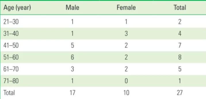
Consequently, 27 patients (17 males, 10 females) ranging in age from 24 to 72 years (mean age, 49.78 years) were included in this study. The patient distribution according to age and intraoral tooth location are as shown in Tables 1 and 2. This study was approved by the Institutional Review Board (IRB) of the Dental Hospital of Yon- sei University of College of Dentistry (IRB number: 2-2012-0035).
Treatment procedure Surgical technique
Under local anesthesia, each hopeless tooth was extracted as atraumatically as possible. If necessary, lingual and buccal flap in- cisions were made and small flap elevation was performed. Patho- logic factors existing on the tooth surface were removed gently using an ultrasonic scaler, and then the tooth surface was planed with a fine diamond bur (Fig. 1). Granulation tissues in the extrac- tion socket were thoroughly removed by curettage, and then saline irrigation was performed for disinfection. The extracted tooth was preserved in medium supplemented with antibiotics (1,000-nm dexamethasone solution) at 4ºC for 10 to 14 days. The average time of extraoral tooth storage was 11.3 days. After that, retrograde root canal treatment was carried out using an ultrasonic scaler and su- per EBA (Harry J. Bosworth Co., Skokie, IL, USA). The tooth was re- positioned in the partially healed extraction socket under local an- Table 1. Patient distribution according to age.
Age (year) Male Female Total
21–30 1 1 2
31–40 1 3 4
41–50 5 2 7
51–60 6 2 8
61–70 3 2 5
71–80 1 0 1
Total 17 10 27
Table 2. Tooth distribution according to intraoral location.
Location Maxilla Mandible Total
Anterior 1 19 20
Posterior 6 1 7
Total 7 20 27
esthesia. The tooth was positioned approximately in line with the adjacent teeth and a resin wire splint was performed in case the stability of the replanted tooth was unstable. When the stability of the replanted tooth was ensured, the patient was instructed to bite down on gauze. Consequently, 18 teeth in total were splinted after delayed replantation. Occlusal adjustment was performed to elimi- nate occlusal interference in centric and eccentric movements.
Postoperative care
After the tooth extraction and tooth replantation procedures, the patients were prescribed oral antibiotic therapy with amoxicil- lin (500-mg thrice per day) for three days. All of the patients were recommended to maintain their routine tooth brushing and to use a 0.12% chlorhexidine solution for 2 weeks. Seven days after tooth replantation, the patients were examined and saline irrigation was performed to clean the replantation sites. One month after replan- tation, the replanted teeth were polished and periapical radiographs were taken. Then patients were recalled every three months for clinical and radiographic examination and maintenance. When there was evidence of the presence of severe inflammation, increasing tooth mobility, or radiographic alveolar bone loss, the replanted tooth was considered a failure and was extracted.
Assessment method Survival criteria
The replanted teeth that were not extracted during the follow-up periods were deemed to have survived. When severe inflammatory signs or increasing mobility were found during the follow-up peri- od, the replanted teeth were defined as failures and extracted.
Radiographic analysis
Periapical radiographs were taken at baseline, one month post- operatively, and at every recall visit. The tooth distribution accord- ing to follow-up period is as shown in Table 3.
The Heliodent MD (Siemens, Fort Madison, IA, USA) was operat-
ed as an x-ray source under 60 kVp, 0.16 mAs conditions, and ra- diographic images were obtained using a charged coupled device sensor (SIGMA, GE Medical System Instrumentarium Co., Tuusula, Finland). Measuring bone loss was performed using the PiView STAR (Infinitt, Seoul, Korea) program and the method described by Schulte et al. [9]. To determine the amount of alveolar bone loss, two types of measurements were used: the total root length (ht) and the intra-alveolar root length (hi) (Fig. 2). The total root length (ht) was defined as the distance from the proximal cementoenamel junction to the apex parallel to the long axis of the tooth. The in- tra-alveolar root length (hi) was defined as the distance from the apex to the highest point on the alveolar margin. In the calcula- tion of bone loss, 1.5 mm was subtracted from the total root length because the distance between the crest of the alveolar bone and the cementoenamel junction ranges between 0.75 and 1.49 mm [10]. The average measurement at the mesial and distal sites was determined, and bone loss (BL) and bone gain (BG) were calculated using the following equations:
BL (%) = [1-hi/(ht-1.5)] × 100
BG (%) = preoperative BL (%) – postoperative BL (%) Table 3. Cumulative survival rates of delayed replanted teeth.
Follow-up
(month) Total
teeth Failed
teeth Cumulative
survival rate (%) Standard error (%)
0–1 27 0 100 -
1–3 27 3 88.9 0.060
3–6 27 2 80.4 0.079
6–9 17 0 80.4 -
9–12 9 1 80.4 -
12–15 9 1 66.4 0.113
15–18 4 0 66.4 -
18–21 3 0 66.4 -
A B
Figure 1. Extraction and preparation of a tooth before storage. (A) The tooth subjected to delayed replantation was extracted atraumatically. (B) Then the tooth surface was debrided and polished with an ultrasonic scaler and fine diamond bur.
Figure 2. Measure- ment for radiographic evaluation. ht: total root length, hi: intra- alveolar root length.
ht hi
Statistical analysis
The radiographic parameters were analyzed using a paired t-test and the cumulative survival rate was analyzed using Kaplan-Meier analysis.
RESULTS
Cumulative survival rate
A total of 27 teeth replanted after a delay were followed for 3–21 months. Seven replanted teeth failed, and the overall cumu- lative survival rate was 66.4%. Five of these teeth were extracted within six months of replantation. The cumulative survival rates for the entire group of replanted teeth by period of time postre- plantation are shown in Table 3 and Fig. 3.
Subcategorial descriptive observation: survival group Twenty teeth were not extracted during the follow-up periods and were defined as the survival cases. In this group, all of the pa- tients tolerated the intentional replantation procedure without any discomfort and were satisfied with the result of the replanta-
tion treatment. Clinically, all of the teeth were functioning without any signs or symptoms. Radiographs taken at every recall check-up showed remarkable bone gain around the replanted tooth (Fig. 4).
The amount of BL was significantly reduced from 68.45% to 34.66%
three months after replantation (P <0.05) (Fig. 5). After that, a consistent level of alveolar bone was maintained and the remodel- ing of new bone was proceeded (Figs. 4 and 6). The amount of BG was 45.02% three months after replantation and maintained a consistent level (Fig. 7). There was radiologic and clinical evidence of ankylosis with 5 teeth. However, root resorption was not found in the entire follow-up period.
Figure 3. Cumulative survival rate of teeth replanted after a delay.
100
80
60
40
20
0
0 3 6 9 12 15 18 21 24 Time (months)
Cumulative survival rate (%)
A B C D E F G
Figure 4. Radiographs taken before (A) and 1 (B), 3 (C), 6 (D), 9 (E), 12 (F), and 15 (G) months after replantation.
Figure 5. Amount of bone loss before and 3 months after tooth replantation,
*P<0.05.
80 70 60 50 40 30 20 10
0 Baseline 3 Months
(%)
Figure 6. The rate of bone loss (%) 1 to 21 months after tooth replantation.
80 70 60 50 40 30 20 10
0 0 1 3 6 9 12 15 18 21
Months
Bone loss (%)
Subcategorial descriptive observation: failure group During the follow-up, 7 teeth were extracted and defined as failure. In the failure group, radiographic measurements (BL, BG) were not investigated because the bone formation adjacent to the root of a replanted tooth was rarely seen in radiographs. After re- plantation, bone formation occurred from the bottom of the sock- et. However, a remarkable radiolucent line existed along the root of each replanted tooth. The line prolonged and expanded as time passed by. Finally, each tooth that had failed was extracted due to inflammatory signs and increased mobility. As a result, 71% of the teeth that had failed were extracted within 6 months after replan- tation (Fig. 8).
DISCUSSION
This study describes the clinical and radiological outcomes of delayed intentional replantation of teeth deemed hopeless due to severe periodontal destruction. We assumed that waiting for im- proved conditions of the extraction socket by delaying the replan- tation procedure could produce more favorable outcomes from in- tentional replantation.
Although it is commonly known that intentional replantation is contraindicated in a periodontally involved tooth [11], several stud- ies have reported successful clinical results and have suggested in- tentional replantation as an alternative treatment technique of last resort for a periodontally involved tooth [6,7]. However, little re- search has addressed this subject. This may be due to the possible risks mentioned below. Intentional replantation includes the fol-
lowing processes: atraumatic tooth extraction, removal of local factors on both the tooth surface and extraction socket, and rein- sertion of the tooth. Through this process, local factors on the tooth surface and extraction socket causing periodontal disease can be eliminated completely. However, forming substantial space between the tooth and socket wall is inevitable as a result of this process.
This space can negatively affect the result of the treatment. First, it is likely to increase the mobility of the replanted tooth. Generally, in case of tooth replantation, if the teeth are fixed in the coronal area, the teeth might be mobile in the apical area. Ferencz [5] re- ported that reduced periodontal regeneration could occur if the treated teeth were not fixed firmly. Mobility can decrease the amount of regenerated periodontal tissue after treatment. Secondly, rein- fection and delayed periodontal regeneration might occur because several microorganisms that can invade through the gap between a tooth root and extraction socket wall. In 2003, Demiralp et al. [7]
performed intentional replantation using teeth that needed to be extracted due to severe periodontal destruction. After 6 months, they found that alveolar bone loss had diminished somewhat, but was still more than 55% of the total tooth length.
In order to overcome the drawbacks of intentional replantation such as increased tooth mobility and the possibility of reinfection, we separated hopeless teeth from their extraction sockets for 10 days and then performed delayed replantation. Woven bone formed in the extraction socket one to two weeks after tooth extraction [12,13], and this bone not only helped each replanted tooth to be fixed firmly but also acted as a scaffold for periodontal tissue re- generation. Furthermore, 10 days of separation was rarely long enough to permit a gap between the replanted tooth and socket wall and thus decreased the risk of reinfection and reduced regen- eration. Additionally, the teeth could be replanted in the healing phase since the inflammatory phase of the extraction socket was converted to the healing phase during that separation period. In a previous immunochemical study [14] regarding the socket from which a periodontally involved tooth was extracted, inflammation persisted in the socket for 3 to 7 days, and was followed by forma- tion of woven bone and angiogenesis. In the same way, we assumed that we could expect a favorable outcome by performing replan- tation to the socket in the healing phase rather than during the inflammatory phase.
In our study, 27 teeth with severe periodontal destruction were intentionally extracted, replanted after a delay, and followed up Figure 7. The rate of bone gain (%) 1 to 21 months after tooth replantation.
60 50 40 30 20 10
0 1 3 6 9 12 15 18 21
Months
Bone gain (%)
Figure 8. Radiographs taken before (A) and 1 (B), 3 (C), 6 (D), and 9 (E) months after replantation. The tooth was removed 9 months after replantation due to loss of tooth stability.
A B C D E
for 3 to 21 months. Among these teeth, 7 teeth were removed and the cumulative survival rate was 66.4%. The radiologic examina- tion was performed to all of the replanted teeth during the follow- up period, and there were remarkably different characteristics of the survival and failure groups.
In the survival group, the amount of BL was reduced from 68.4%
to 34.77% three months after replantation. After that, a consistent level of alveolar bone was maintained. Similarly, the amount of BG was 45.02% three months after replantation and remained steady after that time. The regeneration of alveolar bone occurred rapidly from the bottom of the socket, and the amount of BG was suffi- cient to support the tooth stably. The advantages of delaying the replantation mentioned above might contribute to the rapid and sufficient bone formation we observed in our successful cases.
The healing process after replantation of the failure group pro- duced different radiological findings from that of the survival group.
Similar to the survival group, new bone rapidly formed from the bottom of socket in the failure group. However, an obvious radio- lucent line was commonly exhibited along the replanted root sur- face in the failure group. The radiolucent line prolonged and ex- panded as time passed. Finally, the teeth had to be extracted due to signs of inflammation and loss of stability. Based on the radio- logic findings, it was thought that the failure to maintain and construct vital periodontal structures on the root surface suitable for bone formation was the cause of extraction. Previously, Nasjleti et al. [15] reported that, as along with other factors, maintaining the periodontal structures on the root surface during the delay period was important for periodontal healing after replantation. In our study, the surface of the extracted tooth was thoroughly de- brided to remove pathologic factors completely and stored in dexa- methasone solution. Kum et al. [16] studied the effect of dexameth- asone solution after delayed tooth replantation and concluded that the use of this solution might reduce the degree or rate of root resorption. However, the extracted teeth were stored for ten days, and periodontal structures on the tooth surface could be compromised by these procedures. According to a previous study, the necrotic periodontal membrane remaining on the tooth sur- face could cause ankylosis and root resorption [17,18]. Based on these reports, we debrided and polished the tooth surface by me- chanical methods with an ultrasonic scaler and fine diamond bur.
However, these could produce harmful effects on the periodontal structures needed for periodontal regeneration. In addition, the media used for storing the extracted teeth had no bioactive effect for maintaining and regenerating the periodontal structures. Bal- tacioglu et al. [19] performed intentional replantation with regen- erative techniques using enamel matrix derivative and demineral- ized freeze-dried bone allograft and reported successful results.
Thus alternative methods for treating the tooth surface like using chemical agents and optimal storage media that can maintain re- generative periodontal structures should be developed for im- proved results of delayed intentional replantation.
In conclusion, delayed intentional replantation has many advan-
tages and could serve as an alternative treatment for periodontally involved hopeless teeth. However, techniques and methods for maintaining the vitality of the periodontal structures on the tooth surface should be developed for improved and more predictable results.
CONFLICT OF INTEREST
No potential conflict of interest relevant to this article was re- ported.
ACKNOWLEDGEMENTS
This study was supported by a grant (code #: A101578) of the Ko- rea Healthcare Technology R&D Project, Ministry for Health, Welfare
& Family Affairs, Republic of Korea.
ORCID
Hyun-Chang Lim http://orcid.org/0000-0001-7695-1708 Ui-Won Jung http://orcid.org/0000-0001-6371-4172
REFERENCES
1. Machtei EE, Hausmann E, Dunford R, Grossi S, Ho A, Davis G, et al. Longitudinal study of predictive factors for periodontal dis- ease and tooth loss. J Clin Periodontol 1999;26:374-80.
2. Nieri M, Muzzi L, Cattabriga M, Rotundo R, Cairo F, Pini Prato GP.
The prognostic value of several periodontal factors measured as radiographic bone level variation: a 10-year retrospective multi- level analysis of treated and maintained periodontal patients. J Periodontol 2002;73:1485-93.
3. Wang J, Jiang Y, Chen W, Zhu C, Liang J. Bacterial flora and extr- aradicular biofilm associated with the apical segment of teeth with post-treatment apical periodontitis. J Endod 2012;38:954-9.
4. Siqueira JF Jr. Aetiology of root canal treatment failure: why well-treated teeth can fail. Int Endod J 2001;34:1-10.
5. Ferencz JL. Splinting. Dent Clin North Am 1987;31:383-93.
6. Lu DP. Intentional replantation of periodontally involved and endodontically mistreated tooth. Oral Surg Oral Med Oral Pathol 1986;61:508-13.
7. Demiralp B, Nohutcu RM, Tepe DI, Eratalay K. Intentional replan- tation for periodontally involved hopeless teeth. Dent Traumatol 2003;19:45-51.
8. Miller SC. Textbook of peridontia: oral medicine. 3rd ed. Phila- delphia: Blackistone; 1950.
9. Schulte W, d'Hoedt B, Lukas D, Maunz M, Steppeler M. Periotest for measuring periodontal characteristics: correlation with peri- odontal bone loss. J Periodontal Res 1992;27:184-90.
10. Gargiulo AW, Wentz FM, Orbal B. Dimensions and relations of the dentogingival junction in humans. J Periodontol 1961;32:
261-7.
11. Peer M. Intentional replantation: a 'last resort' treatment or a conventional treatment procedure? Nine case reports. Dent Trau- matol 2004;20:48-55.
12. Cardaropoli G, Araujo M, Hayacibara R, Sukekava F, Lindhe J.
Healing of extraction sockets and surgically produced - augment- ed and non-augmented - defects in the alveolar ridge. An ex- perimental study in the dog. J Clin Periodontol 2005;32:435-40.
13. Cardaropoli G, Araujo M, Lindhe J. Dynamics of bone tissue for- mation in tooth extraction sites. An experimental study in dogs.
J Clin Periodontol 2003;30:809-18.
14. Kim DJ, Cha JK, Yang C, Cho A, Lee JS, Jung UW, et al. Changes in periodontium after extraction of a periodontally-involved tooth in rats. J Periodontal Implant Sci 2012;42:158-65.
15. Nasjleti CE, Castelli WA, Blankenship JR. The storage of teeth be- fore reimplantation in monkeys. A histologic study. Oral Surg Oral
Med Oral Pathol 1975;39:20-9.
16. Kum KY, Kwon OT, Spangberg LS, Kim CK, Kim J, Cho MI, et al.
Effect of dexamethasone on root resorption after delayed replan- tation of rat tooth. J Endod 2003;29:810-3.
17. Lindskog S, Pierce AM, Blomlof L, Hammarstrom L. The role of the necrotic periodontal membrane in cementum resorption and ankylosis. Endod Dent Traumatol 1985;1:96-101.
18. Mahajan SK, Sidhu SS. Periodontal ligament, extra-oral period and use of fluorides in replantation of teeth. Indian J Med Res 1982;75:441-5.
19. Baltacioglu E, Tasdemir T, Yuva P, Celik D, Sukuroglu E. Intention- al replantation of periodontally hopeless teeth using a combina- tion of enamel matrix derivative and demineralized freeze-dried bone allograft. Int J Periodontics Restorative Dent 2011;31:75-81.