www.krspine.org
Potential Risk Factors for Subsequent Fractures according to Treatment of Primary Osteoporotic
Vertebral Fractures
Min-Wook Kim, M.D., Dae-Hyun Yoon, M.D., Sang-Ho Ahn, M.D., Ji-Won Lee, M.D., Cheol-Hwan Kim, M.D., Yong-Soo Choi, M.D.
J Korean Soc Spine Surg 2015 Dec;22(4):146-152.
Originally published online December 31, 2015;
http://dx.doi.org/10.4184/jkss.2015.22.4.146
Korean Society of Spine Surgery
Department of Orthopedic Surgery, Gangnam Severance Spine Hospital, Yonsei University College of Medicine, 211 Eunju-ro, Gangnam-gu, Seoul, 06273, Korea Tel: 82-2-2019-3413 Fax: 82-2-573-5393
©Copyright 2015 Korean Society of Spine Surgery pISSN 2093-4378 eISSN 2093-4386
The online version of this article, along with updated information and services, is located on the World Wide Web at:
http://www.krspine.org/DOIx.php?id=10.4184/jkss.2015.22.4.146
This is an Open Access article distributed under the terms of the Creative Commons Attribution Non-Commercial License (http://
creativecommons.org/licenses/by-nc/3.0) which permits unrestricted non-commercial use, distribution, and reproduction in any medium, provided the original work is properly cited.
Journal of Korean Society of
Spine Surgery
Potential Risk Factors for Subsequent Fractures according to Treatment of Primary Osteoporotic Vertebral Fractures
Min-Wook Kim, M.D., Dae-Hyun Yoon, M.D., Sang-Ho Ahn, M.D., Ji-Won Lee, M.D., Cheol-Hwan Kim, M.D., Yong-Soo Choi, M.D.
Department of Orthopedic Surgery, Kwangju Christian Hospital, Gwangju, Korea Study Design: A retrospective study.
Objectives: To investigate the potential risk factors for subsequent vertebral fractures according to the treatment of primary vertebral fractures.
Summary of Literature Review: Many previous studies have been reported on bone mineral density, bone loss, and mechanical properties as risk factors for osteoporotic vertebral fractures. However, few studies have investigated subsequent osteoporotic vertebral fractures.
Materials and Methods: 57 patients who had undergone follow-up magnetic resonance imaging (MRI) of the spine were divided into two groups depending on the development of subsequent vertebral fractures: the fracture group with 40 cases and the non-fracture group with 17 cases. The patients’ clinical and radiographic data including bone mineral density, medication for osteoporosis, body mass index, vertebroplasty of primary vertebral fractures, thoracic kyphotic angle and lumbar lordotic angle, fat infiltration of the back extensor muscle, and primary multiple fractures were examined.
Results: The subsequent new vertebral fractures occurred at a mean of 24 ± 19 months after primary osteoporotic vertebral fractures.
Vertebroplasty for primary fractures was associated with a higher incidence of subsequent new vertebral fractures (p=0.001). There was a significant increase in fat infiltration of the back extensor muscle after the primary vertebral fractures in the fracture group (p=0.001). A multiple logistic regression analysis showed the significance of vertebroplasty (odds’ ratio: 4.623, 95% confidence interval: 1.145–18.699, p=0.031).
Conclusions: These results suggest that vertebroplasty for primary vertebral fractures and increased fat infiltration of the back extensor muscle could be risk factors related to the development of subsequent osteoporotic vertebral fractures.
Key Words: Vertebral fractures, Osteoporosis, Subsequent fractures, Back extensor muscle
Received: April 25, 2015 Revised: July 21, 2015 Accepted: October 19, 2015 Published Online: December 31, 2015 Corresponding author: Dae-Hyun Yoon, M.D.
Department of Orthopedic Surgery, Kwangju Christian Hospital, 37 Yanrim-ro, Nam-gu, Gwangju, 503-715, Korea
TEL: +82-62-650-5064, FAX: +82-62-650-5066 E-mail: [email protected]
INTRODUCTIOIN
Vertebral fractures are common osteoporotic fractures in postmenopausal women.1,2) Lindsay et al.3) reported that the presence of 1 previous osteoporotic vertebral fracture at the time of the index fracture increased the risk of subsequent vertebral fractures 5-fold over the course of 1 year compared with patients without prevalent vertebral fractures at baseline.
There is still controversy about whether subsequent new vertebral fractures are simply a result of the natural progression of osteoporosis or whether they should be regarded to be related to the global environment such as paravertebral muscle strength and as a consequence of vertebroplasty. Naturally, the primary
osteoporotic vertebral fracture itself is known to increase the risk of subsequent new vertebral fractures by 2- to 12.6-fold during the initial year.3-5)
Many previous studies have been reported on the influence of bone mineral density, bone loss, and mechanical properties
Risk Factors for Subsequent Vertebral Fractures Journal of Korean Society of Spine Surgery
www.krspine.org 147 as risk factors for osteoporotic vertebral fractures.6-8) We
focused whether vertebroplasty for primary vertebral fractures and increased fat infiltration of back extensor muscle had any effect on the development of subsequent osteoporotic vertebral fractures. Fat infiltration seems to be a late stage of muscular degeneration, and can be measured in a noninvasive manner using magnetic resonance imaging. This study was to investigate potential risk factors for subsequent fracture according to treatment of primary osteoporotic vertebral fractures.
MATERIALS and METHODS
This study included 57 patients with osteoporotic vertebral fractures who were examined by radiographic study of the spine and follow-up magnetic resonance imaging (MRI) of the spine in our hospital from November 2006 to June 2013. Patients, who had previously undergone spinal surgery, were excluded from the study. We divided the patients into two groups, depending on the development of subsequent vertebral fractures confirmed on follow-up MRI, the fracture group of 40 patients and the non- fracture group of 17 patients. The mean age of the patients was 71±8.4 years in the fracture group and 70.3±7.7 years in the non-fracture group (p=0.474). The mean body mass index was 23.1±3.4 kg/m2 in the fracture group and 23.1±2.53 kg/m2 in the non-fracture group (p=0.935) (Table 1).
Osteoporotic vertebral fracture was diagnosed using spinal radiographs and MRI. Initial treatment was started with medication and resting on a soft mattress placed on the hard floor for about two days. After acute pain control by using a pain killer, brace for rehabilitation was applied as soon as possible. In patients who had persistent severe pain in spite of conservative treatment, we performed vertebroplasty using PMMA selectively.
All patients wore a brace for three months after primary fracture.
Medication was used to alleviate pain until there was no
inconvenience while performing daily activities. This study was approved by the Institutional Review Board.
For analysis of clinical data, the duration of medication for osteoporosis was classified into 50% or less, 50% to 100%, and 100% regardless of the type of the drug after primary fracture, The degree of injury was categorized into slight sprain with or without memory of trauma, low-energy injury such as slip down, and high-energy injury such as car accidents, fall, etc.
For analysis of radiographic data, the kyphotic angle of the fractured vertebral body was measured by the Cobb method in lateral thoracic and lumbar spine radiography. Using the same method, thoracic kyphotic angle between T4 and T12 and lumbar lordotic angle between L1 and S1 were measured.
Bone mineral density (BMD) was measured with dual- energy X-ray absorptiometry (DXA), and DXA scans were performed and analyzed in accordance with the manufacturer’
s recommendations (Explorer, Hologic Co., Bedford, MA, USA). The MR data were obtained using 1.5-T Signa Excite GE (General Electric, Milwaukee, WI) and the images were analyzed by PiView (Infinitt, Seoul, Korea) using DICOM files stored in the PACS (Picture Archiving and Communication System).
The pseudocoloring technique proposed by Lee et al9) was used for measurement of back extensor muscle volume and fatty infiltration. The T2-weighted axial images of the L3 spine were used because the L3 vertebra was at the center of the lumbar lordotic curvature, so that it may most appropriately reflect the cross sectional area of the paravertebral muscle among the lumbar vertebrae. Pseudocoloring technique is one of the image analyzing tools, which can calculate the ratio of fat to total area of the paravertebral muscles by applying the previously obtained signal intensity of the fat to the histogram of regions of interest (ROI) for the third lumbar vertebra. Both sides, right and left, were calculated, and the mean value was used.
We analyzed the relationship as potential risk factors for
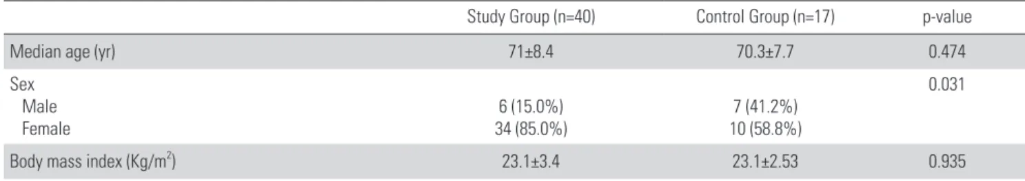
Table 1. Demographic Characteristics of the Study Subjects
Study Group (n=40) Control Group (n=17) p-value
Median age (yr) 71±8.4 70.3±7.7 0.474
Sex Male
Female 6 (15.0%)
34 (85.0%) 7 (41.2%)
10 (58.8%)
0.031
Body mass index (Kg/m2) 23.1±3.4 23.1±2.53 0.935
Values are mean±standard deviation.
subsequent osteoporotic vertebral fractures between the two groups with respect to patient’s clinical and radiographic data, especially vertebroplasty and fat infiltration of back extensor muscles.
Data were entered and analyzed using SPSS version 18.0 for Windows (SPSS, Chicago, Illinois) and Mann-Whitney test was used for continuous variables of the fracture group and the non- fracture group. Wilcoxon signed rank test was conducted to compare continuous variables of primary fracture and variables observed at the final follow up. In addition, the Chi-square test was used for cross-sectional analysis of categorical variables and univariate logistic regression analysis was conducted for variables investigated for analyzing the risk factors of subsequent osteoporotic vertebral fractures. For variables whose p-value was assumed to be 0.35 or lower, multiple logistic regression analysis was performed to analyze the relative risk and confidence interval. If the p-value was lower than 0.05, the result was considered to be significant.
RESULTS
The subsequent new vertebral fractures occurred at a mean of 24±19 months after primary osteoporotic vertebral fractures.
On comparison of baseline characteristics between both group, there was no statistically significant difference in bone mineral density (p=0.761), number of fractures (p=0.423), back
extensor muscle volume at L3 (p=0.329), thoracic kyphotic angle (p=0.704), and lumbar lordotic angle (p=0.669) between the two groups at the time of primary fractures. There was no difference in osteoporosis medication (p=0.060) and the interval of MRI examination (p=0.868) between the two groups until the final follow up (Table 2).
Twenty-nine cases (72.5%) in the fracture group underwent vertebroplasty for primary fractures compared with 4 cases in the non-fracture group (23.5%). Vertebroplasty for primary fractures was associated with higher incidence of subsequent new vertebral fractures (p=0.001).
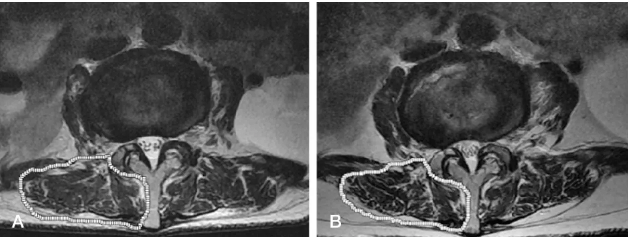
The mean muscle volume of the back extensor on MRI at the final follow up was decreased from 1981.8 mm3 to 1871.3 mm3 in the fracture group and from 1788 mm3 to 1605.1 mm3 in the non-fracture group. There was a significant decrease in muscle volume of the back extensor muscle after primary vertebral frac- tures in both groups (Fracture group, p=0.006; Non-fracture group, p=0.001). In addition, there was a significant increase in fat infiltration of back extensor muscle after primary vertebral fractures in the fracture group (p=0.001, Fig. 1) (Table 3).
The lumbar T- score of bone mineral density was decreased in both groups, and there was no difference in the mean change between the two groups (Fracture group:-0.24±0.16, p=0.089, Non-fracture group:-0.12±0.08, p=0.600).
The variables with p-value < 0.35 of univariate logistic re- gression analysis showed vertebroplasty, sex, the muscle volume
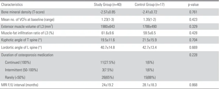
Table 2. Baseline Characteristics of the Study Subjects
Characteristics Study Group (n=40) Control Group (n=17) p-value
Bone mineral density (T-score) -2.57±0.85 -2.41±0.72 0.761
Mean no. of VCFs at baseline (range) 1.23(1-3) 1.35(1-2) 0.423
Extensor muscle volume of L3 (mm2) 1980±643 1788±490 0.329
Muscle-fat infiltration ratio of L3 (%) 61.6±9.6 59.5±6.5 0.428
Kyphotic angle of T spine (°) 19.5±11.6 21.5±15.9 0.704
Lordortic angle of L spine (°) 40.7±14.8 42.7±13.4 0.669
Duration of osteoporosis medication 0.228
Continued (100%) 11(27.5%) 1(6%)
Intermittent (50-100%) 3(7.5%) 1(6%)
Rarely (<50%) 26(65%) 15(88%)
MRI F/U interval (months) 24±19.2 28.1±18.3 0.868
Values are mean±standard deviation.
Risk Factors for Subsequent Vertebral Fractures Journal of Korean Society of Spine Surgery
www.krspine.org 149 Table 3. Changes from Baseline to Final Follow up of Both Groups
Variables Study Group Control Group
1st 2nd P 1st 2nd P
Extensor muscle volume of L3 (mm2) 1980.8 1871.3 0.006* 1788.5 1605.1 <0.001*
Muscle-fat infiltration ratio of L3 (%) 61.6 57.72 0.001* 59.5 57.4 0.394
BMD (T-score) -2.57 -2.81 0.089 -2.41 -2.53 0.600
BMI (Kg/m2) 23.1 22.9 0.331 23.1 22.9 0.612
T-Kyphotic angle(°) 19.5 21.2 0.310 21.5 22.7 0.715
L-Lordortic angle (°) 40.7 39.6 0.401 42.7 41.5 0.381
* Significantly variables, 1st means baseline, 2nd means final follow up.
Table 4. Univariate Logistic Regression Analysis for New Vertebral Fracture
Variables B OR 95% Cl p-value
Primary factured age (yr) 0.023 1.024 0.955-1.096 0.509
Sex 1.378 4.325 1.082-14.533 0.038*
Primary multiple fracture 0.511 1.667 0.454-6.112 0.441
Vertebroplasty 2.148 10.201 2.293-32.015 0.001*
ΔBMD -0.900 0.995 0.069-2.381 0.318*
ΔBMI (Kg/m2) 0.002 0.995 0.723-1.389 0.986
ΔThoracic kyphosis (°) 0.017 1.018 0.946-1.095 0.641
ΔLumbar lordorsis (°) 0.002 1.003 0.934-1.076 0.940
ΔExtensor muscle of L3 (mm2) 0.001 1.054 0.998-1.004 0.304*
ΔExtensor muscle of L3 (%) -0.044 0.911 0.867-1.055 0.378
Δ: Changed variables during follow up, B: regression coefficient, OR: odds ratio, Cl: Confidence interval.
* Included in the multiple logistic model.
A B
Fig. 1. (A) An 80- years -old female patient with a primary L2 osteoporoti vertebral fracture., The paravertebral muscle sity was measured by using a pseudocoloring tool on the L3 axial image. Muscle densty: 1111.87 (74.6%)., In this case, vertebroplas- trformed. (B) After 18Eighteen months after the primary fracture, a subsequent L4 vertebral frcture occurred. The Pparavertebral muscle density was measured by ug the same method on atthe same level. The Mmuscle denity had decreased to: 924.01 (64.0%).
of back extensor and bone mineral density (Table 4). Multiple logistic regression analysis showed significance of vertebroplasty (OR 4.623, 95% CI 1.145-18.699, p=0.031). But there was no significance of the muscle volume of back extensor (p=0.174) and bone mineral density (p=0.648) (Table 5).
DISCUSSION
The subsequent fractures after primary osteoporotic vertebral fractures compromise an additional public health burden and several important questions arise. Why do osteoporotic vertebral fractures apparently lead to subsequent new fractures? How does vertebroplasty with bone cement affect subsequent new vertebral fractures?
To understand why osteoporotic vertebral fractures increases the risk for a subsequent vertebral fractures, in physical principle of normal sagittal alignment, the erect human’s center of gravity is anterior to the spine in the trunk. In order to hold human body erect, the posterior muscles of the spine exert an equal erecting force on the posterior elements of the spine. When an osteoporotic vertebral fracture occurs, the spine becomes kyphosis. As stress in the compression fracture of vertebral body is increased by kyphosis, so are stresses in adjacent vertebral bodies. The additional stresses on adjacent vertebral bodies increase the risk of subsequent fracture on weakened osteoporotic vertebral body. The osteoporotic vertebral fracture itself increases the risk of subsequent vertebral fractures.3-5,10) Other risk factors for subsequent fractures after primary osteoporotic vertebral fractures relate to both underlying disease such as low bone mineral density and the number of prevalent fractures.11)
The effect of vertebroplasty on the potential risk of subsequent vertebral fractures has not been well established. Various studies have reported the 1-year subsequent new vertebral fractures rate
after vertebroplasty to be 20.5%,12) 21.7%,4) 7.8%,13) 7.9%,14) and 15.5%.15) It is probable that vertebroplasty may increase the risk of adjacent vertebral fractures by imposing greater stress on the untreated levels. The increased stiffness of the vertebroplasty- treated vertebra alters the biomechanics of load transfer to the adjacent vertebra by the stress-riser effect.16) Ma et al17) reported that there are three strong risk factors for subsequent new vertebral fractures as a lower bone mineral density, intradiscal cement leakage, and kyphosis. On the other hand, the incidence of subsequent vertebral fractures is approximately the same as that in patients with osteoporosis without prior vertebroplasty.3,18) In this study, vertebroplasty for primary fractures was associated with higher incidence of subsequent new vertebral fractures (p=0.001). We think that it is related to the stress-riser effect according to a difference of stiffness of the vertebroplasty- treated vertebra. In osteoporotic vertebral fractures, a reason for the interest in the back extensor muscle is that it is not only considered as a mobilizer but also as a stabilizer for the spine.
Briggs et al 19) also mentioned about the role of global environ- ment such as paravertebral muscle strength in preventing verte- bral fractures. Cunha et al20) reported that a reduction in the ex- tensor muscle of the lumbar spine increased the risk of vertebral fractures. So et al21) showed that dysfunction of back extensor muscles with fat infiltration weakens the stabilizing ability and this results in increased vulnerability to osteoporotic vertebral fractures. Sinaki et al22) suggested that strengthening of back ex- tensor muscle may prevent osteoporotic vertebral fractures and they reported that the relative risk for compression fracture was 2.7 times lower in the back-exercise group than in the control group in a prospective 10 year follow up study.23) In the present study, the mean muscle volume of the back extensor on MRI at the final follow up was decreased in the both group, but there was a significant increase in fat infiltration of the back exten- sor muscle after primary vertebral fracture in the fracture group Table 5. Multiple Logistic Regression Analysis for New Vertebral Fracture
Variables B OR 95% Cl p-value
Vertebroplasty 1.532 4.623 1.145-18.699 0.031
Sex (Female) 1.144 2.221 0.697-14.142 0.136
ΔExtensor muscle of L3 (mm2) 0.003 1.846 0.998-1.008 0.174
ΔBMD -0.589 0.207 0.05-6.448 0.648
Δ: Changed variables during follow up, B: regression coefficient, OR: odds ratio, Cl: Confidence interval.
* Significantly variables.
Risk Factors for Subsequent Vertebral Fractures Journal of Korean Society of Spine Surgery
www.krspine.org 151 (p=0.001). These results mean that the fracture group show
more dystrophy of the back extensor muscle than the non- fracture group. Therefore, to decrease subsequent osteoporotic vertebral fractures, physician should explain to the patient about the importance of back extensor muscle strengthening exercise.
Age, bone mineral density, and body mass index may all re- flect the consistency in the natural progression of osteoporosis for evaluation of subsequent new vertebral fractures.24) The bone mineral density tends to decrease with increasing age because of progressive bone resorption. Body mass index is positively asso- ciated with estrogen activity, and estrogen stimulates osteoblasts to increase bone mass through increased secretion of osteoid. In our study, there was no significant difference between the two groups. We think that these results were attributed to the increase in age as well as the decrease in bone mineral density at the time of diagnosis of primary fractures in both groups. Therefore, the important risk factor for subsequent new vertebral fractures is osteoporosis itself although age, bone mineral density, and body mass index were not statistically significant. In addition, the proportion of patients who took a drug for less than half of the therapeutic medication period for osteoporosis was 72% until the final follow up in both groups although there was no significant difference between the two groups (p=0.228). This result sug- gests that it is important to have a regular follow up for continu- ous medication for osteoporosis.
This study has some limitations. First, we could not select the patients randomly for dividing them into the fracture group and the non-fracture group. Second, we performed a retrospective study to investigate the risk factors for fractures.
Third, the number of patients was too small to evaluate the multifactorial risk. However, despite these limitations, this study provides guidance for future studies in this area. To provide more reliability, a prospective cohort study is needed in the future to compare the risk factors between the subsequent new vertebral fracture group and the non-fracture group after primary osteoporotic vertebral fractures.
CONCLUSION
These results suggest that vertebroplasty for primary vertebral fractures and increased fat infiltration of the back extensor muscle could be a risk factor related to the development of subsequent osteoporotic vertebral fractures.
REFERENCES
1. Johnell O, Kanis JA. An estimate of the worldwide preva- lence and disability associated with osteoporotic fractures.
Osteoporos Int. 2006;17:1726-33.
2. Melton LJ, 3rd. Epidemiology worldwide. Endocrinol Metab Clin North Am. 2003;32:1-13.
3. Lindsay R, Silverman SL, Cooper C, et al. Risk of new vertebral fracture in the year following a fracture. JAMA.
2001;285:320-3.
4. Syed MI, Patel NA, Jan S, et al. New symptomatic vertebral compression fractures within a year following vertebro- plasty in osteoporotic women. AJNR Am J Neuroradiol.
2005;26:1601-4.
5. Klotzbuecher CM, Ross PD, Landsman PB, et al. Patients with prior fractures have an increased risk of future frac- tures: a summary of the literature and statistical synthesis. J Bone Miner Res. 2000;15:721-39.
6. Hayes WC, Piazza SJ, Zysset PK. Biomechanics of fracture risk prediction of the hip and spine by quantitative com- puted tomography. Radiol Clin North Am. 1991;29:1-18.
7. Hayes WC, Myers ER. Biomechanical considerations of hip and spine fractures in osteoporotic bone. Instr Course Lect.
1997;46:431-8.
8. Myers ER, Wilson SE. Biomechanics of osteopo- rosis and vertebral fracture. Spine (Phila Pa 1976).
1997;22(Suppl):25S-31S.
9. Lee JC, Cha JG, Kim Y, et al. Quantitative analysis of back muscle degeneration in the patients with the degenera- tive lumbar flat back using a digital image analysis: com- parison with the normal controls. Spine (Phila Pa 1976).
2008;33:318-25.
10. Yuan HA, Brown CW, Phillips FM. Osteoporotic spinal deformity: a biomechanical rationale for the clinical conse- quences and treatment of vertebral body compression frac- tures. J Spinal Disord Tech. 2004;17:236-42.
11. Zhang Z, Fan J, Ding Q, et al. Risk factors for new osteo- porotic vertebral compression fractures after vertebroplasty:
a systematic review and meta-analysis. J Spinal Disord Tech. 2013;26:E150-7.
12. Lin WC, Cheng TT, Lee YC, et al. New vertebral osteopo- rotic compression fractures after percutaneous vertebroplas- ty: retrospective analysis of risk factors. J Vasc Interv Radiol.
2008;19:225-31.
13. Lee WS, Sung KH, Jeong HT, et al. Risk factors of devel- oping new symptomatic vertebral compression fractures after percutaneous vertebroplasty in osteoporotic patients.
Eur Spine J. 2006;15:1777-83.
14. Kim SH, Kang HS, Choi JA, et al. Risk factors of new compression fractures in adjacent vertebrae after percutane- ous vertebroplasty. Acta Radiol. 2004;45:440-5.
15. Moon ES, Kim HS, Park JO, et al. The incidence of new vertebral compression fractures in women after kyphoplasty and factors involved. Yonsei Med J. 2007;48:645-52.
16. Berlemann U, Ferguson SJ, Nolte LP, et al. Adjacent verte- bral failure after vertebroplasty. A biomechanical investiga- tion. J Bone Joint Surg Br. 2002;84:748-52.
17. Ma X, Xing D, Ma J, et al. Risk factors for new vertebral compression fractures after percutaneous vertebroplasty.
Spine (Phila Pa 1976). 2013;38:E713-22.
18. Sanfelix-Genoves J, Reig-Molla B, Sanfelix-Gimeno G, et al. The population-based prevalence of osteoporotic vertebral fracture and densitometric osteoporosis in post- menopausal women over 50 in Valencia, Spain (the FRAVO study). Bone. 2010;47:610-6.
19. Briggs AM, Greig AM, Wark JD, et al. A review of ana- tomical and mechanical factors affecting vertebral body integrity. Int J Med Sci. 2004;1:170-80.
20. Cunha-Henriques S, Costa-Paiva L, Pinto-Neto AM, et al. Postmenopausal women with osteoporosis and muscu- loskeletal status: a comparative cross-sectional study. J Clin Med Res. 2011;3:168-76.
21. So KY, Kim DH, Choi DH, et al. The influence of fat infil- tration of back extensor muscles on osteoporotic vertebral fractures. Asian Spine J. 2013;7:308-13.
22. Sinaki M, Wollan PC, Scott RW, et al. Can strong back extensors prevent vertebral fractures in women with osteo- porosis? Mayo Clin Proc. 1996;71:951-6.
23. Sinaki M, Itoi E, Wahner HW, et al. Stronger back muscles reduce the incidence of vertebral fractures: a prospec- tive 10 year follow-up of postmenopausal women. Bone.
2002;30:836-41.
24. Nevitt MC, Thompson DE, Black DM, et al. Effect of alen- dronate on limited-activity days and bed-disability days caused by back pain in postmenopausal women with exist- ing vertebral fractures. Fracture Intervention Trial Research Group. Arch Intern Med. 2000;160:77-85.
원발성 골다공증 척추 골절 치료 후에 속발된 골절의 잠재적 위험인자
김민욱 • 윤대현 • 안상호 • 이지원 • 김철환 • 최용수 광주기독병원 정형외과
연구 계획: 후향적 연구
목적: 골다공증성 척추 골절 치료에 따라 속발된 인접 부위 척추골절에 대한 잠재적 위험인자에 대하여 조사하고자 하였다.
선행 문헌의 요약: 기존의 연구들에서 골밀도 및 골소실, 척추 주위 근육 등 일차 골다공증성 척추 골절 위험인자 연구가 보고되었으나 속발된 인접부위 척추골절에 대한 연구는 아직 부족하다.
대상 및 방법: 골다공증성 척추 골절이 있었던 환자들 중 골절 이후 추적 자기 공명 영상 검사를 시행한 57명의 환자들을 대상으로 인접 부위 척추골절 이 발생한 40명과 발생하지 않은 17명을 두 군으로 나누어 연구를 진행하였다. 흉추 후만각, 요추 전만각, 요추 신전근의 지방 침윤 등 환자들의 방사선 학적 요인들과 골밀도 수치, 체질량 지수, 골다공증 치료 약제 복용력, 척추 성형술 기왕력 등 전신 인자 및 임상적 요인들에 대하여 조사하였다.
결과: 인접 분절 척추골절은 선행한 골다공증성 척추 골절후 평균 24±19 개월 후에 발생하였다. 선행한 골절을 척추 성형술로 치료한 경우에 인접 부 위 척추골절의 발생률이 유의하게 증가하였으며(p=0.001) 척추골절이 발생한 군에서 요추 신전근의 지방 침윤 정도가 골절이 발생하지 않은 군에 비 하여 유의하게 증가되어있음을 확인할 수 있었다(p=0.001). 로지스틱 회귀 분석 결과에서도 척추성형술과 척추골절의 연관성이 높게 나타났다(OR 4.623, 95% CI 1.145-18.699, p=0.031).
결론: 선행한 골다공증성 척추 골절을 척추 성형술로 치료와 척추 골절 후 요추 신전근의 지방 침윤 증가는 인접 부위 척추 골절의 잠재적 위험인자의 하나로 고려될 수 있다.
색인 단어: 척추 골절, 골다공증, 속발성 척추 골절, 요추 신전근 약칭 제목: 속발성 척추골절의 위험인자