Clinical Application of Electrogastrography in Patients with Stomach Cancer Who Undergo Distal Gastrectomy
Ho Yeun Kim, Sun Jin Park, and Yong Ho Kim
Department of Surgery, Kyung Hee University School of Medicine, Seoul, Korea
Purpose: Electrogastrography is a method of measuring action potentials of the stomach. The purpose of this study was to investigate early postoperative changes in the electrogastrography and determine the correlation between electrogastrography and quality of life of patients with stomach cancer who underwent distal gastrectomy.
Materials and Methods: This study analyzed 20 patients with stomach cancer who underwent electrogastrography and quality of life was measured 1, 12, and 24 weeks after the operation. Quality of life-C30 version 3.0 and quality of life-STO22, were used.
Results: Fasting and postprandial mean dominant frequency at 1 week after the operation was 2.7 and 2.7 cycles per minute, and 2.8 and 2.7 cycles per minute at 12 weeks, 2.6 and 2.8 cycles per minute at 24 weeks. Fasting and postprandial mean dominant power at 1 week was 36.5 and 36.4 dB, 36.3 and 40.1 dB at 12 weeks and 40.9 and 42.3 dB at 24 weeks. The percentage of tachygastria was increased whereas the percentage of bradygradia was decreased during the postoperative periods (P<0.05). Global health, physi- cal, emotional and social functioning scales were improved, but role and cognitive functioning were not changed. Pain, insomnia, diar- rhea and financial difficulties were significantly improved according to the postoperative periods (P<0.05). The correlation between the STO22 and electrogastrography parameters was not significant (P>0.05).
Conclusions: These may suggest that electrogastrography is a simple and noninvasive method and may be applicated for evaluating mo- tility and autonomic functions of the remnant stomach.
Key Words: Electrogastrography; Stomach neoplasms; Distal gastrectomy
Correspondence to: Yong Ho Kim
Department of Surgery, Kyung Hee University Medical Center, 23 Kyungheedae-ro, Dongdaemun-gu, Seoul 130-872, Korea
Tel: +82-2-958-8246, Fax: +82-2-966-9366 E-mail: kyjho@khmc.or.kr
Received March 3, 2014 Revised March 24, 2014 Accepted March 26, 2014
Copyrights © 2014 by The Korean Gastric Cancer Association www.jgc-online.org
This is an open-access article distributed under the terms of the Creative Commons Attribution Non-Commercial License (http://creativecommons.org/
licenses/by-nc/3.0) which permits unrestricted noncommercial use, distribution, and reproduction in any medium, provided the original work is properly cited.
Introduction
The primary functions of the stomach are to mix secreted en- zymes and peptides and store, grind, digest, and empty food into the duodenum. Multiple levels of neuronal mechanisms of the cen- tral, spinal, and enteric nervous systems are in place to coordinate the stomach’s ordered movement.1 The stomach muscle itself also exhibits myoelectrical activity that mediates stomach motility. Both
slow waves (electrical control activity) and spikes (electrical re- sponse activity) are well-known components of stomach myoelec- tricity.2 In 1922, Alvarez3 recorded a three-cycles per minute (cpm) sinusoidal wave using electrodes placed on the abdominal skin.
Noninvasiveness is the greatest advantage of electrogastrographies (EGGs), which use surface-placed electrodes to record stomach myoelectricity.2,4-6 Since Alvarez’s report, a number of studies concerning EGG have determined that gastric electrical potentials break out from a pacemaker located on the upper gastric body along the greater curvature.7,8 These potentials are related to an electrical pacing function and the power of gastric contraction.9-11 The EGG result is a combination of gastric signals and noise. The gastric signal consists of normal slow waves (regular frequency, 2~4 cpm), tachygastria (regular frequency, 4~9 cpm), bradygastria
(regular frequency, 0.5~2 cpm), and arrhythmia (irregular rhythmic activities). The noise is composed of respiratory and motion arti- facts, electrocardiography, and electrical interference of the small intestine.9 Gastric emptying (GE) is a function of the net inhibitory and excitatory results of antropyloroduodenal coordination among all portions of the stomach, pylorus, and duodenum.1 Partial or to- tal removal of the upper gastrointestinal organs impairs GE. In the present study, we investigated postoperative changes on the EGGs of patients with stomach cancer and analyzed the relationship be- tween the EGG system and the quality of life (QOL) of patients with stomach cancer who were treated with distal gastrectomy.
Materials and Methods
1. Patients
Twenty individuals who underwent distal gastrectomy with
gastroduodenostomy for the treatment of stomach cancer at Kyung Hee University Medical Center between May and December 2010 were enrolled. Abnormal GE and associated dyspeptic symptoms are common in the early period after the operation, while GE may return to normal 12 months after surgery. In addition, some changes in EGG parameters occur in the early days after subtotal gastrectomy. Therefore, we enrolled patients with stomach cancer who were treated with distal gastrectomy to investigate postopera- tive EGG changes and analyzed the correlation between EGG findings and QOL.
The elapsed time after distal gastrectomy was obtained from chart reviews. However, we excluded subjects with distal gastrec- tomy who showed evidence of diabetes, tumor metastasis or recur- rence, tumor-related terminal stages, dumping syndrome, obvious dyspepsia, or the current use of any medications known to alter gastrointestinal motility. An EGG was recorded for each of the
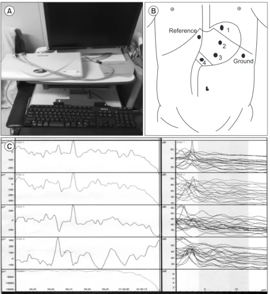
Fig. 1. Electrogastrography (EGG) system (POLYGRAM NET™) (A), elec- trode position (B), and EGG graphs (C).
20 patients with gastric cancer who underwent gastrectomy (15 men, five women; mean age±standard deviation [SD], 59.8 years;
range, 28~74 years). All subjects were advised to accept endos- copy to confirm their distal gastrectomy and the absence of tumor recurrence. Our hospital’s ethics committee approved this project.
Informed consent was obtained from all patients before the study was conducted.
2. Electrogastrography system
Each EGG was recorded with a POLYGRAM NETTM (Medtronic A/S; Alpine Biomed ApS, Skovlunde, Denmark) (Fig.
1). Active electrodes for a four-channel EGG study were placed to follow the antral axis of the stomach. The electrodes were placed under abdominal ultrasonographic guidance. Pre-gelled electrodes were applied to prepared spots in the following sequence:
1. The channel 3 electrode was placed approximately 2 cm left of the midway point between the umbilicus and xiphoid process on the patient’s ventral midline.
2. The channel 4 active electrode was placed approximately 5 cm right of the channel 3 electrode at the same level.
3. The channel 2 active electrode was placed approximately 5 cm left of and 45o to the channel 3 active electrode at the same level.
4. The channel 1 active electrode was placed approximately an- other 5 cm away and 45o to the channel 2 electrode.
5. A common reference electrode was placed just below the xi- phoid process.
6. A ground electrode was placed approximately 10 cm left of ventral midline at the same level of the channel 3 electrode.
3. Electrogastrography measurement
After an overnight fast, the subjects rested quietly in the supine position and were asked not to move, sleep, or talk throughout the recording. A baseline EGG recording was obtained for 45 minutes in the supine position. After the basal recording was taken, the subjects were asked to consume a standard light meal including 180 ml of orange juice and a piece of cake (347 kcal) in a sitting posi- tion. We then recorded a postprandial EGG for 45 minutes.
4. Analysis
The results are expressed as mean±SD. Numerical data were analyzed with paired Student’s t-test or an analysis of covariance test. Statistical analyses were performed with SPSS statistical soft- ware, version 17.0 (SPSS Inc., Chicago, IL, USA). Values of P<0.05 were considered statistically significant.
Results
Table 1 illustrates the characteristics of the patients with gastric cancer. According to the tumor location, there were 10 cases (50%) in the body and 10 cases (50%) in the antrum. According to the Union for International Cancer Control 7th TNM classification (2010), there were seven cases (35%) of stage Ia, five cases (25%) of stage Ib, two cases (10%) of stage IIa, two cases (10%) of stage IIb, and four cases (20%) of stage IIIa. Open distal gastrectomy was performed in 11 cases (55%) and laparoscopic-assisted distal gastrectomy was performed in nine cases (45%). Twelve cases (60%) received adjuvant chemotherapy.
Table 2 illustrates the effect of the meal on EGG parameters during the postoperative period (~24 weeks). According to the ef- fect of the meal on EGG parameters during the early postoperative period, between the fasting and postprandial recordings, there were no significance differences in dominant frequency (DF), dominant power (DP), and percentage of normal frequency, but there were significant decreases in the percentage of bradygastria and increases in the percentage of tachygastria (P<0.05). The percentage of bradygastria decreased significantly on the postprandial recordings between 1 week and 3 months, between 1 week and 6 months, but not between 3 months and 6 months. The percentage of tachy- gastria increased between 1 week and 6 months but not between 3 months and 6 months. The percentage of normal frequency was
Table 1. Characteristics of patients with gastric cancer Gender (male : female) 14 : 6 (2.3 : 1)
Age (yr) 59.8 (28~74)
Stage*
Ia Ib IIa IIb IIIa
7 (35) 5 (25) 2 (10) 2 (10) 4 (20) Tumor location
Body
Antrum 10 (50)
10 (50) Surgery
DG
LADG 11 (55)
9 (45) Chemotherapy
Yes
No 12 (60)
8 (40)
Values are presented as number, median (range), or number (%). DG
= distal gastrectomy; LAGD = laparoscopic-assisted distal gastrectomy.
*Union for International Cancer Control 7th TNM classification.
slightly increased in the postprandial recordings, but there were no significant differences except in postoperative week 1.
Global health status and physical, emotional, and social func- tioning scales were improved, but role and cognitive functioning were not changed. Most symptom scales were slightly improved, whereas pain, insomnia, diarrhea, and financial difficulties were significantly improved according to the postoperative periods re- garding the European Organisation for Research and Treatment of Cancer (EORTC) quality of life questionnaire QOL-C30 version 3.0 (P<0.05) (Table 3). Comparison of QOL with the EORTC QLQ- STO22 questionnaire during the postoperative periods (~12 weeks) revealed no statistically significant differences (P>0.05) (Fig. 2), and the correlation between QOL and EGG parameters was not statistically significant (P>0.05) (Table 4).
Discussion
Stomach surgery may influence gastric motor function. Even gastric restrictive surgery without resection in obese subjects leads to altered DF and DP but not power ratio.12 Truncal vagotomy with
gastric resection for peptic ulcer disease impairs esophageal sphinc- ter tone, food accommodation, and the grinding and emptying ability.13 Distal gastrectomy is severely destructive to the stomach anatomy and its neighboring organs and may lead to poor motil- ity.14-16
Debate persists regarding when to record postoperative EGG parameters. Abnormal GE and associated dyspeptic symptoms are common in the early days after an operation, while GE may require 12 months after stomach surgery to return to normal.1,15 Some changes in EGG parameters occur in the early days after subtotal gastrectomy.17 Lee et al.18 enrolled subjects for distal robotic sleeve gastrectomy (RSG) who had undergone stomach surgery at least 1 year previously. Homma et al.19 reported that a series of regular peaks of 3 cpm activity were not clearly visible in the EGGs running spectra recorded from patients who had undergone a distal gastrectomy with a short postoperative period (13~71 days in 13 subjects). Imai and Sakita20 recorded postoperative EGGs in gastrectomy patients 3 to 5 weeks after surgery. Hayashi et al.21 recorded EGGs at least 1 year after surgery. In our study, we re- corded and analyzed EGGs in patients who had undergone distal Table 2. Effect of the meal on EGG parameters during the postoperative period (~24 weeks)
EGG parameter Fasting Postprandial P-value
Dominant frequency (cpm) 1 week
12 weeks 24 weeks
2.7±0.1 2.8±0.4 2.6±0.4
2.7±0.3 2.7±0.5 2.8±0.5
0.432 0.704 0.101 Dominant power (dB)
1 week 12 weeks 24 weeks
36.5±2.4 36.3±3.2 40.9±6.5
36.4±2.7 40.1±4.9 42.3±8.4
0.107 0.647 0.498 Percentage of normal frequency (2.7~3.4 cpm)
1 week 12 weeks 24 weeks
18.1±13.4 19.4±5.6 21.9±9.1
23.6±14.2 22.5±8.8 23.3±10.2
0.003 0.077 0.170 Percentage of bradygastria (<2.7 cpm)
1 week 12 weeks 24 weeks
58.5±15.9 48.2±14.8 48.0±14.9
41.8±13.9 37.0±11.0 36.5±9.3
0.000 0.000 0.001 Percentage of tachygastria (>3.4 cpm)
1 week 12 weeks 24 weeks
21.9±7.4 28.9±10.7 26.6±9.8
33.8±11.5 37.9±10.1 37.7±12.2
0.000 0.001 0.000 Instability coefficient (DF)
1 week 12 weeks 24 weeks
0.82±0.34 0.75±0.29 0.98±0.39
0.86±0.45 0.88±0.32 1.1±0.45
0.660 0.042 0.335 Valuse are presented as mean±standard deviation. EGG = electrogastrography; cpm = cycles per minute; DF = dominant frequency.
gastrectomy over postoperative periods from 1 to 24 weeks to investigate postoperative changes according to the various time in- tervals.
Bradygastria (regular frequency, 0.5~2.0 cpm) is currently de- fined as values below the normal range (regular frequency, 2~4 cpm), whereas tachygastria (regular frequency, 4~9 cpm) is defined as values above the normal range.9,22,23 Unfortunately, the normal
Fig. 2. Changes in mean quality of life score scales on the STO22 ques- tionnaire during the postoperative period (~12 weeks). POD#1 = post- operative 1 week; POD#12 = postoperative 12 weeks.
Table 3. Comparison with the European Organisation for Research and Treatment of Cancer QOL questionnaire (QOL-C30 version 3.0) according to postoperative period
Postoperative
1 week Postoperative 12 weeks P-value Global health status 49.2±38.0 60.8±30.1 0.000 Functional scales
Physical functioning 63.9±26.9 76.8±13.4 0.046 Role functioning 67.1±33.4 81.6±21.8 0.118 Emotional functioning 78.5±18.5 90.4±12.2 0.025 Cognitive functioning 85.1±14.6 90.4±17.0 0.268 Social functioning 66.7±18.4 80.7±22.4 0.025 Symptom scales
Fatigue 32.2±15.2 25.7±20.0 0.243
Nausea/vomiting 7.0±11.5 2.6±6.2 0.135
Pain 38.6±24.2 7.9±14.0 0.000
Dyspnea 14.0±20.2 8.8±15.1 0.187
Insomnia 21.1±25.4 5.3±12.5 0.008
Appetite 14.0±20.2 5.3±16.7 0.056
Constipation 12.3±22.8 15.8±17.1 0.494
Diarrhea 45.6±33.7 17.5±20.4 0.003
Financial difficulties 42.1±24.4 24.6±33.0 0.047 Valuse are presented as mean±standard deviation. QOL = quality of life.
Table 4. Correlation between QOL questionnaires (QOL-C30 version 3.0 and ST022) and EGG parameters by postoperative period QOL
EGG parameters (P-value)
Postoperative 1 week Postoperative 12 weeks
DF DP BR NO TA DF DP BR NO TA
Global health status 0.44 0.40 0.59 0.91 0.38 0.95 0.61 0.88 0.62 0.55
Functional scales 0.13 0.04 0.01 0.13 0.39 0.63 0.18 0.46 0.40 0.35
Symptom scales 0.88 0.77 0.58 0.45 0.72 0.77 0.41 0.65 0.70 0.87
Fatigue 0.19 0.87 0.00 0.24 0.20 0.58 0.60 0.65 0.30 0.19
Nausea/vomiting 0.57 0.82 0.27 0.36 0.96 0.31 0.78 0.34 0.47 0.95
Pain 0.40 0.68 0.14 0.66 0.31 0.27 0.42 0.36 0.25 0.55
Dyspnea 0.05 0.63 0.12 0.18 0.98 0.84 0.72 0.59 0.27 0.33
Insomnia 0.70 0.22 0.07 0.01 0.10 0.76 0.28 0.04 0.05 0.18
Appetite 0.31 0.36 0.14 0.52 0.40 0.01 0.43 0.50 0.40 0.74
Constipation 0.30 0.99 0.78 0.39 0.27 0.58 0.13 0.17 0.68 0.99
Diarrhea 0.85 0.98 0.71 0.27 0.02 0.58 0.27 0.54 0.12 0.22
Financial difficulties 0.12 0.24 0.78 0.90 0.78 0.10 0.23 0.53 0.85 0.99
STO22 0.88 0.77 0.68 0.45 0.72 0.77 0.41 0.65 0.70 0.87
QOL = quality of life; EGG = electrogastrography; DF = dominant frequency; DP = dominant power; BR = bradygastria; NO = normogastria; TA
= tachygastria.
range is very difficult to define due to variations in the record- ing system and a lack of standard analysis methods.24 Moreover, the occurrence of tachygastria is influenced by electrode position and configuration.25 However, the association between tachygastria and gastric motor disorders has been substantiated in other studies.
For example, You et al.26 observed tachygastria in a 26-year-old woman with persistent nausea, vomiting, and abdominal pain and severely impaired antral motor function. The correlation between bradygastria and gastric motility is not yet as clear. van der Schee and Grashuis27 observed bradygastria in dogs and found that it was correlated with strong antral contractions.
In the present study, comparison of temporal changes in EGG parameters revealed no significant temporal differences for DF, DP, percentage of normal frequency, or percentage of tachygastria.
However, it did reveal significant temporal differences in the per- centage of bradygastria for postprandial recordings between 1 week and 3 months and between 1 week and 6 months but not between 3 months and 6 months. Unlike other studies that obtained a DF of ~3 cpm in the gastric remnant, Lee et al.18 found that distal RSG patients had a lower DF value with an increased percentage of bradygastria (<2 cpm) irrespective of meal ingestion. Bracci et al.28 observed the less common 3 cpm with an increased chance of bradygastria in children who underwent partial gastrectomy to aug- ment bladder function. Although they lacked a clear definition of bradygastria, they suggested that this is the effect of the removal of stomach pacemaker cells.
Roux-en-Y reconstruction in previous gastrectomy patients also led to bradygastria, which was closely related to the time after surgery.29 Hayashi et al.21 pointed out that proximal subtotal gas- trectomy subjects with more than half of the stomach resected had lower percentages of normal rhythm, DF, and even DP compared with those with greater stomach preservation. Lee et al.18 suggested that removal of part of the main pacemaker in distal RSG pa- tients leads to an increased percentage of bradygastria and that the ectopic pacemaker of the rest of the stomach in lower frequency values would replace the originally removed main pacemaker cells to dominate the gastric SW rhythm. Murakami et al.30 reported that longer periods of normal gastric function (normogastria, 2.0~4.0 cycles min-1) were detected in channel 1 in the vagus nerve- preserving distal gastrectomy group (VP-DG) than in the standard distal gastrectomy without vagus nerve preservation group (DG) in either the fasted or fed state (P<0.05). The VP-DG group showed better preserved gastric myoelectric activity than the DG group.
Homma et al.19 recognized serial 3 cpm peaks in the running spectra of EGGs recorded in patients who had undergone distal
gastrectomy 15 to 20 years previously and suggested that the reor- ganization of gastric pacemaker activity represents reorganization of the interstitial cells of Cajal in the remnant stomach of distally gastrectomized patients. According to the effect of the meal on EGG parameters during the early postoperative period, comparison of fasting to postprandial recordings revealed significant decreases in the percentage of bradygastria and increases in the percentage of tachygastria. According to time intervals, there was a significant decrease in the percentage of bradygastria in fasting and postpran- dial recordings between 1 and 12 weeks and between 1 and 24 weeks. The percentage of normal frequency was increased in the postprandial recording between 1 and 24 weeks. These results sug- gest that the decreased percentage of bradygastria and the increased percentage of tachygastria indicate a change in EGG parameters that represents reorganization of the interstitial cells of Cajal in the remnant stomach of distally gastrectomized patients.
In conclusion, these findings suggest that EGG may be a nonin- vasive method for evaluating the motility and autonomic functions of the remnant stomach after distal gastrectomy. However, the present study has shortcomings including its lack of a control group and preoperative baseline EGGs and its small number of patients.
References
1. Quigley EMM. Gastric motor and sensory function and motor disorders of the stomach. In: Feldman M, Friedman LS, Brandt LJ, eds. Sleisenger & Fordtran’s Gastrointestinal and Liver Dis- ease Pathophysiology/Diagnosis/Management. 8th ed. Phila- delphia: WB Saunders/Elsevier, 2006:999-1028.
2. Parkman HP, Hasler WL, Barnett JL, Eaker EY; American Motility Society Clinical GI Motility Testing Task Force. Elec- trogastrography: a document prepared by the gastric section of the American Motility Society Clinical GI Motility Testing Task Force. Neurogastroenterol Motil 2003;15:89-102.
3. Alvarez WC. The electrogastrogram and what it shows. JAMA 1922;78:1116-1119.
4. Yin J, Chen JD. Roles of interstitial cells of Cajal in regulating gastrointestinal motility: in vitro versus in vivo studies. J Cell Mol Med 2008;12:1118-1129.
5. Smout AJPM, Jebbink HJA, Samson JM. Acquisition and analysis of electrogastrographic data, the Dutch experience. In:
Chen JZ, McCallum RW, eds. Electrogastrography, Principles and Applications. New York: Raven Press, 1994:3-30.
6. Chang FY. Electrogastrography: basic knowledge, recording, processing and its clinical applications. J Gastroenterol Hepatol
2005;20:502-516.
7. Hinder RA, Kelly KA. Human gastric pacesetter potential.
Site of origin, spread, and response to gastric transection and proximal gastric vagotomy. Am J Surg 1977;133:29-33.
8. Sarna SK, Bowes KL, Daniel EE. Gastric pacemakers. Gastro- enterology 1976;70:226-231.
9. Chen JD, McCallum RW. Clinical applications of electrogastro- graphy. Am J Gastroenterol 1993;88:1324-1336.
10. Chen JD, Richards RD, McCallum RW. Identification of gastric contractions from the cutaneous electrogastrogram. Am J Gas- troenterol 1994;89:79-85.
11. Sun WM, Smout A, Malbert C, Edelbroek MA, Jones K, Dent J, et al. Relationship between surface electrogastrography and antropyloric pressures. Am J Physiol 1995;268:G424-G430.
12. van Dielen FM, de Cock AF, Daams F, Brummer RJ, Greve JW. Gastric myoelectrical activity in morbidly obese patients before and 3 months after gastric restrictive surgery. Obes Surg 2003;13:721-727.
13. Richter HM 3rd. Physiologic consequences of vagotomy and gastric resection. Gastroenterol Clin North Am 1994;23:193- 213.
14. Mackay S, Hayes T, Yeo A. Management of gastric cancer. Aust Fam Physician 2006;35:208-211.
15. Houghton J, Wang TC. Tumors of the stomach. In: Feldman M, Friedman LS, Brandt LJ, eds. Sleisenger & Fordtran’s Gas- trointestinal and Liver disease, Pathophysiology/Diagnosis/
Management. 8th ed. Philadelphia: WB Saunders/Elsevier, 2006:1139-1170.
16. Carter DC. Carcinoma of the stomach and other tumors. In:
Shearman DJC, Finlayson N, Camilleri M, Carter SD, eds. Dis- eases of the Gastrointestinal Tract and Liver. 3rd ed. New York:
Churchill Livingstone, 1997:293-322.
17. Zhang Q, Yu JC, Ma ZQ, Kang WM, Ke MY, Qian JM. The ef- fects of enteral nutrition vs parenteral nutrition on gastric mo- tility and gastroenteric hormones after subtotal gastrectomy:
a perspective randomized compared clinical trial. Zhonghua Wai Ke Za Zhi 2006;44:728-732.
18. Lee HF, Chang FY, Lu CL, Luo JC, Chen CY, Wu HC. Electro- gastrographic characteristics in subjects with stomach rem- nant. J Gastroenterol Hepatol 2010;25:339-344.
19. Homma S, Kobayashi Y, Kosugi S, Ohashi M, Kanda T, Oka-
moto H, et al. Postoperative reorganization of gastric pacemak- er activity in patients after an extended period following distal gastrectomy. J Smooth Muscle Res 2008;44:113-122.
20. Imai K, Sakita M. Pre- and postoperative electrogastrogra- phy in patients with gastric cancer. Hepatogastroenterology 2005;52:639-644.
21. Hayashi T, Kinami S, Fushida S, Fujimura T, Miwa K, Inoue K. Evaluation of residual stomach motility after proximal gas- trectomy for gastric cancer by electrogastrography. Dig Dis Sci 2006;51:268-273.
22. Koch KL. Electrogastrography: physiological basis and clini- cal application in diabetic gastropathy. Diabetes Technol Ther 2001;3:51-62.
23. Camilleri M, Hasler WL, Parkman HP, Quigley EM, Soffer E.
Measurement of gastrointestinal motility in the GI laboratory.
Gastroenterology 1998;115:747-762.
24. Riezzo G, Chiloiro M, Guerra V. Electrogastrography in healthy children: evaluation of normal values, influence of age, gender, and obesity. Dig Dis Sci 1998;43:1646-1651.
25. Cheung B, Vaitkus P. Perspectives of electrogastrography and motion sickness. Brain Res Bull 1998;47:421-431.
26. You CH, Chey WY, Lee KY, Menguy R, Bortoff A. Gastric and small intestinal myoelectric dysrhythmia associated with chronic intractable nausea and vomiting. Ann Intern Med 1981;95:449-451.
27. van der Schee EJ, Grashuis JL. Contraction-related, low- frequency components in canine electrogastrographic signals.
Am J Physiol 1983;245:G470-G475.
28. Bracci F, Matarazzo E, Mosiello G, Caione P, Cianchi D, Ponti- celli A. Preliminary report of electrogastrography in pediatric gastroresection: can it be predictive of alteration of gastric mo- tility? J Pediatr Surg 2001;36:1157-1159.
29. Bures J, Kabelác K, Kopácová M, Vorísek V, Siroký M, Palicka V, et al. Electrogastrography in patients with Roux-en-Y recon- struction after previous Billroth gastrectomy. Hepatogastroen- terology 2008;55:1492-1496.
30. Murakami H, Matsumoto H, Kubota H, Higashida M, Naka- mura M, Hirai T. Evaluation of electrical activity after vagus nerve-preserving distal gastrectomy using multichannel elec- trogastrography. J Smooth Muscle Res 2013;49:1-14.