INTRODUCTION
Non-cutaneous melanoma often develops in areas shielded from ultraviolet rays, and its incidence is less than that of cutaneous melanoma. Vaginal malignant melanoma is a rare form of non-cutaneous melanoma, and accounts for only 1.6%
of female genital tract melanoma cases. Furthermore, vaginal melanoma accounts for 2.4%-2.8% of all vaginal cancers and
0.3%-0.8% of all malignant melanomas [1], and is the second most common female genital tract melanoma. The age of onset of vaginal melanoma has been reported to range from 38 to 90 years, with most patients being diagnosed between the ages of 60 and 80 years. Most vaginal melanoma patients are postmenopausal, and their primary complaints tend to include vaginal bleeding and vaginal discharge. Unlike melanoma of the skin, vaginal melanomas can be staged according to the International Federation of Gynecology and Obstetrics (FIGO) staging criteria [2]. Unfortunately, vaginal melanomas are often only diagnosed at an advanced stage [3], and treatment options include local excision with wide margins, radical surgery, radiotherapy, chemotherapy, and im- munotherapy. Currently, no standard treatment protocol has been established, and the prognosis remains poor, with the 5-year survival rate for vaginal malignant melanoma reported
Clinical outcome of 31 patients with primary malignant melanoma of the vagina
Qidan Huang*, He Huang*, Ting Wan, Ting Deng, Jihong Liu
State Key Laboratory of Oncology in South China & Department of Gynecologic Oncology, Sun Yat-sen University Cancer Center, Guangzhou, China
Received Oct 23, 2012, Revised May 10, 2013, Accepted May 16, 2013
*Both authors contributed equally.
Correspondence to Jihong Liu
Department of Gynecologic Oncology, Sun Yat-sen University Cancer Center, 651 Dongfengdong Road, 510060, Guangzhou, China. E-mail:
Objective: To investigate the clinical characteristics of and prognostic factors for primary malignant melanoma of the vagina.
Methods: Clinical data from 31 patients treated for primary malignant melanoma of the vagina at the Sun Yat-sen University Cancer Center between March 1970 and June 2005 were retrospectively analyzed.
Results: The median age was 58 years (range, 18 to 73 years), and the main symptoms reported were vaginal bleeding and vaginal discharge. Most tumors were of the nodular type and classified as stage I according to International Federation of Gynecology and Obstetrics staging criteria. Surgery was performed on 22 patients, chemotherapy was administered to 7 patients, and immunotherapy was administered to 19 patients. Recurrent tumors developed in 11 patients (35.5%) during a median follow-up period of 20.2 months (range, 1 month to 18 years). The 5-year overall survival rate was 32.3%. Univariate analysis revealed that macroscopic tumor growth and the treatment method significantly affected survival outcome (p=0.039 and p<0.001, respectively), whereas the radicality of surgery did not (p=0.296). Multivariate analysis revealed that macroscopic tumor growth (hazard ratio [HR], 4.1; 95% confidence interval [CI], 1.4 to 12.1; p=0.010) and treatment method (HR, 0.3; 95% CI, 0.1 to 0.9; p=0.025) were independent prognostic factors for overall survival.
Conclusion: Patients with primary vaginal melanoma have a poor prognosis. Macroscopic tumor growth and treatment method are prognostic factors for primary malignant melanoma of the vagina.
Keywords: Immunotherapy, Malignant melanoma, Surgical treatment, Vaginal malignancies
to range from 0% to 25% [4-6]. The risk factors for vaginal melanoma also remain unclear.
In this study, records from 31 patients with vaginal malignant melanoma diagnosed between March 1970 and June 2005 were retrospectively analyzed to investigate the factors that affect patient prognosis and the most effective treatment strategy for this disease.
MATERIALS AND METHODS 1. Patient enrollment
The clinical records of all patients diagnosed with vaginal malignant melanoma who were treated at the Sun Yat-sen University Cancer Center between March 1970 and June 2005 were reviewed for this study. The histologic type of each case was reassessed by pathologists according to World Health Organization criteria prior to inclusion in the present study.
Prior patient consent and approval from the Institutional Research Ethics Committee of Sun Yat-sen University Cancer Center were obtained for the use of clinical materials for this study.
2. Factors for analysis
Macroscopic tumor growth was divided into 3 categories:
cauliflower-like, nodular, and ulcerative. Tumor size was evaluated as both a continuous variable and a binary variable (<4 cm or ≥4 cm). Disease was staged according to the FIGO staging criteria (2009) for vaginal carcinoma. Four types of treatment were administered to patients in this study: surgery followed by immunotherapy, surgery alone, chemotherapy plus immunotherapy, and chemotherapy alone. Radical surgery included hysterectomy, whereas conservative surgery did not.
3. Surveillance after completion of treatment
After primary treatment, patients were followed up every 3 months for the first 2 years, every 6 months for the next 3 years, and annually thereafter. Pelvic ultrasonography was performed annually, and a chest radiograph was also obtained.
Other imaging studies (e.g., chest/abdominal/pelvic com- puted tomography and magnetic resonance imaging) were performed, as clinically indicated.
4. Statistical analysis
SPSS ver. 16.0 (SPSS Inc., Chicago, IL, USA) was used for analy- sis of data. Patient age, macroscopic tumor growth, tumor size, FIGO staging, radicality of surgery, lymphadenectomy, and treatment method were analyzed using one-way analysis
of variance. Two-sided probability values were calculated and were considered significant at the level of p=0.05. Overall survival (OS) was the primary endpoint evaluated, and was calculated from the first day of treatment. When information on survival was not available, an attempt was made to obtain death certificate information. If such information could not be obtained, data after the last contact were censored. OS was calculated using the Kaplan-Meier method. Tumor stage, macroscopic tumor growth, tumor size, radicality of surgery, treatment method, and patient age were also assessed using univariate analysis to determine their impact on OS. Multivari- ate analysis was performed using the Cox regression method.
Hazard ratios with their 95% confidence intervals are reported.
RESULTS 1. Clinical data
1) Patient characteristics
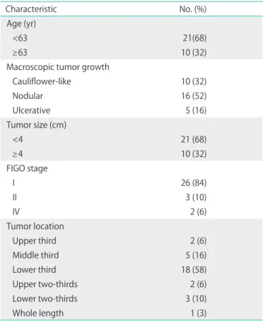
A total of 31 patients with stage I (n=26), stage II (n=3), or
Table 1. Characteristics of 31 consecutive patients with primary malignant melanoma of the vagina
Characteristic No. (%)
Age (yr)
<63 21(68)
≥63 10 (32)
Macroscopic tumor growth
Cauliflower-like 10 (32)
Nodular 16 (52)
Ulcerative 5 (16)
Tumor size (cm)
<4 21 (68)
≥4 10 (32)
FIGO stage
I 26 (84)
II 3 (10)
IV 2 (6)
Tumor location
Upper third 2 (6)
Middle third 5 (16)
Lower third 18 (58)
Upper two-thirds 2 (6)
Lower two-thirds 3 (10)
Whole length 1 (3)
FIGO, International Federation of Gynecology and Obstetrics.
stage IV (n=2) primary vaginal melanoma were enrolled in this study. Table 1 summarizes the characteristics of these patients and their tumors. The median age of the patients was 58 years (range, 18 to 73 years), and most patients presented with vaginal bleeding (n=19), vaginal discharge (n=6), or a vaginal mass (n=4). The median time interval between the onset of symptoms and diagnosis was 3 months (range, 1 month to 36 years). The median duration of follow-up was 20.2 months from the initial treatment (range, 1 month to 18 years).
2) Tumor characteristics
All of the vaginal lesions examined were black or grey in color, and most were nodular in shape. Tumor diameters ranged from 0.5 to 7.0 cm, and the majority were localized in the lower third of the vagina. FIGO stage I tumors were diag- nosed in 26 cases. For treatment, 9 patients underwent pelvic lymphadenectomy, and 4 of these patients (44%) developed pelvic lymph node (LN) metastases. Five patients underwent resection of the inguinal LNs, and inguinal LN metastasis was found in 2 patients (40%).
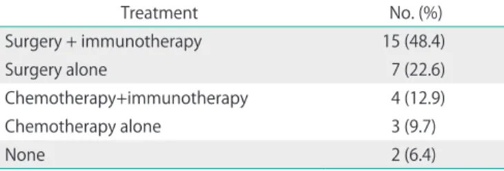
2. Treatment methods
Although 2 patients did not receive any treatment, 29 re- ceived 4 different types of treatment (Table 2). Surgery-based
treatment was performed for 22 patients, with 11 patients undergoing radical surgery and the other 11 undergoing con servative surgery. LN dissection or biopsy (pelvic and/
or inguinal LN) was performed in 12 cases. Neoadjuvant chemotherapy was administered to 6 patients, and 1 patient underwent brachytherapy prior to surgery. Following surgery, 16 patients received chemotherapy and 2 patients underwent chemoradiation therapy. Chemotherapy alone without surgery was used to treat 7 patients, 4 of whom received immuno- therapy together with chemotherapy.
Immunotherapy was administered to 19 patients, including regimens of interferon-α (IFN-α), interleukin-2 (IL-2), bacillus Calmette-Guérin (BCG), dendritic cells (DC-cells), lymphokine- activated killer cells (LAK cells), and a measles vaccine.
3. Survival
The disease recurred in 11 cases, 3 of which (27.3%) involved local recurrence, 7 involved (63.6%) distant recurrence, and 1 involved (9.1%) both local and distant recurrence. However, it should be noted that 2 types of follow-up methods were employed in this study. One group of patients underwent physical examination and/or imaging examination for recur- rent disease as scheduled. In contrast, the other group of patients did not come back for examination. These patients were contacted by phone, and their survival was confirmed.
However, the exact onset of disease recurrence is unknown.
Overall, 8 patients survived and 23 patients died due to tumor progression. The 5-year OS rate was 32.3% for this cohort, and the median survival time was 20.1 months. Characteristics of the 8 patients who survived are listed in Table 3.
4. Prognostic factors for survival
Kaplan-Meier survival analysis revealed that macroscopic tumor growth and the treatment method were factors signifi- Table 2. Treatment methods
Treatment No. (%)
Surgery + immunotherapy 15 (48.4)
Surgery alone 7 (22.6)
Chemotherapy+immunotherapy 4 (12.9)
Chemotherapy alone 3 (9.7)
None 2 (6.4)
Table 3. Characteristics of the 8 survivors Age (yr)
Tumor location Tumor size
(cm) Macroscopic
tumor growth Treatment method Radicality
of surgery FIGO
stage Recurrence
Segment Wall
65 Whole length Anterior NA Cauliflower-like Chemotherapy NA I No
49 Lower third All 2.0 Cauliflower-like Surgery alone Conservative I Yes*
64 Lower third Anterior 3.5 Cauliflower-like Surgery + immunotherapy Conservative I No
18 Lower third Posterior 3.0 Nodular Surgery + immunotherapy Conservative I Yes*
63 Lower third Anterior 1.0 Nodular Surgery alone Conservative I Yes*
64 Lower third Anterior 2.5 Nodular Surgery + immunotherapy Radical I Yes*
67 Upper third All 1.0 Ulcerative Surgery + immunotherapy Radical II No
20 Lower two-thirds Left 5.0 Ulcerative Surgery + immunotherapy Radical I No
FIGO, International Federation of Gynecology and Obstetrics; NA, not available.
*Local recurrence.
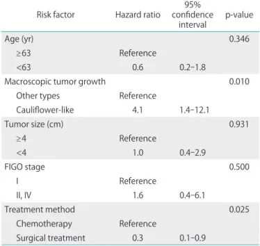
cantly associated with OS (p=0.039 and p<0.001, respectively) (Table 4). The 5-year OS rates for patients with cauliflower- like, nodular, and ulcerative tumors were 20%, 38%, and 40%, respectively. The 5-year OS rates for patients who were treated with surgery plus immunotherapy versus surgery alone were 47% and 29%, respectively. All patients who were treated with non-surgical therapy plus immunotherapy or non-surgical therapy alone died within 5 years of the diagnosis. Other prognostic factors assessed for an impact on survival included patient age at diagnosis, tumor size, FIGO stage, radicality of surgery, and lymphadenectomy performed during surgery.
However, these factors were not significantly associated with survival in univariate analysis. Multivariate analysis identified cauliflower-like macroscopic tumor growth (hazard ratio
[HR], 4.1; 95% confidence interval [CI], 1.4 to 12.1; p=0.010) and surgical treatment (HR, 0.3; 95% CI, 0.1 to 0.9; p=0.025) as significant poor and favorable prognostic factors for OS, respectively. These analyses are summarized in Table 5.
DISCUSSION
Malignant melanoma of the vagina is a rare disease, especial- ly in Asian women. In this study, the majority of patients were postmenopausal and their median age at diagnosis was 58 years. There were no cases of amelanotic vaginal melanoma.
Of the vaginal melanomas, 18 (58%) were located in the lower third of the vagina, and most were nodular under gross observation. Furthermore, 84% were of FIGO stage I, whereas 10% and 7% were of stage II and stage IV, respectively. Overall, the characteristics of the subjects in this study are consistent with the patient characteristics described in previous reports of vaginal melanoma [3,7-11].
In this study, we found that patients with cauliflower-like vaginal melanomas had the worst prognosis, whereas in con- trast, those with ulcerative type tumors survived the longest.
In previous studies, melanoma ulceration was associated with a worse prognosis due to the thickness and aggressiveness of the tumors [12]. However, ulceration has also been identified as a predictive marker for response to adjuvant IFN therapy Table 4. Univariate analysis of overall survival (OS) based on risk
factors
Risk factor No. 5-year OS rate (%)
Median survival
(mo) p-value
Age (yr) 0.148
<63 21 29 18.2
≥63 10 40 60.0
Macroscopic tumor growth 0.039
Cauliflower-like 10 20 6.5
Nodular 16 38 21.0
Ulcerative 5 40 27.0
Tumor size (cm) 0.115
<4 21 33 22.4
≥4 10 30 9.7
FIGO stage 0.175
I 26 31 21.0
II 3 67 31.1
IV 2 0 5.5
Radicality of surgery 0.296
Radical 11 27 18.2
Conservative 11 55 60.6
Lymphadenectomy 0.469
Yes 12 25 22.4
No 10 33 32.2
Treatment method <0.001
S+I 15 47 31.1
S 7 29 26.7
C+I 4 0 5.8
C 3 0 3.8
C, chemotherapy; FIGO, International Federation of Gynecology and Obstetrics; I, immunotherapy; S, surgical treatment.
Table 5. Multivariate analysis of overall survival based on risk factors Risk factor Hazard ratio 95%
confidence
interval p-value
Age (yr) 0.346
≥63 Reference
<63 0.6 0.2-1.8
Macroscopic tumor growth 0.010
Other types Reference
Cauliflower-like 4.1 1.4-12.1
Tumor size (cm) 0.931
≥4 Reference
<4 1.0 0.4-2.9
FIGO stage 0.500
I Reference
II, IV 1.6 0.4-6.1
Treatment method 0.025
Chemotherapy Reference
Surgical treatment 0.3 0.1-0.9
FIGO, International Federation of Gynecology and Obstetrics.
[13]. In the present study, cauliflower-like tumors were typi- cally large, and ulcerative type tumors had a better treatment response to immunotherapy. The sample size of the present study, especially for the ulcerative group, was relatively small.
Therefore, these results need to be confirmed in a study with a larger sample size.
More aggressive surgeries have not been found to alter the prognosis of patients with vaginal melanomas [6,10,14,15].
However, a high rate of local recurrence or local disease progression has been associated with conservative treatment approaches [11,16]. In this study, no significant survival ad- vantage was conferred by radical surgery, and indeed patients who underwent more conservative surgery survived longer than patients who underwent radical surgery. Therefore, on this basis, we suggest that a radical surgical approach including hysterectomy should be avoided for patients with early-stage vaginal melanoma, as it is unlikely to improve their prognosis. However, additional cases need to be examined to confirm this hypothesis because of the small sample size of the present study.
Currently, there is no general recommendation for the treatment of primary vaginal melanoma [6,8,14]. Melanoma is frequently considered a radioresistant tumor, and most re- ports favor surgery over primary radiotherapy [8]. Surgery has been associated with better clinical outcomes compared to chemotherapy approaches. In the present study, only a small number of patients received radiotherapy as their primary treatment, and this is consistent with previous reports [3,7-11].
Immunotherapies recommended for stage II-IV melanoma according to the classification criteria of The American Joint Committee on Cancer (AJCC) include IFN-α, anti-GM2 antibod- ies, and IL-2 [17-19]. In the current study, IL-2, IFN-α, BCG, DC- cells, LAK cells, and a measles vaccine were administered to patients as immunotherapy agents. Patients who underwent surgery followed by immunotherapy attained the longest OS on average, among the 4 treatment groups. Further to these results, surgery followed by a postoperative immunotherapy regimen is recommended as the treatment protocol for patients with primary vaginal melanoma.
The 5-year OS rate for the 31 patients in this study was 32%.
For patients who underwent surgical treatment, the 5-year OS rate was as high as 47%. This value is in the upper range of the 5-year survival rates previously reported for vaginal mela- nomas [1,4-7]. In conclusion, the prognosis of patients with primary malignant melanoma of the vagina is poor. However, for patients with vaginal melanomas that are not cauliflower- like in shape, wide local excision followed by immunotherapy may improve prognosis.
CONFLICT OF INTEREST
No potential conflict of interest relevant to this article was reported.
REFERENCES
1. Weinstock MA. Malignant melanoma of the vulva and vagina in the United States: patterns of incidence and population-based estimates of survival. Am J Obstet Gynecol 1994;171:1225-30.
2. FIGO (International Federation of Gynecology and Obstetrics) 26th annual report on the results of treatment in gynecological cancer. Int J Gynaecol Obstet 2006;95 Suppl 1:S1-257.
3. Gupta D, Malpica A, Deavers MT, Silva EG. Vaginal melanoma: a clinicopathologic and immunohistochemical study of 26 cases.
Am J Surg Pathol 2002;26:1450-7.
4. Frumovitz M, Etchepareborda M, Sun CC, Soliman PT, Eifel PJ, Levenback CF, et al. Primary malignant melanoma of the vagina.
Obstet Gynecol 2010;116:1358-65.
5. Cobellis L, Calabrese E, Stefanon B, Raspagliesi F. Malignant mela- noma of the vagina: a report of 15 cases. Eur J Gynaecol Oncol 2000;21:295-7.
6. Reid GC, Schmidt RW, Roberts JA, Hopkins MP, Barrett RJ, Morley GW. Primary melanoma of the vagina: a clinicopathologic analysis.
Obstet Gynecol 1989;74:190-9.
7. Ragnarsson-Olding B, Johansson H, Rutqvist LE, Ringborg U.
Malignant melanoma of the vulva and vagina: trends in incidence, age distribution, and long-term survival among 245 consecutive cases in Sweden 1960-1984. Cancer 1993;71:1893-7.
8. Van Nostrand KM, Lucci JA 3rd, Schell M, Berman ML, Manetta A, DiSaia PJ. Primary vaginal melanoma: improved survival with radical pelvic surgery. Gynecol Oncol 1994;55:234-7.
9. Borazjani G, Prem KA, Okagaki T, Twiggs LB, Adcock LL. Primary malignant melanoma of the vagina: a clinicopathological analysis of 10 cases. Gynecol Oncol 1990;37:264-7.
10. Neven P, Shepherd JH, Masotina A, Fisher C, Lowe DG. Malignant melanoma of the vulva and vagina: a report of 23 cases presenting in a 10-year period. Int J Gynecol Cancer 1994;4:379- 83.
11. Raber G, Mempel V, Jackisch C, Schneider HP. Clinical aspects of primary malignant melanoma of the vagina. Zentralbl Gynakol 1993;115:416-22.
12. Edge SB, Byrd DR, Compton CC, Fritz AG, Greene FL, Trotti A. AJCC cancer staging manual. 7th ed. New York: Springer; 2010.
13. McMasters KM, Edwards MJ, Ross MI, Reintgen DS, Martin RC 2nd, Urist MM, et al. Ulceration as a predictive marker for response to adjuvant interferon therapy in melanoma. Ann Surg 2010;252:
460-5.
14. Davidson T, Kissin M, Westbury G. Vulvo-vaginal melanoma: should radical surgery be abandoned? Br J Obstet Gynaecol 1987;94:473-6.
15. Konstadoulakis MM, Ricaniadis N, Driscoll DL, Karakousis CP.
Malignant melanoma of the female genital system. Eur J Surg Oncol 1994;20:141-5.
16. Miner TJ, Delgado R, Zeisler J, Busam K, Alektiar K, Barakat R, et al.
Primary vaginal melanoma: a critical analysis of therapy. Ann Surg Oncol 2004;11:34-9.
17. Gray RJ, Pockaj BA, Kirkwood JM. An update on adjuvant inter- feron for melanoma. Cancer Control 2002;9:16-21.
18. Atkins MB, Lotze MT, Dutcher JP, Fisher RI, Weiss G, Margolin K, et al. High-dose recombinant interleukin 2 therapy for patients with metastatic melanoma: analysis of 270 patients treated between 1985 and 1993. J Clin Oncol 1999;17:2105-16.
19. Schadendorf D, Vaubel J, Livingstone E, Zimmer L. Advances and perspectives in immunotherapy of melanoma. Ann Oncol 2012;23 Suppl 10:x104-8.
Standards for Different Types of Articles
Guidelines for different types of articles have been adopted by the Journal of Gynecologic Oncology:
1. CONSORT (Consolidated Standards of Reporting Trials) standards for reporting randomized trials 2. PRISMA (Preferred Reporting Items for Systematic Reviews and Meta-analyses) guidelines for
reporting systematic reviews and meta-analyses
3. MOOSE (Meta-analysis of Observational Studies in Epidemiology) guidelines for meta-analyses and systematic reviews of observational studies
4. STROBE (Strengthening the Reporting of Observational Studies in Epidemiology) guidelines for the reporting of observational studies
5. STARD (Standards for Reporting of Diagnostic Accuracy) standards for reporting studies of diagnostic accuracy
6. REMARK (Reporting of Tumor Markers Studies) guidelines for reporting tumor marker prognostic studies
7. SQUIRE (Standards for Quality Improvement Reporting Excellence) guidelines for quality improvement in health care
8. CHEERS (Consolidated Health Economic Evaluation Reporting Standards) statement for eco- nomic evaluations of health interventions
9. COREQ (Consolidated criteria for Reporting Qualitative research) for qualitative research inter- views and focus groups
10. SAMPL (Statistical Analyses and Methods in the Published Literature) guidelines for basic statistical reporting for articles published in biomedical journals
Investigators who are planning, conducting, or reporting randomized trials, meta-analyses of ran- domized trials, meta-analyses of observational studies, observational studies, studies of diagnostic accuracy, or tumor marker prognostic studies should be familiar with these sets of standards and follow these guidelines in articles submitted for publication.
NOW AVAILABLE ONLINE at http://www.ejgo.org