10.3988/jcn.2010.6.3.127 J Clin Neurol 2010;6:127-137
Melatonin Potentiates the Neuroprotective Properties
of Resveratrol Against Beta-Amyloid-Induced Neurodegeneration by Modulating AMP-Activated Protein Kinase Pathways
Kyoung Ja Kwon, PhDa*; Hee-Jin Kim, MD, PhDb*; Chan Young Shin, PhDc; Seol-Heui Han, MD, PhDa
aDepartments of Neurology and cPharmacology, Center for Geriatric Neuroscience Research, Institute of Biomedical Science and Technology, School of Medicine, Konkuk University, Seoul, Korea
bDepartment of Neurology, Hanyang University College of Medicine, Seoul, Korea
Received February 10, 2010 Revised April 15, 2010 Accepted April 15, 2010
*These authors equally contributed.
Correspondence Seol-Heui Han, MD, PhD Departments of Neurology, Center for Geriatric Neuroscience Research,
Institute of Biomedical Science and Technology, School of Medicine, Konkuk University,
1 Hwayang-dong, Gwangjin-gu, Seoul 143-701, Korea Tel +82-2-2030-7561 Fax +82-2-2030-7899 E-mail [email protected]
Background and PurposezzRecent studies have demonstrated that resveratrol (RSV) reduces the incidence of age-related macular degeneration, Alzheimer’s disease (AD), and stroke, while mel- atonin (MEL) supplementation reduces the progression of the cognitive impairment in AD pa- tients. The purpose of this investigation was to assess whether the co-administration of MEL and RSV exerts synergistic effects on their neuroprotective properties against β-amyloid (Aβ)-induced neuronal death.
MethodszzThe neuroprotective effects of co-treatment with MEL and RSV on Aβ1-42 -induced cell death, was measured by MTT reduction assay. Aβ1-42 caused an increase in intracellular lev- els of reactive oxygen species (ROS), as assessed by H2-DCF-DA dye, and a reduction of total glu- tathione (GSH) levels and mitochondrial membrane potential, as assessed using monochlorobi- mane and rhodamine 123 fluorescence, respectively. Western blotting was used to investigate the intracellular signaling mechanism involved in these synergic effects.
ResultszzWe treated a murine HT22 hippocampal cell line with MEL or RSV alone or with both simultaneously. MEL and RSV alone significantly attenuated ROS production, mitochondrial mem- brane-potential disruption and the neurotoxicity induced by Aβ1-42. They also restored the Aβ1- 42-induced depletion of GSH, back to within its normal range and prevented the Aβ1-42-induced activation of glycogen synthase kinase 3β (GSK3β). However, co-treatment with MEL and RSV did not exert any significant synergistic effects on either the recovery of the Aβ1-42-induced de- pletion of GSH or on the inhibition of Aβ1-42-induced GSK3β activation. Aβ1-42 treatment in- creased AMP-activated protein kinase (AMPK) activity, which is associated with subsequent neu- ronal death. We demonstrated that MEL and RSV treatment inhibited the phosphorylation of AMPK.
ConclusionszzTogether, our results suggest that co-administration of MEL and RSV acts as an ef- fective treatment for AD by attenuating Aβ1-42-induced oxidative stress and the AMPK-depen-
dent pathway. J Clin Neurol 2010;6:127-137
Key Wordszz melatonin, resveratrol, neuroprotection, reactive oxygen species, glycogen synthase kinase 3β, AMP-activated protein kinase.
Introduction
Alzheimer’s disease (AD) is a progressive and age-related neurodegenerative disorder, that is, characterized clinically by irreversible cognitive dysfunction, memory loss, and behav- ioral changes.1 These features are accompanied by specific pathologic changes in the brain, which manifest as the extra-
cellular deposition of a fibrillar form of amyloid-β1-42 peptide (Aβ1-42) constituted senile plaques. Aβ1-42 is the cleavage fragment made by proteinases from the amyloid precursor protein, which exerts neurotoxic effects concerned with neu- roinflammation, immune activation, and oxidative stress and thus has been considered to play a critical role in the patho- genesis of AD.2 However, the mechanism underlying this
β-amyloid peptide (Aβ) neurotoxicity remains to be fully elu- cidated. There is increasing evidence that Aβ alters Ca2+ ho- meostasis, mitochondrial dysfunction, and apoptosis, and in- creases the intracellular level of reactive oxygen species (ROS) in the AD brain.
It is well known that the neuronal cell death induced by Aβ1- 42 is effected via several pathogenic mechanisms, such as the induction of glycogen synthase kinase-3β (GSK3β).3 There are several recent lines of evidence that neurodegeneration, such as in AD and stroke, is associated with the activation of AMP-activated protein kinase (AMPK).4 However, the signal mechanisms underlying Aβ neurotoxicity have not been elu- cidated. Thus, in this investigation, we explored the relation- ship between GSK3β and AMPK signaling in Aβ1-42-induced neurotoxicity.
One of the potential candidates, that can regulate the Aβ-in- duced neuroinflammatory response, is melatonin (N-acetyl-5- methoxy-tryptamine, MEL), which is the main secretory pro- duct of the pineal gland,5 and participates in various physio- logical functions including the control of seasonal repro- duction, regulation of circadian rhythms and body tempera- ture via receptors,6 and acts a potent free-radical scavenger and antioxidant.7 The special features of MEL include its role in the regulation of the immune response and exertion of cyto- protective properties in various neurodegeneration models, both in vivo and in cell cultures.8,9 It was recently suggested that MEL plays a critical role as an antioxidant and neuropro- tector in aging and AD. Levels of MEL decrease with age, and patients with AD experience an even greater reduction in this hormone. Clinical reports indicate that MEL supplementation improves sleep and slows down the progression of cognitive impairment in patients with AD. It also effectively prevents Aβ-mediated neuronal cell death via its antioxidant and anti- amyloid properties. Interest in MEL has grown following st- udies in which it was established that 1) MEL can easily cross the blood brain barrier;10 2) the endogenous production of MEL falls dramatically with increasing age;11 3) high doses of MEL do not present any harmful side-effects;12 and 4) MEL acts as a free-radical scavenger in most tissues.13 On the basis of these data, MEL has been proposed as a preventive treat- ment against neurodegenerative disorders; however, the pro- tective mechanism underlying its actions remains to be eluci- dated.
Another plausible and wide-studied candidate is resvera- trol (trans-3,4,5’-Trihydroxystilbene, RSV), which is a natural polyphenolic compound found mainly in the skin of grapes and nuts, pomegranates, and Polygonum cuspidatum, a com- ponent of Chinese herbal medicines, and is present at high levels in red wine. RSV is a competent estrogenic product, ex- hibiting protective effects in cardiovascular diseases, cancer,
and neurodegenerative diseases,14 partly as a result of its anti- oxidative, anti-inflammatory, and anti-mutagenic activities.15 There is evidence for beneficial effects of RSV in neurodegen- erative conditions, such as cerebral ischemia,16 Parkinson’s disease (PD),17 AD,18 and normal aging.19 The action of RSV ma- nifests as the regulation of the activities and expression levels of enzymes and proteins associated with the survival signal, regulation of ion channels, and anti-oxidative actions.20 RSV also activates the sirtuin categorized class III histone deacety- lases. Sirtuins play a key role in various cellular processes, in- cluding lifespan extension in response to caloric restriction (CR).21,22 However, the mechanisms involved in these protec- tive effects of RSV on neurons are not fully understood.
Based on these observations, we investigated whether com- bination with MEL and RSV can attenuate Aβ1-42-induced neuronal death. In particular, we focused on the effects of com- bined treatment on oxidative stress-induced parameters, such as mitochondrial membrane potential (Δψm), GSK3β, and AM- PK signaling associated with energy homeostasis and cell survival.
Methods
Materials
Pregnant Sprague -Dawley (SD) rats were obtained from Ori- ent Bio (Seoul, Korea) and minimum essential medium (MEM) was purchased from JBI (Seoul, Korea). Dulbecco’s modified Eagle’s medium (DMEM), fetal bovine serum, and antibiotics were purchased from Gibco (Gaithersburg, MD, USA), and Aβ1-42 peptide was purchased from BACHEM (Bubendorf, Switzerland). RSV, N-acetyl cysteine (NAC), 5-aminoimi- dazole-4-carboxamide-1-β-D-ribofuranoside (AICAR), ade- nine 9-β-D-arabinofuranoside (Ara-A), and MEL were purchas- ed from Sigma Chemical (St. Louis, MO, USA). PD98059 (PD) and SB203580 (SB) were purchased from Calbiochem, EMD Biosciences (La Jolla, CA, USA), and SP600125 (SP) and Tro- lox were purchased from Biomol (Plymouth Meeting, PA, USA). Monochlorobimane (mBCl) was purchased from Fluka (St. Louis MO, USA) and H2-2’7’ dichlorofluorescein diacetate dye (H2DCF-DA), dihydroethidium (DHE), and rhodamine 123 were purchased from Molecular Probes (Eugene, OR, USA).
Cell culture
All experimental procedures were carried out using protocols approved by the Institutional Animal Care and Use Commit- tee of Konkuk University. Murine HT22 hippocampal cells were cultured with DMEM supplemented with 10% fetal bo- vine serum, 100 U/mL penicillin, and 100 mg/mL streptomy- cin. The cells were rinsed twice with serum-free medium and then detached with 0.2% trypsin with ethylenediaminetet-
raacetic acid, and re plated at a low density (5,000 cells/cm2) in a 24-well or 6-well plate (Becton-Dickinson, Franklin Lakes, NJ, USA). The re-plated cells were incubated for 24 -h before the experiment.
Primary hippocampal neuronal cultures were obtained from the dissociated embryonic day-18 hippocampus of pregnant SD rats. Briefly, the hippocampus, freed of meninges, was me- chanically dissociated and gently triturated thrice with a flame- polished Pasteur pipette in the culture medium (Eagle’s MEM supplemented with 20 mM glucose). The cells were seeded onto plates coated with 100 μg/mL poly-D-lysine and 200 μg/mL laminin in the culture medium supplemented with 5% fetal bovine serum, 5% horse serum, and 2 mM glutamine. The cul- tures were maintained at 37ºC in a humidified incubator con- taining 5% CO2. For pure neuron cultures, 5 μM cytosine-β- arabinofuranoside was added after 3 days. The cultured cells were used after 7 days, by which time the neurons had differ- entiated from neuronal precursor cells.
Preparation of the Aβ1-42 oligomer
Aβ1-42 was dissolved to a 10- mM stock solution in dimeth- ylsulfoxide (DMSO, Sigma Chemical, SIGMA, St. Louis, USA) and diluted in phosphate-buffered saline (PBS) to a final Aβ concentration of 1 mM. Aggregation of the Aβ1-42 peptide- was achieved by incubating the cells with the diluted Aβ for 3- h at 37ºC, as described previously.23
Experimental procedure
MEL and RSV were dissolved in ethanol and DMSO, respec- tively, at final ethanol and DMSO concentrations of 0.1% and 0.01%, respectively. The effects of MEL and RSV on neuro- nal cell death were determined by evaluating the following:
1) viability of the cells, assessed using a 3-(4,5-dimethylthi- azxol-2-yl)-2,5-diphenyl-tetrazolium bromide (MTT) assay;
2) measurement of intracellular ROS; 3) measurement of to- tal glutathione (GSH); 4) change in the Δψm; and, 5) activa- tion of the intracellular signaling molecules, such as mito- gen-activated protein kinase (MAPKs), GSK3β, and AMPK.
The concentration-dependency of the effects of MEL and RSV on Aβ1-42-induced neuronal death was evaluated in HT22 hippocampal cells, incubated for 24-h and 48 -h with various concentrations of MEL (1, 10, 50, 100, and 500 μM) and RSV (0.1, 1, 5, 10, and 20 μM).
Cells were pretreated with MEL for 1 -h before Aβ1-42, and RSV was added along with Aβ1-42. To establish the participa- tion of AMPK in the neuroprotective effects of MEL and RSV, the cells were incubated for 1-h with the AMPK activator;
AICAR (10, 100, or 1,000 μM) or AMPK inhibitor; Ara-A (10, 100, or 1,000 μM), followed by incubation with Aβ1-42, MEL, and RSV for 24-h.
Assessment of cell viability
Cell viability was also assessed by the MTT assay. MTT is a water-soluble tetrazolium salt that is reduced by metabolically viable cells to a colored, water-insoluble formazan salt. MTT (5 mg/mL) was added to the cell-culture medium. After incu- bating the plates at 37ºC for 2- h in a 5% CO2 atmosphere, the assay was suspended and the MTT-containing medium was replaced with DMSO. The absorbance was read at 570 nm with a microplate reader (Molecular Devices, Palo Alto, CA, USA).
The percentage of surviving neurons was measured relative to control values (untreated cells, 100%).
Measurement of intracellular ROS
Intracellular ROS formation was measured by fluorescence using H2DCF-DA.24 This non-fluorescent dye freely perme- ates into cells, where it de-esterifies to form the ionized free acid (dichlorofluorescein), which reacts with ROS to form the fluorescent 2’,7’- dichlorofluorescein (DCF). After the drug treatment, cultures were washed with Hank’s balanced aque- ous salts solution (HBSS) containing 120 mM NaCl, 5 mM KCl, 1.6 mM MgCl2, 2.3 mM CaCl2, 15 mM glucose, 20 mM HEPES, and 10 mM NaOH, loaded with 20 μM H2DCF-DA and 20% Pluronic F-127 for 30 min at 37ºC, and then washed again with HBSS and kept at room temperature for an addi- tional 30 min to allow for the complete de-esterification of the dye.
DCF fluorescence was analyzed using a fluorescence plate reader (Spectramax Gemini EM, Molecular Devices) at exci- tation and emission wavelengths of 490 and 530 nm, respec- tively, with a fluorescence microscope.
Measurement of Δψm
Changes in Δψm were estimated by the uptake of a cell-per- meant, lipophilic, cationic, fluorescent dye, rhodamine 123, which enters the mitochondria as a result of the highly nega- tive Δψm. Depolarization of Δψm results in the loss of rho- damine 123 from the mitochondria, resulting in a decrease in the intracellular fluorescence. Treated cells were incubated with rhodamine 123 (10 μM) at 37ºC for 20 min, after which they were washed twice with PBS and then observed under a fluorescence microscope (LSM10, Carl Zeiss, Dublin, CA, USA).
Determination of GSH
To measure the total GSH content, the cells cultured in six- well plates were first washed with PBS, and 20 μM mBCl with PBS was added and incubated for 20 min at 37ºC, and then washed again with PBS. Fluorescence was monitored at excitation and emission wavelengths of 390 and 480 nm, re- spectively, using a fluorescence plate reader (Spectramax Ge- mini EM, Molecular Devices).
Western blot analysis
After treatment with Aβ1-42, MEL, and RSV for a specific time period, the cells were harvested and homogenized in 100 μL/well sodium dodecylsulfate (SDS) sample buffer contain- ing 62.5 mM Tris-HCl (pH 6.8), 2% (w/v) SDS, 10% glycer- ol, 50 mM dithiothreitol, 0.1% (w/v) bromophenol blue, and 1 mM sodium orthovanadate. After boiling for 5 min, equal amounts of protein were subjected to 10% SDS polyacrylamide gel electrophoresis for 140 min, after which the separated proteins were transferred electrophoretically to nitrocellulose membranes (Whatman, Dassel, Germany) for 20 min. The blot was blocked with 5% non-fat, dried milk at room tempera- ture and subsequently incubated overnight at 4ºC with the following antibodies: anti-p38, anti-p44/42, anti-c Jun N ter- minal kinases (JNKs), anti-GSK3β, anti-AMPKα, anti-phos- pho-p38 (Thr180/Tyr182), anti-phospho-p44/42 (Thr202/Tyr- 204), anti-phospho-JNK (Thr183/Tyr185), anti-phospho- GSK3β (Ser9), anti-phospho-AMPKα (Thr172) (1:1,000), and anti -β-actin. After incubation with horseradish peroxidase- conjugated secondary antibodies at room temperature for 1-h, the bands were detected with an enhanced chemiluminescence detection system (Amersham Biosciences, Piscataway, NJ, USA) and analyzed using an LAS-3000 image detection sys- tem (Fuji, Tokyo, Japan).
Statistical analysis
Data are expressed as the mean±S.E.M values. Differences between groups were examined for statistical significance using one-way analysis of variance with Student’s t-test and the level of statistical significance was set at p<0.05.
Results
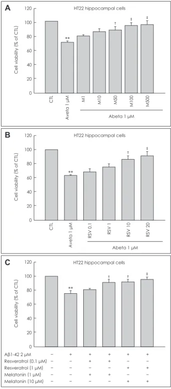
Effects of MEL and RSV on Aβ1-42-induced cytotoxicity in HT22 hippocampal neuronal cells We investigated the synergistic effects of combined treatment with MEL and RSV against Aβ1-42-induced neurotoxicity, us- ing an HT22 hippocampal neuronal cell line, primary hippo- campal neurons, and an MTT assay. Aβ1-42 treatment induced neuronal cell death in a concentration-dependent manner (data not shown). We tested the protective effects of MEL and RSV at a fixed Aβ1-42 concentration of 2 μM. The concentra- tion ranges of MEL and RSV were 1-500 μM and 0.1-20 μM, respectively. MEL and RSV prevented Aβ1-42-induced neu- rotoxicity in HT22 hippocampal neuronal cell line in a con- centration-dependent manner (Fig. 1A and B). The protective effects of MEL and RSV were also observed in rat primary hippocampal neurons (data not shown). Maximum protection against Aβ1-42-induced neurotoxicity was achieved with 500 μM of MEL and 20 μM of RSV. Low concentrations of MEL
A
‡ ‡
†
**
Cell viability
(% of CTL
) CTL Aveta 1 μM M1 M10 M50 M100 M500
120 100 80 60 40 20 0
Abeta 1 μM HT22 hippocampal cells
B
† ‡
**
Cell viability
(% of CTL
) CTL Aveta 1 μM RSV 0.1 RSV 1 RSV 10 RSV 20
120 100 80 60 40 20 0
Abeta 1 μM HT22 hippocampal cells
C
† ‡
†
**
Cell viability
(% of CTL
)
120 100 80 60 40 20
0
Aβ1-42 2 μM − + + + + +
Resveratrol (0.1 μM) − − + + − −
Resveratrol (1 μM) − − − − + +
Melatonin (1 μM) − − + + − −
Melatonin (10 μM) − − − − + +
HT22 hippocampal cells
Fig. 1. Protective effects of MEL and RSV on Aβ1-42-induced cyto- toxicity in HT22 hippocampal neuronal cells. A and B: HT22 cells were treated with the indicated concentrations of MEL (1, 10, 50, 100, and 500 μM) and RSV (0.1, 1, 10, and 20 μM) in the absence or presence of Aβ1-42 for 24 h at 37°C. The viability of HT22 cells was determined by the MTT reduction assay after treatment with MEL, RSV, and Aβ1-42. MEL was administered 1- hr before Aβ1-42 treatment, while RSV was added to the medium along with Aβ1-42.
C: Co treatment effects of MEL (1 and 10 μM) and RSV (0.1 and 1 μM). Data are expressed as the mean±S.E.M values of three ex- periments. The data shown are representative of five independent experiments that gave similar results. **p<0.01 vs. control, †p<0.05,
‡p<0.01 vs. Aβ1-42 alone. MEL: melatonin, RSV: resveratrol, Aβ: β- amyloid.
(1-10 μM) and RSV (0.1-1 μM) did not prevent Aβ1-42-induc- ed cytotoxicity.
However, co-treatment with MEL and RSV at concentra- tions that when treated via monotherapy provided no protec- tion synergistically prevented Aβ1-42-induced cytotoxicity (Fig. 1C). These results suggest that co treatment with MEL and RSV is more effective in preventing Aβ1-42 induced cy- totoxicity than either MEL or RSV alone.
Effects of MEL and RSV on Aβ1-42-induced oxidative stress in HT22 hippocampal neuronal cells
It is relatively well known that the toxicity of Aβ1-42 is medi- ated through the production of ROS and oxidative stress-in- duced damage. Hence, we investigated whether the neuro- protective effects of MEL and RSV blocked Aβ1-42-induced oxidative stress and increased the anti-oxidative properties in the hippocampal neuronal cells. Cellular oxidative stress was determined spectrophotometrically with excitation at 490 nm and emission at 530 nm emission, based on ROS-mediated conversion of DCFH to fluorescent DCF. Aβ1-42 at about 2 μM increased the intracellular ROS production by about 1.8-folds after 6 -h in the hippocampal neuronal cells. MEL and RSV at the tested concentrations profoundly reduced the Aβ1-42-induced production of ROS to control levels (Fig. 2A) without affecting basal levels of ROS production. Trolox (100 μM, a water soluble vitamin E analog), which was used as a positive control, reduced the Aβ1-42 induced increase in ROS by about 40%. This indicates that MEL and RSV have potent antioxidant properties even compared with Trolox in Aβ1-42-stimulated HT22 hippocampal cells. In this study, even co-treatment with MEL and RSV did not induce a reduc- tion of the ROS production below control levels, suggesting that MEL and RSV do not perturb the normal range of ROS production, or even probably ROS-mediated physiological intracellular signaling. The Aβ1-42 induced increase in ROS as measured by DHE was blocked by cotreatment with MEL and RSV (data not shown).
Anti-oxidative properties of MEL and RSV on Aβ1-42-induced oxidative stress in HT22 hippocampal neurons
It has been suggested that the antioxidative properties of MEL and RSV are mediated by the prevention of GSH depletion.
The effects of MEL and RSV on the level of GSH, an essen- tial endogenous antioxidant, were examined by studying the effects of MEL and RSV on Aβ1-42-induced GSH depletion.
After 12- h of Aβ1-42 treatment, mBCl was applied to the medium and the total GSH levels were determined spectro- photometrically with excitation and emission wavelengths of
390 and 485 nm, respectively. In the hippocampal neuronal cells, treatment with Aβ1-42 led to a decrease of about 20%
in intracellular GSH levels within 12- h. Treatment with MEL and RSV restored that GSH depletion to at least the control levels (Fig. 2B). As a result of ROS production, either MEL or RSV was sufficient to prevent GSH depletion; co treatment with MEL and RSV did not further increase intracellular GSH levels.
Effects of MEL and RSV on Δψm
It has been reported that both MEL and RSV, prevent mito- chondrial oxidative stress. Thus, we examined the effects of MEL, RSV, and a combination of both on Δψm as one of the markers of mitochondrial function. Δψm was assessed using rhodamine 123 fluorescence dye. Aβ1-42 treatment induced a loss of about 20 % of Δψm at 6 h, MEL and RSV prevented that loss (Fig. 2C and D). MEL and RSV alone and co treatment with MEL and RSV together did not elevate Δψm over con- trol values.
Effects of MEL and RSV on Aβ1-42-induced MAPK signaling
Oxidative stress is one of the major stimuli for MAPK signal- ing cascades, such as extracellular signal related kinase (ERK) 1/2 and p38. It has been shown that Aβ1-42 treatment leads to activation of ERK1/2, which is related to cell death. The ef- fects of MEL and RSV on the MAPK activation induced by Aβ1-42, the cell lysates, with Aβ1-42 peptide with or without MEL and RSV for 30 min. The results were analyzed by West- ern blotting using anti-phospho-ERK1/2 (Thr202/Tyr204), anti-phospho-p38 (Thr180/Tyr182), and anti-phospho-JNK (Thr183/Tyr185) antibodies. Aβ1-42 treatment resulted in an increase in phosphorylated ERK1/2 and p38 MAPK; this ac- tivation was prevented by MEL and RSV in hippocampal neu- ronal cells (Fig. 3A).
Combination treatment with MEL and RSV inhibited p38 phosphorylation, but did not prevent ERK activation. Aβ1-42- induced neurotoxicity was inhibited by 20 μM PD and 20 μM SB, which are inhibitors of ERK and p38 MAPK, respective- ly. Activation of ERK was blocked by its inhibitor, 20 μM PD, and phosphorylation of p38 MAPK was prevented by the in- hibitor of p38 MAPK, 20 μM SB (Fig. 3B), but not by a JNK in- hibitor, SP. These results provide further evidence that ERK1/2 and p38 MAPK are associated with cell death in the presence of Aβ1-42. Moreover, our data demonstrate that the neuropro- tective effects of MEL and RSV are mediated through the in- hibition of ERK1/2 and p38 activity, and that the inhibitory ef- fects of combination treatment with MEL and RSV on Aβ1- 42-induced neurotoxicity are, at least partially regulated, by p38 MAPK.
A
‡ ‡
‡
†
**
DCF fluorescence
(% of CTL
) CTL RSV 20 MEL 500 UM Tro 100 Abeta 1 μM RSV MEL MR Tro
250 200 150 100 50 0
Abeta 1 μM
HT22 hippocampal cells B
†
† †
†
*
Total GSH
(% of CTL
) CTL RSV 20 MEL 500 Abeta 1 μM RSV MEL MR NAC
160 140 120 100 80 60 40 20 0
Abeta 1 μM HT22 hippocampal cells
D
**
Rhodamine fluorescence
(% of CTL
) 140
120 100 80 60 40 20 0
† ‡ ‡ †
Aβ1-42 2 μM − + + + + +
Resveratrol (1 μM) − − + − + −
Melatonin (10 μM) − − − + + −
Trolox (100 μM) − − − − − +
Fig. 2. Anti oxidative properties of MEL and RSV on Aβ1-42-induced oxidative stress in HT22 hippocampal neuronal cells. MEL was adminis- tered 1-h before Aβ1-42 treatment, while RSV was added to the medium along with the Aβ1-42. A: Effects of MEL and RSV on Aβ1-42- induced increase in DCF fluorescence. RSV at the indicated concentration was applied concomitantly with 1 μM Aβ1-42 for 6-h in HT22 cells. Intracellular ROS was evaluated by measuring H2DCF-DA fluorescence (490/530 nm), as described in the Materials and Methods. B:
Effects of MEL and RSV on Aβ1-42-induced depletion of GSH. After 12-h of Aβ1-42 treatment, mBCl was applied to the medium and total GSH levels were determined spectrophotometrically at 390/480 nm. C: Effects of MEL and RSV on Δψm, as assessed by fluorescence mi- croscopy using the fluorescence dye rhodamine 123, as described in the Materials and Methods. D: Measurement of Δψm by fluorospec- trophotometry: MEL (100 μM), RSV (20 μM), M10R1 (MEL 10 μM+RSV 1 μM), and Tro (Trolox, 100 μM). Data are expressed as mean±S.
E.M values. The data shown are representative of five independent experiments that gave similar results. *p<0.05, **p<0.01 vs. control, †p<
0.05, ‡p<0.01 vs. Aβ1-42 alone. MEL: melatonin, RSV: resveratrol, Aβ: β- amyloid, DCF: dichlorofluorescein.
CTL Abeta 1 μM ARSV AMEL AMR ATro
C
Effects of MEL and RSV on Aβ1-42-induced GSK3β activation
Activation of GSK3β (Ser9) has been shown to be a key com- ponent in the signaling pathways that underlie neurodegenera- tive diseases. It is presumed that Aβ1-42 peptides are involved in the activation of GSK3β. Conversely, inactivation of GSK 3β by phosphoinositide 3-kinase (PI3K)/Akt is an important neuroprotective mechanism. Hence, we examined whether MEL, RSV, and co treatment with both induced inactivation of GSK 3β. Aβ1-42 activated GSK3β, which could be prevented by MEL and RSV at 3 h after Aβ1-42 treatment (Fig. 4A). Either
MEL or RSV alone was sufficient to inactivate GSK3β; com- bination treatment with MEL and RSV did not show further inhibition of GSK3β activation. Aβ1-42-induced neuronal death was inhibited by treatment with LiCl (100 and, 1,000 μM), an inhibitor of GSK3β (Fig. 4B). These results suggest that the neuroprotective effects of MEL and RSV are mediated th- rough an increase in inhibitory GSK3β phosphorylation.
Effects of MEL and RSV on AMPK signal activation
AMPK, a master sensor of energy balance in the peripheral tis- Fig. 3. Effects of MEL and RSV on Aβ1-42-induced MAPK signaling. A: HT22 cells were incubated with 1-μM Aβ1-42 for 30-min in the pres- ence or absence of MEL and RSV at the indicated concentrations, and then harvested for Western blot analysis. Immunoblots of lysates from treated HT22 cells were probed with phospho-ERK (Thr202/Tyr204), phospho-p38 (Thr180/Tyr182), and phospho-JNK (Thr183/Tyr185) anti- bodies. As a loading control, total ERK/p38/JNK levels were also measured (lower panel). MEL and RSV attenuated Aβ1-42-induced activa- tion of ERK and p38 MAPK. B: HT22 cells were treated with the indicated concentrations of PD (inhibitor of ERK), SB (inhibitor of p38 MAPK), and SP (inhibitor of JNK) in the presence of Aβ1-42 for 24-h at 37°C. The viability of HT22 cells was determined by the MTT reduc- tion assay after treatment with PD, SB, and SP with Aβ1-42. Treatment with these MAPK inhibitors blocked the Aβ1-42–induced activation of ERK/p38 MAPK. Data are expressed as the mean±S.E.M values of four independent experiments. **p<0.01 vs. control, †p< 0.05, ‡p<0.01 vs.
Aβ1-42 alone. MEL: melatonin, RSV: resveratrol, Aβ: β- amyloid, MAPK: mitogen-activated protein kinase, ERK: extracellular signal related kinase.
A
B
† †
**
Cell viability
(% of CTL
)
140 120 100 80 60 40 20 0
Aβ1-42 2 μM − + + + +
Resveratrol (20 μM) − − + − +
Melatonin (100 μM) − − − + +
Aβ1-42 2 μM − + + + +
PD95059 (20 μM) − − + − −
SB203580 (20 μM) − − − + −
SP600125 (2 μM) − − − − +
Aβ1-42 2 μM − + + + +
Resveratrol (20 μM) − − + − +
Melatonin (100 μM) − − − + +
HT22 hippocampal cells P-ERK
ERK
P-p38
p38
P-JNK
JNK
pERK/ERK ratiopp38/p38 ratio
1.2 1.0 0.8 0.6 0.4 0.2 0.0 1.0 0.8 0.6 0.4 0.2 0.0
‡
†
†
‡ †
**
**
sues, is phosphorylated and activated when the energy balance is low. RSV exerts multiple beneficial effects similar to those associated with CR, which improves neuronal health; AMPK is thought to be activated during CR. The neuroprotective pro-
perties of MEL and RSV led us to hypothesize that the neuro- nal activation of AMPK is an important component of MEL and RSV activity. We therefore investigated whether AMPK was activated in HT22 hippocampal neuronal cells after Aβ1- 42 treatment and whether MEL or RSV alone or their co treat- ment inhibited AMPK activation. We found a robust increase in the pAMPK (Thr172) level 24-h after Aβ1-42 treatment (Fig.
5A); that elevation was maintained for 48 h. This phosphory- lation of AMPK was blocked by treatment with either MEL or RSV; the inhibitory effects of MEL and RSV were augment- ed by their co treatment. Ara-A (100 μM, a pharmacological inhibitor of AMPK) prevented Aβ1-42-induced neurotoxicity in HT22 hippocampal neuronal cells (Fig. 5B), and the AMPK- activating compound AICAR (100 μM) induced neuronal cell death in HT22 hippocampal cells (Fig. 5C). These findings demonstrate that neuronal inactivation of AMPK by MEL and RSV could affect neuronal energy homeostasis and contrib- ute to the neuroprotective effects of MEL and RSV.
Discussion
The reported results show that MEL and RSV prevent the ac- tivation of ERK, decrease ROS production, and restore GSH levels, and eventually attenuate neuronal cell death in hippo- campal neuronal cells. However, although combination ther- apy with MEL and RSV did not inhibit the activation of ERK, the production of ROS, or the depletion of GSH, it did exert a synergistic effect in reducing Aβ1-42-induced neurotoxicity.
Aβ1-42 reduced the Δψm in the hippocampal neuronal cells, an action that was restored by cotreatment with MEL and RSV.
The Aβ1-42 induced potentiation of GSK3β activity was in- hibited by, either MEL or RSV alone. Similarly, the Aβ1-42- induced activation of AMPK, a well-known regulator of ener- gy homeostasis, was effectively prevented by treatment with either MEL or RSV. Importantly, combined treatment with MEL and RSV exerted a synergic reduction in AMPK activation.
The aggregation of soluble Aβ1-42 into oligomers/fibrils is one of the key pathological features of AD. We used Aβ1-42 of oligomers/fibrils made by incubation for 3 h at 37ºC, as desc- ribed in the Materials and Methods. Although some batch to- batch variation was observed, the manufactured Aβ1-42 in- duced neurotoxicity at a rate of 20-40% in HT22 hippocampal cells. Since MEL and its metabolites act as radical scavengesr, they can stimulate the activity of the mitochondrial enzyme (NA- DH-coenzyme Q reductase, and cytochrome C oxidase) asso- ciated with oxidative phosphorylation,25 and subsequently in- crease ATP production. RSV, a polyphenolic compound, is known to be a potent activator of the sirtuin pathway and mimics the CR, both of which are associated with life-span expansion and aging. As a sirtuin activator, RSV is associated with the induc- Fig. 4. Inhibitory effects of MEL and RSV on Aβ1-42-induced GSK-
3β activation. A: HT22 cells were incubated with 2 μM Aβ1-42 for 6- h in the presence or absence of the indicated concentrations of MEL and RSV, and harvested for Western blot analysis. Immunob- lots of lysates from treated HT22 cells were probed with phospho- GSK3β (Ser9) and GSK3β antibodies. Actin levels were measured for the confirmation of equal amount of protein loading (lower pan- el). MEL and RSV inhibited the Aβ1-42-induced activation of GSK- 3β. B: HT22 cells were treated with indicated concentrations of LiCl (inhibitor of GSK3β) in the presence of Aβ1-42 for 24- h at 37°C. The viability of HT22 cells was determined by the MTT reduction assay after treatment of LiCl with Aβ1-42. Data are expressed as the mean
±S.E.M values of four independent experiments. **p<0.01 vs. con- trol, †p<0.05, ‡p<0.01 vs. Aβ1-42 alone; RSV (20 μM), MEL (100 μM), MR (MEL 10 μM+RSV 1 μM), Li (LiCl, 1 mM). MEL: melatonin, RSV:
resveratrol, Aβ: β- amyloid.
B
† †
**
Cell viability
(% of CTL
)
140 120 100 80 60 40 20 0
HT22 hippocampal cells
CTL Aveta 1 μM Li 100 μM Li 1 mM
Abeta 1 uM p-GSK 3b (Ser9)
GSK 3b β-actin
A
‡
† †
†
pGSK3b/GSK33b ratio **
4
3
2
1
0
Aβ1-42 2 μM − + + + + +
Resveratrol (20 μM) − − + − + −
Melatonin (100 μM) − − − + + −
LiCI (1 mM) − − − − − +
tion of genes for oxidative phosphorylation and mitochon- drial biogenesis,26 and can stimulate the preservation of brain mitochondrial function after hypoxia- reperfusion. We expect- ed that co- treatment with MEL and RSV would be more pro- tective against Aβ-induced neurotoxicity than either treat- ment alone. Our data indicate that the neuroprotective effects of MEL and RSV are mediated via a decrease in ROS and an up regulation of the antioxidant system, and that combination treatment with these two factors has a synergic effect on neu- roprotection. Therefore, the actions of combined treatment with MEL and RSV could be beneficial for energy preserva- tion and prevention of neuronal cell death.
Combination treatment is a method of treating disease th- rough the simultaneous use of a variety of drugs to eliminate or control the biochemical cause of the disease. The rationale for combination treatment is to use drugs that work via differ- ent mechanisms of action without intolerable side effects.
However, there have been few studies on combination treat- ments. It was recently reported that combination therapy may be better than monotherapy in bipolar disorder in patients with bipolar disorder needing long-term treatment, the com-
bination of lithium plus valproate is more effective than val- proate alone.27 In the present study, we investigated the effects of combination treatment with MEL and RSV, which are well- known neuroprotectors.
Functionally, RSV exhibits histone deacetylase activity as a sirtuin activator (SIRT1), which is associated with CR and energy preservation, and so could have neuroprotective quali- ties and promote longevity. SIRT1 is a class III histone deacet- ylase and is essential for maintaining silent chromatin through the deacetylation of histones. MEL also is known to be an inducer of SIRT1.28 It is possible that a combination of MEL and RSV exerts a synergic effect on SIRT1 activity, and that such combination treatment could thus induce a synergic ef- fect on neuroprotection. Further study is needed to elucidate the exact mechanism underlying this potential effect.
Our results show that ERK activation is related to Aβ1- 42- induced neurotoxicity in HT22 hippocampal neurons. Previ- ous studies have shown that ERK activation is associated with cell survival and synaptic sprouting. However, it has been re- ported recently that Aβ-induced neurotoxicity is regulated by the inhibition of ERK activation in rat organotypic hippocam- Fig. 5. Effects of MEL and RSV on the activation of AMPK signaling. A: HT22 cells were incubated with 2 μM Aβ1-42 for 24-h in the presence or absence of the indicated concentrations of MEL and RSV, and then harvested for Western blot analysis. Immunoblots of lysates from treated HT22 cells were probed with phospho-AMPK (Thr172) and total AMPK antibodies. Actin levels were also measured to confirm equal amounts of protein loading (lower panel). MEL and RSV inhibited the Aβ1-42-induced activation of AMPK. B: HT22 cells were treated with Ara-A (inhibitor of AMPK) indicated concentrations, and Aβ1-42, or (C) the cells were treated with AICAR (activator of AMPK) without Aβ1-42 for 24- h at 37°C for the MTT assay. The viability of HT22 cells was determined by the MTT reduction assay. The inhibitor of AMPK blocked Aβ1-42-induced neuronal cell death and the activator of AMPK augmented Aβ1-42-induced neuronal cell death. Data are expressed as the mean±S.E.M values of three independent experiments. **p<0.01 vs. control, †p<0.05, ‡p<0.01 vs. Aβ1-42 alone, §p<0.01 vs. drug only; RSV (20 μM), MEL (100 μM), MR (MEL 10 μM+RSV 1 μM), AIC (AICAR, 10-100 μM), Ara-A (10-100 μM). MEL: melatonin, RSV: resveratrol, Aβ: β- amyloid, ERK: extracellular signal related kinase, AMPK: AMP-activated protein kinase.
A
B
†
†
†
§
** §
pAMPK/AMPK ratio
1.8 1.6 1.4 1.2 1.0 0.8 0.6 0.4 0.2 0.0
Aβ1-42 2 μM − + + + +
Resveratrol (20 μM) − − + − +
Melatonin (100 μM) − − − + +
HT22 hippocampal cells p-AMPKα
(Thr172) AMPK β-actin
‡
‡
**
Cell viability
(% of CTL
) 120
100 80 60 40 20 0
AMPK inhibitor: AraA
CTL Aveta 1 μM AraA 10 AraA 100
Abeta 1 μM
C
** **
**
Cell viability **
(% of CTL
) 120
100 80 60 40 20 0
AICAR: AMPK Activator
CTL Aveta 1 μM AIC 10 AIC 100 AIC 1000
pal slice cultures,29 and that glutamate-induced toxicity is re- lated to ERK activation.30 Although co treatment with MEL and RSV did not exert a synergistic effect in reducing ERK activation, our results demonstrate that Aβ1-42 induced neu- rotoxicity could be mediated through ERK activation.
AMPK has emerged as a master sensor of energy balance, and is phosphorylated and activated when energy balance is low. In other words, AMPK is activated by increasing the cel- lular AMP: ATP ratio, whereupon it functions to help preserve cellular energy.4 RSV is associated with CR, increasing the lifespan, and delaying the onset of diseases associated with aging. CR improves neuronal health, and a key enzyme th- ought to be activated during CR is AMPK. Recent reports suggest that AMPK is highly expressed in cortical and hippo- campal neurons under both normal and ischemic conditions.
Pharmacological inhibition of AMPK reduces stroke damage, whereas the activation of AMPK by AICAR exacerbates that damage. We hypothesized that AMPK activation is an impor- tant mediator of MEL and RSV actions in hippocampal neu- ronal cells. Our results suggest that MEL and RSV manipulate the neuronal energy balance during Aβ1-42 treatment.
It was shown recently, that RSV activates AMPK signaling and that this activation of AMPK lowers extracellular Aβ ac- cumulation.31 However, MEL did not influence AMPK acti- vation in C2C12 murine skeletal muscle cells.32 Our results show that the Aβ1-42-induced activation of AMPK was in- hibited by both monotherapy with either MEL or RSV and by their co- treatment in the murine HT22 hippocampal cell line.
Neuronal damage induced by Aβ1-42 activates AMPK, and combination treatment with MEL and RSV synergistically pre- vented neuronal death by inhibiting the activation of AMPK.
However, this effect on AMPK activation needs to be studied further.
In this study, MEL and RSV reversed the Aβ1-42 induced al- teration in GSK3β, ERK, and AMPK activity. One obvious question pertains to the inter relationship of the activities of th- ese signaling molecules. For example, activation of the PI3K- Akt-GSK3β pathway can regulate the AMPK pathway, and we reported that inhibition of GSK3β can activate ERK1/2 in cultured rat astrocytes.33 It is clear that understanding the nature of the extensive interplay between these diverse signal- ing systems may help us to understand the mechanism under- lying Aβ-dependent neurotoxicity as well as the protective mechanism induced by MEL and RSV.
In conclusion, our data demonstrate clearly that MEL and RSV have strong neuroprotective properties against Aβ1-42- induced cytotoxicity in HT22 hippocampal neuronal cells, by attenuating oxidative stress through the induction of anti- oxidants, such as GSH, and the inhibition of GSK3β activity.
We also found that combined treatment with MEL and RSV
exerts beneficial effects on neuronal survival by inhibiting the activation of AMPK. Our data suggest that co treatment with MEL and RSV provides a useful therapeutic strategy against Aβ1-42-induced neurotoxicity.
Conflicts of Interest
The authors have no financial conflicts of interest.
Acknowledgements
This work was supported by WCU (World Class University) program through the National Research Foundation of Korea funded by the Min- istry of Education, Science and Technology (R33-2008-000-10090-0).
REFERENCES
1. Kelley BJ, Petersen RC. Alzheimer’s disease and mild cognitive im- pairment. Neurol Clin 2007;25:577-609.
2. Selkoe DJ. The molecular pathology of Alzhiemer’s disease. Neuron 1991;6:487-498.
3. Bhat RV, Budd Haeberlein SL, Avila J. Glycogen synthase kinase 3:
a drug target for CNS therapies. J Neurochem 2004;89:1313-1317.
4. Williams KW, Coppari R, Elmquist JK. “AMPing up” our understand- ing of the hypothalamic control of energy balance. J Clin Invest 2007;
117:2089-2092.
5. Reiter RJ. Pineal melatonin: cell biology of its synthesis and of its physiological interactions. Endocr Rev 1991;12:151-180.
6. Witt-Enderby PA, Radio NM, Doctor JS, Davis VL. Therapeutic treat- ments potentially mediated by melatonin receptors: potential clinical uses in the prevention of osteoporosis, cancer and as an adjuvant ther- apy. J Pineal Res 2006;41:297-305.
7. Reiter RJ, Tan DX, Qi W, Manchester LC, Karbownik M, Calvo JR.
Pharmacology and physiology of melatonin in the reduction of oxida- tive stress in vivo. Biol Signals Recept 2000;9:160-171.
8. Antolín I, Mayo JC, Sainz RM, del Brío Mde L, Herrera F, Martín V, et al. Protective effect of melatonin in a chronic experimental model of Parkinson’s disease. Brain Res 2002;943:163-173.
9. Shen YX, Xu SY, Wei W, Wang XL, Wang H, Sun X. Melatonin blocks rat hippocampal neuronal apoptosis induced by amyloid beta-peptide 25-35. J Pineal Res 2002;32:163-167.
10. Costa EJ, Lopes RH, Lamy-Freund MT. Permeability of pure lipid bi- layers to melatonin. J Pineal Res 1995;19:123-126.
11. Pierpaoli W, Regelson W. Pineal control of aging: effect of melatonin and pineal grafting on aging mice. Proc Natl Acad Sci U S A 1994;91:
787-791.
12. Reiter RJ, Pablos MI, Agapito TT, Guerrero JM. Melatonin in the con- text of the free radical theory of aging. Ann N Y Acad Sci 1996;786:
362-378.
13. Reiter RJ, Melchiorri D, Sewerynek E, Poeggeler B, Barlow-Walden L, Chuang J, et al. A review of the evidence supporting melatonin’s role as an antioxidant. J Pineal Res 1995;18:1-11.
14. Baur JA, Sinclair DA. Therapeutic potential of resveratrol: the in vivo evidence. Nat Rev Drug Discov 2006;5:493-506.
15. Zhuang H, Kim YS, Koehler RC, Doré S. Potential mechanism by which resveratrol, a red wine constituent, protects neurons. Ann N Y Acad Sci 2003;993:276-286.
16. Gao D, Zhang X, Jiang X, Peng Y, Huang W, Cheng G, et al. Resve- ratrol reduces the elevated level of MMP-9 induced by cerebral isch- emia-reperfusion in mice. Life Sci 2006;78:2564-2570.
17. Okawara M, Katsuki H, Kurimoto E, Shibata H, Kume T, Akaike A.
Resveratrol protects dopaminergic neurons in midbrain slice culture from multiple insults. Biochem Pharmacol 2007;73:550-560.
18. Marambaud P, Zhao H, Davies P. Resveratrol promotes clearance of Alzheimer’s disease amyloid-beta peptides. J Biol Chem 2005;280:
37377-37382.
19. Valenzano DR, Terzibasi E, Genade T, Cattaneo A, Domenici L, Cel- lerino A. Resveratrol prolongs lifespan and retards the onset of age-re- lated markers in a short-lived vertebrate. Curr Biol 2006;16:296-300.
20. Baur JA, Sinclair DA. Therapeutic potential of resveratrol: the in vivo evidence. Nat Rev Drug Discov 2006;5:493-506.
21. Han SH. Potential role of sirtuin as a therapeutic target for neurode- generative diseases. J Clin Neurol 2009;5:120-125.
22. Baur JA, Pearson KJ, Price NL, Jamieson HA, Lerin C, Kalra A, et al.
Resveratrol improves health and survival of mice on a high-calorie diet. Nature 2006;444:337-342.
23. Mook-Jung I, Joo I, Sohn S, Kwon HJ, Huh K, Jung MW. Estrogen blocks neurotoxic effects of beta-amyloid (1-42) and induces neurite extension on B103 cells. Neurosci Lett 1997;235:101-104.
24. LeBel CP, Ali SF, McKee M, Bondy SC. Organometal-induced in- creases in oxygen reactive species: the potential of 2',7'-dichlorofluo- rescin diacetate as an index of neurotoxic damage. Toxicol Appl Phar- macol 1990;104:17-24.
25. Martín M, Macías M, León J, Escames G, Khaldy H, Acuña-Castro- viejo D. Melatonin increases the activity of the oxidative phosphoryla- tion enzymes and the production of ATP in rat brain and liver mitochon- dria. Int J Biochem Cell Biol 2002;34:348-357.
26. Lagouge M, Argmann C, Gerhart-Hines Z, Meziane H, Lerin C, Dau- ssin F, et al. Resveratrol improves mitochondrial function and protects against metabolic disease by activating SIRT1 and PGC-1alpha. Cell 2006;127:1109-1122.
27. BALANCE investigators and collaborators, Geddes JR, Goodwin GM, Rendell J, Azorin JM, Cipriani A, et al. Lithium plus valproate
combination therapy versus monotherapy for relapse prevention in bipolar I disorder (BALANCE): a randomised open-label trial. Lancet 2010;375:385-395.
28. Tajes M, Gutierrez-Cuesta J, Ortuño-Sahagun D, Camins A, Pallàs M.
Anti-aging properties of melatonin in an in vitro murine senescence model: involvement of the sirtuin 1 pathway. J Pineal Res 2009;47:
228-237.
29. Chong YH, Shin YJ, Lee EO, Kayed R, Glabe CG, Tenner AJ. ERK- 1/2 activation mediates Abeta oligomer-induced neurotoxicity via caspase-3 activation and tau cleavage in rat organotypic hippocampal slice cultures. J Biol Chem 2006;281:20315-20325.
30. Lee EO, Park HJ, Kang JL, Kim HS, Chong YH. Resveratrol reduces glutamate-mediated monocyte chemotactic protein-1 expression via inhibition of extracellular signal-regulated kinase 1/2 pathway in rat hippocampal slice cultures. J Neurochem 2010;112:1477-1487.
31. Vingtdeux V, Giliberto L, Zhao H, Chandakkar P, Wu Q, Simon JE, et al. AMP-activated protein kinase signaling activation by resveratrol modulates amyloid-beta peptide metabolism. J Biol Chem 2010;285:
9100-9133.
32. Ha E, Yim SV, Chung JH, Yoon KS, Kang I, Cho YH, et al. Melatonin stimulates glucose transport via insulin receptor substrate-1/phosphati- dylinositol 3-kinase pathway in C2C12 murine skeletal muscle cells. J Pineal Res 2006;41:67-72.
33. Kim SD, Yang SI, Kim HC, Shin CY, Ko KH. Inhibition of GSK-3be- ta mediates expression of MMP-9 through ERK1/2 activation and tr- anslocation of NF-kappaB in rat primary astrocyte. Brain Res 2007;1186:
12-20.