Effect of biofilm formation, and biocorrosion on denture base fractures
Cem Sahin1*, DDS, PhD, Alper Ergin2, PhD, Simel Ayyildiz3, DDS, PhD, Erdal Cosgun4, MS, Gulay Uzun5, DDS, PhD
1School of Health Services, Dental Prosthetics Technology, Hacettepe University, Ankara, Turkey
2School of Health Services, Hacettepe University, Ankara, Turkey
3Department of Prosthodontics, Center for Dental Sciences, Gulhane Military Medical Academy, Ankara, Turkey
4Department of Biostatistics, Faculty of Medicine, Hacettepe University, Ankara, Turkey
5School of Health Services, Dental Prosthetics Technology, Hacettepe University, Ankara, Turkey
PURPOSE. The aim of this study was to investigate the destructive effects of biofilm formation and/or biocorrosive activity of 6 different oral microorganisms. MATERIALS AND METHODS. Three different heat polymerized acrylic resins (Ivocap Plus, Lucitone 550, QC 20) were used to prepare three different types of samples. Type “A”
samples with “V” type notch was used to measure the fracture strength, “B” type to evaluate the surfaces with scanning electron microscopy and “C” type for quantitative biofilm assay. Development and calculation of biofilm covered surfaces on denture base materials were accomplished by SEM and quantitative biofilm assay.
According to normality assumptions ANOVA or Kruskal-Wallis was selected for statistical analysis (α=0.05).
RESULTS. Significant differences were obtained among the adhesion potential of 6 different microorganisms and there were significant differences among their adhesion onto 3 different denture base materials. Compared to the control groups after contamination with the microorganisms, the three point bending test values of denture base materials decreased significantly (P<.05); microorganisms diffused at least 52% of the denture base surface. The highest median quantitative biofilm value within all the denture base materials was obtained with P. aeruginosa on Lucitone 550. The type of denture base material did not alter the diffusion potential of the microorganisms significantly (P>.05). CONCLUSION. All the tested microorganisms had destructive effect over the structure and composition of the denture base materials. [J Adv Prosthodont 2013;5:140-6]
KEY WORDS: Biocorrosion; Microorganisms; Biofilm; Denture base fractures
INTRODUCTION
Polymethyl methacrylate (PMMA) is primarily used for removable partial and complete denture fabrication as a base material. Reinforcements such as fibers, glass1 and
Corresponding author:
Cem Sahin
School of Health Services, Dental Prosthetics Technology, Hacettepe University, Sihhiye 06100, Ankara, Turkey
Tel. 905053353092: e-mail, [email protected]
Received November 27, 2012 / Last Revision April 16, 2013 / Accepted April 23, 2013
© 2013 The Korean Academy of Prosthodontics
This is an Open Access article distributed under the terms of the Creative Commons Attribution Non-Commercial License (http://creativecommons.
org/licenses/by-nc/3.0) which permits unrestricted non-commercial use, distribution, and reproduction in any medium, provided the original work is properly cited.
polyethylene or activation and processing techniques such as injection-molding and microwave2 activation provided new benefits in polymer knowledge. However, this material still has some limitations about mechanical properties such as dimensional stability, color and flexural strength.
Eventually fracture of acrylic resin denture base is known to be the common clinical problem in prosthodon- tic practice. Causes of such fractures are known to be relat- ed to porosity, residual monomer, poor fit of denture base, occlusal imbalance, design and fabrication failures, material choices, stress after long clinical usage and accidents.3,4 Besides, in certain circumstances before complete fracture, a crack formation may be propagated bound to different types of stresses that denture base materials are subjected to. It is known that rough surfaces present suitable condi- tions for microorganism colonisation and/or biofilm for- mation,5,6 denture base cracks may become to be one of the best sites for microorganism propagation.
Biofilm is a microbial community that has dense and complex structure and may represent multiple organisms.7 They are often encapsulated within a matrix of exopoly- meric material that consists of intricate networks of cells attached to biotic surfaces. They resist antimicrobials and immune cell challenge8 and are deeply embedded into cracks and porosities of dental materials as mentioned before. Metallic and non-metallic medical devices like cath- eters, implants, dental materials are suitable sites for coloni- zation of various types of microorganisms.9 Corrosion at the interfacial surfaces of non-metallic materials usually starts with swelling after infiltration of little molecules or microorganisms. The chemical bonds are often subjected to corrosion with physiochemical process. Interfacial electro- chemical process may be activated with the formation of biofilm on metallic and non-metallic surfaces resulting in an increased corrosion of colonized substratum. This develop- ment can detoriorate the materials with the presence of biofilm and is termed as biocorrosion.9
Three-dimensional structure of biofilm is known to provide a highly complex arrangement of microorgan- isms.10 Several studies regarding the developments and structures of biofilms on different dental materials includ- ing denture bases and their effects over oral health have been constituted.8-13 However the relationship between the biofilm related biocorrosion and crack and/or fracture for- mation still remains complicated even undefined.
The aim of this study was to investigate the destructive effects of biofilm formation and/or biocorrosive activity of 6 different oral microorganisms by evaluating the diffu- sion potential of the microorganisms, fracture propagation and scanning electron microscope (SEM) images (calculat- ing biofilm covered surfaces) on three different denture base materials.
MATERIALS AND METHODS
Three different heat polymerized acrylic resins (Table 1) were used to prepare 50 × 15 × 4 mm (Type A; for three point bending (TPB) test, n=210), 8 × 8 × 1 mm (Type B;
for SEM analysis, n=54) and 2 × 2 × 2 mm (Type C; for spectrophotometer analysis, n=180) samples (Table 2).
Negative molds of the metal masters were obtained with a medium viscosity impression material. This technique was used to fabricate all types of the specimens. Wax patterns were invested in metal dental flasks. Acrylic resins were polymerized according to manufacturers’ instructions.
Flasks were left for 180 minutes cooling. Next, each speci- men were deflasked and finished with 320, 400 and 600-grit silicone carbid papers. To simulate a crack line on the den- ture base, “V” type notch was carved in the middle of each specimen of impact test groups (A type) along with the 15 mm surface by using a milling machine and a milling tool as shown in the Fig. 1. The depth was 2 ± 0.2 mm. All type of specimens were ultrasonically cleaned for 20 minutes and immersed in distilled water for 48 hours at 37℃ before tests.
S. aureus, E. faecalis, P. aeruginosa, E. coli, S. mutans and C. albicans strains were inoculated in trypticase soy broth media and grown to stationary phase overnight. The sam- ples were diluted 1:100 and each diluted bacterial culture (200 µl) was inoculated into each well in a fresh 96-well flat- bottom microtiter polystyrene plates, which also contain
“C” type acrylic resin samples. Plates were incubated for 48 hours at 37℃ and visualized by staining with 0.5% crystal violet for five minutes after washing with water. The bio- film was quantified in duplicate, after adding 100 µL of 95% ethanol and the contents were transferred to new wells of microtiter plate. Optical density (OD) of stained adher- ent bacteria was determined with a micro ELISA auto read- er at wavelength of 620 nm spectrophotometrically.14,15
specimen 15 mm
50 mm milling tool 4 mm
Fig. 1. Schematic view of ‘V’ type notch carving.
Table 1. Description of denture base materials
Acrylic resin Lot number Manufacturer Description Polymerization type
Ivocap Plus M38730 Ivoclar Vivadent, Schaan, Liechtenstein High impact denture base material Heat polymerized Injection method
Lucitone 550 1002V7 Dentsply International Inc., Chicago, IL, USA High impact denture base material Heat polymerized Conventional method
QC 20 64015503 Dentsply International Inc., Chicago, IL, USA Denture base material Heat polymerized Conventional method
These OD values were calculated as: “OD-control” and considered as an index of bacteria adhering to acrylic sur- faces and forming biofilms.
Ethylene oxide was used to sterilize the specimens. Ten specimens of type “A”, 3 specimens of type “B” and 10 specimens of type “C” denture base materials from 3 dif- ferent brands (Table 1) were randomly inoculated into one of; Staphylococcus aureus (ATCC 6538), Streptococcus mutans (ATCC 35688), Enterococcus faecalis (ATCC 10541), Escherichia coli (ATCC 25922), Pseudomonas aeruginosa (ATCC 2327) or Candida albicans (ATCC 18804) culture as indicated in table 2 at 0.5 McFarland scale which corresponds to 108 cfu/mL for 168 hours at 37℃. A previously described method was modified and performed for biofilm forma- tion.11 In brief, 1 mL aliquot of the bacterial and yeast cul- tures were introduced into 500 mL of brain heart infusion broth media in conical flasks and prepared denture base materials were inoculated into media. For the maintenance of bacterial and yeast density near the steady-state growth phase, 50% of the media were drained and replaced with the equal amount of a fresh sterile medium every two days and on day seven, the denture base materials were retrieved from the inoculated media for SEM examination. The con- trol group was composed of 10 non-contaminated “A” type
samples of each denture base material (30 total) and was kept in distilled water at 37℃ until the TPB test.
The contaminated samples of type “A”, were rinsed with PBS and kept in running water for 15 minutes. The impact values of the specimens were measured under 2500 N maximum load (1 N preload) with 1 mm/min cross-head speed with TPB test by using an universal testing machine (Lloyd LRX, West Sussex, UK). The specimens were then supported on the jigs with a diameter of 3.2 mm with span length 50 mm. The “V notch” was placed face down on the jigs and the load was applied to the centre of the speci- mens. The data of the measurements were transferred to a personal computer, and the results were recorded.
After incubation, each specimen of type “B” was removed and kept in an ultrasonic cleaner for 1 minute and rinsed with PBS to remove non or weak adhered microor- ganisms. The samples were fixed in 2.5% glutaraldehyde solution for 1h at room temperature, then rinsed with PBS.
Ethanol solutions with concentrations graded from 75% to 95% were used in 5 steps to dehydrate the specimens.
Specimens were then dried and placed on stubs to coat with 20 A0 gold/palladium for SEM (JEOL 6400, JEOL Corp., Tokyo, Japan) analysis operating at 10 kV. Digital photo- graphs as TIFF files at ×5,000 magnification were obtained Table 2. Types of samples, names of microorganisms and denture base materials
Sample Microorganism Denture base material
Lucitone 550 (n) QC 20 (n) Ivocap Plus (n)
Type A S. aureus 10 10 10
for three point bending test S. mutans 10 10 10
50 x 15 x 4 mm E. faecalis 10 10 10
E. coli 10 10 10
P. aeruginosa 10 10 10
C. albicans 10 10 10
Control 10 10 10
Total 70 70 70
Type B S. aureus 3 3 3
for SEM analysis S. mutans 3 3 3
8 x 8 x 1 mm E. faecalis 3 3 3
E. coli 3 3 3
P. aeruginosa 3 3 3
C. albicans 3 3 3
Total 18 18 18
Type C S. aureus 10 10 10
for spectrophotometric analysis S. mutans 10 10 10
2 x 2 x 2 mm E. faecalis 10 10 10
E. coli 10 10 10
P. aeruginosa 10 10 10
C. albicans 10 10 10
Total 60 60 60
from three different regions of each sample surface (Fig. 2).
The images were transferred to a personal computer to cal- culate total area and area fractions of biofilms using Image J software.16 Since cleaning and dehydrating processes were performed accurately, the area aspects apart from denture surface were all accepted as biofilm surface of that micro- organism. Area fraction of each image was recorded as the data of that sample.
Descriptive data were expressed as median, maximum, minimum and mean ± standard deviation. Statistical analy- sis was performed using “PASW 18.0 Statistics” and
“STATISTICA-7” statistical software. When normality assumptions were satisfied Analysis of Variance (ANOVA) otherwise the equivalent non-parametric test: Kruskal- Wallis was used for group comparisons. When significant differences found between groups, we used Tukey and Dunnett’s test (after ANOVA) and Dunn’s test (after Kruskall Wallis) for multiple group comparisons. Results were considered statistically significant at α=0.05.
RESULTS
Table 3 shows the mean quantitative biofilm values and sta- tistical significances according to microorganism and den-
ture base material. There were significant differences among the adhesion potential of 6 different microorgan- isms and there were significant differences among their adhesion onto 3 different denture base materials. The high- est median value within all the denture base materials was obtained with P. aeruginosa (0.095 ± 0.018) followed by S.
aureus (0.085 ± 0.014), C. albicans (0.081 ± 0.017), S. mutans (0.079 ± 0.010), E. faecalis (0.070 ± 0.013) and E. coli (0.070 ± 0.012). E. faecalis and E. coli were found to be the least adherent microorganisms. The difference of adhesion between P. aeruginosa and S. aureus was not significant how- ever there were significant differences between P. aeruginosa and other 4 microorganisms. The adhesion potential of the microorganisms over Lucitone denture base material was higher than the other materials. The difference was not sig- nificant compared with QC-20 but it was significant when compared with Ivocap Plus. P. aeruginosa exhibited the high- est median value on Lucitone 550 denture base material surface (0.105 ± 0.020) while C. albicans exhibited the low- est value on Ivocap Plus (0.067 ± 0.021).
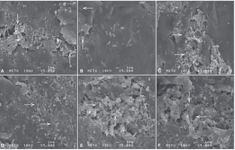
SEM images revealed the regions of typical biofilm for- mation of each selected microorganism on denture base materials (Fig. 2). Table 4 shows the mean biofilm covered regions (%) on denture base materials and statistical signifi- Fig. 2. SEM images of biofilm formations of C. albicans (A), E. coli (B), E. faecalis (C), P. aeruginosa (D), S. aureus (E), S.
mutans (F).
A B C
D E F
cances according to microorganism and denture base mate- rial. The type of denture base material did not alter the dif- fusion potential of the microorganisms significantly. The percentages of biofilm covered areas of denture base mate- rials ranged from 52.57% to 70.96%. S. aureus and P. aerugi- nosa had significantly higher (≥68.66%) diffusion potential than the other tested microorganisms, but the difference
among them was not significant. E. coli had the least diffu- sion potential and was significantly different from all the tested groups.
Table 5 shows the mean and standard deviation values of TPB test and statistical significances according to micro- organism and denture base material. Biofilm formation of the tested microorganisms decreased the TPB test values Table 3. Quantitative biofilm values: median and standard deviation (Kruskal Wallis)
Microorganism Denture base materials
Median ± SD
Lucitone 550 QC-20 Ivocup Plus
S. aureus 0.093 ± 0.012 0.086 ± 0.017 0.075 ± 0.018 0.085 ± 0.014a,b,c
P. aeruginosa 0.105 ± 0.020 0.095 ± 0.014 0.085 ± 0.010 0.095 ± 0.018a
C. albicans 0.087 ± 0.015 0.082 ± 0.013 0.067 ± 0.021 0.081 ± 0.017b,c,d
S. mutans 0.081 ± 0.008 0.078 ± 0.010 0.073 ± 0.011 0.079 ± 0.010c,d,e
E. faecalis 0.073 ± 0.015 0.071 ± 0.013 0.068 ± 0.013 0.070 ± 0.013d,e
E. coli 0.074 ± 0.013 0.068 ± 0.014 0.068 ± 0.010 0.070 ± 0.012e
Median ± SD 0.085 ± 0.018x 0.079 ± 0.014x,y 0.073 ± 0.015y
No statistically significant differences with same letters
Table 4. The mean percentages of biofilm covered regions and standard deviation of 6 different microorganisms on 3 different denture base materials (ANOVA)
Microorganism Denture base materials
Mean ± SD (%)
Lucitone 550 (%) QC-20 (%) Ivocup Plus (%)
S. aureus 70.58 ± 2.49 69.97 ± 1.94 68.98 ± 3.52 69.85 ± 2.71a
P. aeruginosa 70.96 ± 2.70 69.96 ± 2.70 68.66 ± 3.33 69.86 ± 2.97a
C. albicans 67.61 ± 2.86 64.83 ± 2.54 65.78 ± 2.52 66.07 ± 2.80b
S. mutans 64.45 ± 1.75 64.45 ± 2.11 64.43 ± 1.56 64.44 ± 1.75b
E. faecalis 59.13 ± 2.22 58.93 ± 1.85 57.64 ± 1.33 58.57 ± 1.89c
E. coli 52.57 ± 14.44 57.00 ± 1.81 56.45 ± 1.41 55.34 ± 8.35d
Mean ± SD 64.21 ± 8.94x 64.19 ± 5.40x 63.66 ± 5.51x
No statistically significant differences with same letters
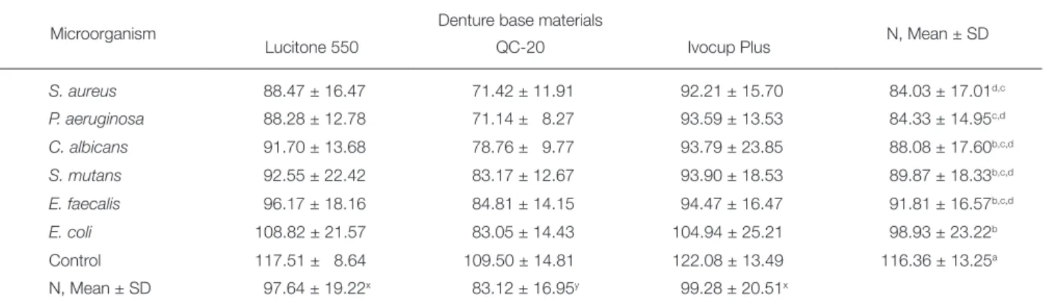
Table 5. The mean and standard deviation values of three-point bending test (ANOVA)
Microorganism Denture base materials
N, Mean ± SD
Lucitone 550 QC-20 Ivocup Plus
S. aureus 88.47 ± 16.47 71.42 ± 11.91 92.21 ± 15.70 84.03 ± 17.01d,c
P. aeruginosa 88.28 ± 12.78 71.14 ± 8.27 93.59 ± 13.53 84.33 ± 14.95c,d
C. albicans 91.70 ± 13.68 78.76 ± 9.77 93.79 ± 23.85 88.08 ± 17.60b,c,d
S. mutans 92.55 ± 22.42 83.17 ± 12.67 93.90 ± 18.53 89.87 ± 18.33b,c,d
E. faecalis 96.17 ± 18.16 84.81 ± 14.15 94.47 ± 16.47 91.81 ± 16.57b,c,d
E. coli 108.82 ± 21.57 83.05 ± 14.43 104.94 ± 25.21 98.93 ± 23.22b
Control 117.51 ± 8.64 109.50 ± 14.81 122.08 ± 13.49 116.36 ± 13.25a
N, Mean ± SD 97.64 ± 19.22x 83.12 ± 16.95y 99.28 ± 20.51x
No statistically significant differences with same letters
compared to the control group and this was statistically sig- nificant. Lucitone 550 and Ivocup Plus denture base materi- als were significantly more resistant than QC-20 when mean TPB test values were consulted. The difference between Lucitone 550 and Ivocup Plus were not significant.
E. coli produced the minimum destructive effect to the base materials when mean TPB test values of materials were evaluated, however this was not significantly different from E. faecalis (P=.699>.05), S. mutans and C. albicans.
DISCUSSION
This study investigated the effects of biofilm formation and/or biocorrosive activity of 6 different oral microorgan- isms on three different denture base materials. All tested microorganisms significantly decreased the TPB test values of the tested denture base materials.
After contamination with microorganisms, the TPB test values of denture base materials were decreased significant- ly compared with the control groups. It may be speculated that the structure of all the denture base materials were decomposed by the biofilm formation and/or biocorrosive activity of microorganisms.
Several studies have investigated the fracture resistance of denture materials.17-20 In this study Ivocap Plus appears to be the most resistant base material when the peak data of control group and mean values at TPB tests were evalu- ated. This was compatible with the results from the study of Hedzelek and Gajdus.21 However, when the percentages of TPB test value reduction were examined, Lucitone 550 was the most resistant material. The mean TPB test value of samples exhibited 18% resistance reduction at Lucitone 550 samples, 19% at Ivocap Plus and 24% at QC-20 den- ture base materials compared with control group after con- tamination processes of 6 different microorganisms.
Evaluating the subgroups data of denture base materials and microorganisms of this study, the highest TPB test val- ue reduction was observed in P. aeruginosa-QC group with 36%. Even E coli, known to have the least degenerative bio- activity,12 decreased the TPB test values of Lucitone 550 by 8%. Eventually considering all the mean TPB test values of three denture bases of our study it can be affirmed that microorganisms had biocorrosive activity and deteriorated at least 15% of the initial physical composition of tested denture base materials.
However, the difference of mean biofilm covered regions on denture base materials at SEM display were not statistically significant the mean quantitative biofilm values of microorganisms on Ivocap Plus was significantly differ- ent from Lucitone 550. This may be due to the dissimilar accumulation of microorganisms on the material surface (Fig. 2) that varies according to the production of extracel- lular polysaccharides which can only be demonstrated using advanced techniques like three-dimensional confocal scan- ning laser microscopy.13 Consequently, the two-dimensional SEM display may have inhibited quantitative estimation of this phenomenon.
Serrano-Granger et al.22 claimed that there was no rela- tionship between the microorganism adhesion and acrylic resin type and/or composition. This was particularly com- patible with our study; when biofilm covered regions were evaluated when we observed that microorganisms showed minimal adhesion to Ivocap Plus. This result was not statis- tically significant when compared with QC but significant compared with Lucitone 550. This may be attributed to the special fabrication technique of this material (injection molding) which may form samples with smoother surface and lesser microporosities. The other reason for minimal adhesion may be the higher residual monomer release after using injection molding technique.23 It is known that the PMMA monomer is toxic for living cells.23,24 During the experimental process, with the mentioned destructive effect, PMMA monomer may have inhibited the survival and/or adhesion of microorganisms tested on denture base materials. On the contrary, the higher adhesion to Lucitone 550 may be attributed to the reinforcing materials that poorly adhere to the polymer matrix forming microporosi- ties that become suitable for microorganism accumula- tion.25
C. albicans was shown to have higher adhesion potential to denture base materials in most of the studies.10 Conversely, in our study C. albicans-Ivocap Plus group exhibited the least adhesion potential when all the sub- groups of quantitative biofilm values were examined.
Nevertheless, P. aeruginosa appeared to be significantly the most adherent microorganism. This result was compatible with the literature that P. aeruginosa and S. aureus have higher survival success over the plastic and/or metallic devices used in medicine.26 Additionally the mean biofilm covered regions on denture base materials at SEM display indicated the highest percentages for P. aeruginosa and S. aureus con- firming the results of mean quantitative biofilm values of our study.
Within the limitations of this study it was shown that microorganisms diffused at least 52% of the denture base surface that cannot be neglected in dental practice.
However it was a remarkable result that this rate did not exceed 71% (Table 4) which may be attributed to the envi- ronmental or local conditions of this study or the floral equilibrium of microorganisms in certain circumstance.
CONCLUSION
It can be reported that all the tested microorganisms had destructive effect over the structure and composition of the denture base materials.
REFERENCES
1. van Heumen CC, Kreulen CM, Bronkhorst EM, Lesaffre E, Creugers NH. Fiber-reinforced dental composites in beam testing. Dent Mater 2008;24:1435-43.
2. Keenan PL, Radford DR, Clark RK. Dimensional change in complete dentures fabricated by injection molding and mi-
crowave processing. J Prosthet Dent 2003;89:37-44.
3. Rached RN, Powers JM, Del Bel Cury AA. Efficacy of con- ventional and experimental techniques for denture repair. J Oral Rehabil 2004;31:1130-8.
4. Stipho HD. Repair of acrylic resin denture base reinforced with glass fiber. J Prosthet Dent 1998;80:546-50.
5. Berger JC, Driscoll CF, Romberg E, Luo Q, Thompson G.
Surface roughness of denture base acrylic resins after pro- cessing and after polishing. J Prosthodont 2006;15:180-6.
6. Verran J, Maryan CJ. Retention of Candida albicans on acryl- ic resin and silicone of different surface topography. J Prosthet Dent 1997;77:535-9.
7. Nikawa H, Hamada T, Yamamoto T. Denture plaque-past and recent concerns. J Dent 1998;26:299-304.
8. Ramage G, Tomsett K, Wickes BL, López-Ribot JL, Redding SW. Denture stomatitis: a role for Candida biofilms. Oral Surg Oral Med Oral Pathol Oral Radiol Endod 2004;98:53-9.
9. Beech IB, Sunner JA, Arciola CR, Cristiani P. Microbially- influenced corrosion: damage to prostheses, delight for bac- teria. Int J Artif Organs 2006;29:443-52.
10. Busscher HJ, Rinastiti M, Siswomihardjo W, van der Mei HC.
Biofilm formation on dental restorative and implant materi- als. J Dent Res 2010;89:657-65.
11. Yuan SJ, Pehkonen SO. Microbiologically influenced corro- sion of 304 stainless steel by aerobic Pseudomonas NCIMB 2021 bacteria: AFM and XPS study. Colloids Surf B Biointerfaces 2007;59:87-99.
12. Paranhos HF, Silva-Lovato CH, de Souza RF, Cruz PC, de Freitas-Pontes KM, Watanabe E, Ito IY. Effect of three methods for cleaning dentures on biofilms formed in vitro on acrylic resin. J Prosthodont 2009;18:427-31.
13. da Silva WJ, Seneviratne J, Samaranayake LP, Del Bel Cury AA. Bioactivity and architecture of Candida albicans biofilms developed on poly(methyl methacrylate) resin surface. J Biomed Mater Res B Appl Biomater 2010;94:149-56.
14. Wakimoto N, Nishi J, Sheikh J, Nataro JP, Sarantuya J, Iwashita M, Manago K, Tokuda K, Yoshinaga M, Kawano Y.
Quantitative biofilm assay using a microtiter plate to screen for enteroaggregative Escherichia coli. Am J Trop Med Hyg 2004;71:687-90.
15. Christensen GD, Simpson WA, Younger JJ, Baddour LM, Barrett FF, Melton DM, Beachey EH. Adherence of coagu- lase-negative staphylococci to plastic tissue culture plates: a quantitative model for the adherence of staphylococci to medical devices. J Clin Microbiol 1985;22:996-1006.
16. Sahin C, Cehreli ZC, Yenigul M, Dayangac B. In vitro perme- ability of etch-and-rinse and self-etch adhesives used for im- mediate dentin sealing. Dent Mater J 2012;31:401-8.
17. Zappini G, Kammann A, Wachter W. Comparison of frac- ture tests of denture base materials. J Prosthet Dent 2003;
90:578-85.
18. Bural C, Bayraktar G, Aydin I, Yusufoğlu I, Uyumaz N, Hanzade M. Flexural properties of repaired heat-polymeris- ing acrylic resin after wetting with monomer and acetone.
Gerodontology 2010;27:217-23.
19. Takahashi T, Gonda T, Maeda Y. Influence of reinforcing materials on strain of maxillary complete denture. Acta
Odontol Scand 2013;71:307-11.
20. Takahashi Y, Yoshida K, Shimizu H. Fracture resistance of maxillary complete dentures subjected to long-term water immersion. Gerodontology 2012;29:1086-91.
21. Hedzelek W, Gajdus P. Comparison of mechanical strength of palatal denture bases made from various plastic materials.
Int J Prosthodont 2006;19:193-4.
22. Serrano-Granger C, Cerero-Lapiedra R, Campo-Trapero J, Del Río-Highsmith J. In vitro study of the adherence of Candida albicans to acrylic resins: relationship to surface en- ergy. Int J Prosthodont 2005;18:392-8.
23. Kedjarune U, Charoenworaluk N, Koontongkaew S. Release of methyl methacrylate from heat-cured and autopolymer- ized resins: cytotoxicity testing related to residual monomer.
Aust Dent J 1999;44:25-30.
24. Waltimo T, Vallittu P, Haapasalo M. Adherence of Candida species to newly polymerized and water-stored denture base polymers. Int J Prosthodont 2001;14:457-60.
25. Narva KK, Lassila LV, Vallittu PK. The static strength and modulus of fiber reinforced denture base polymer. Dent Mater 2005;21:421-8.
26. Bak J, Begovic T, Bjarnsholt T, Nielsen A. A UVC device for intra-luminal disinfection of catheters: in vitro tests on soft polymer tubes contaminated with Pseudomonas aeruginosa, Staphylococcus aureus, Escherichia coli and Candida albi- cans. Photochem Photobiol 2011;87:1123-8.