Cortical and cancellous bone thickness
on the anterior region of alveolar bone in Korean:
a study of dentate human cadavers
Heung-Joong Kim1, DDS, PhD, Sun-Kyoung Yu1, DDS, MS, Myoung-Hwa Lee1, MS, Hoon-Jae Lee2, DDS, PhD, Hee-Jung Kim2, DDS, PhD, Chae-Heon Chung2*, DDS, PhD
1Department of Anatomy and Orofacial Development, 2Department of Dental Prosthetics, School of Dentistry, Chosun University, Gwangju, Korea
PURPOSE. The cortical bone thickness on the anterior region is important for achieving implant stability. The purpose of this study was to examine the thickness of the cortical and cancellous bones on the anterior region of the maxilla and mandible. MATERIALS AND METHODS.
Twenty-five cadaver heads were used (16 male and 9 female; mean death age, 56.7 years). After the long axis of alveolar process was set up, it was measured in 5 levels starting from 2 mm below the cementoenamel junction (L1) at intervals of 3 mm. All data was analysed statistically by one-way ANOVA at the .05 significance level. RESULTS. The cortical bone thickness according to measurement levels in both the labial and lingual sides increased from L1 to L5, and the lingual side below L3 was significantly thicker than the labial side on the maxilla and mandible.
In particular, the labial cortical bone thickness in the maxilla was the thinnest compared to the other regions. The cancellous bone thickness according to measurement levels increased from L1 to L5 on the maxilla, and on the mandible it was the thinnest at the middle level of the root.
CONCLUSION. For implant placement on the anterior region, a careful evaluation and full knowledge on the thickness of the cortical and cancellous bone are necessary, therefore, these results may provide an anatomic guideline to clinicians. [J Adv Prosthodont 2012;4:146-52]
KEY WORDS: Cortical bone thickness; Cancellous bone thickness; Anterior region; Implant placement
Corresponding author: Chae-Heon Chung
Department of Dental Prosthetics, School of Dentistry, Chosun University, 375 Seosuk-dong, Dong-gu, Gwangju, 501-759, Korea
Tel. 82 62 220 3826: e-mail, [email protected]
Received February 28, 2012 / Last Revision June 8, 2012 / Accepted June 28, 2012
ⓒ 2012 The Korean Academy of Prosthodontics
This is an Open Access article distributed under the terms of the Creative Commons Attribution Non-Commercial License (http://creativecommons.org/licenses/by- nc/3.0) which permits unrestricted non-commercial use, distribution, and reproduction in any medium, provided the original work is properly cited.
INTRODUCTION
The anterior teeth are an important factor in dental and facial esthetics.1At this time, in the anterior region they are dif- ficult for both stabilization of the implant fixture and aesthesis of the restoration because they have narrower alveolar ridge and thinner cortical bone than in the posterior region.2,3In addi- tion, the thickness of the cortical bone has a larger influ- ence on the initial stabilization than the length of the implant fixture in an edentulous region. Therefore, information on the cortical and cancellous bone thickness in the labial and lingual sides, especially the anterior part, is the key to successful den- tal implantation.4
Many researchers have evaluated the thickness of labial cortical bone using various methods in the anterior region. Above all, computed tomography (CT) is used widely to measure the
alveolar bone for preoperative evaluation before dental pros- thetic and orthodontic treatment because it can measure a large number of the samples and various age groups. Using a CT, Flanagan5compared the thickness of the labial and lingual cor- tical bone on the edentulism, and Swasty et al.6measured the thickness of the mandibular alveolar bone in various age groups. In addition, Deguchi et al.7and Lim et al.8evaluated the thickness of the buccal and lingual cortical bone on the pos- terior region using CT for the implantation of mini-screws..
Recently, micro-CT is often used for measuring the bone struc- tures because it is convenient, noninvasive tool and has the high- er resolution compared to conventional CT image.9However, until now, most of these researches have been done by using the radiographic methods such as CT. Despite the limited num- ber of the cadaver samples, few studies by direct manual measurement have evaluated the thickness of cortical and can-
*This research was supported by Basic Science Research Program through the National Research Foundation of Korea (NRF) funded by the Ministry of Education, Science and Technology (KRF-2008-313-E00549).
cellous bone on cadavers.
Therefore, this study was carried out to evaluate the cortical and cancellous bone thickness at the midline and interdental areas of the tooth in the anterior region to provide anatomic infor- mation for dental implantation on dentate Korean cadavers.
MATERIALS AND METHODS
Twenty-five cadaver heads were used (16 male and 9 female), whose age at death ranged from 40 to 90 years (mean age: 56.7 years). To measure the cortical and cancellous bone thickness of the anterior region in dentate Korean cadav- ers, the maxilla and mandible with all anterior teeth including canine were chosen.
All specimens were decalcified in a decalcification solution (a mixture of 8 N formic acid + 1 N sodium formate) for 1 month, and then neutralized in distilled water for 12 hours. They were cut along the midline and interdental (distal surface) areas of each anterior tooth from the labial side to the lingual side using a microtome blade (Feather Co., Osaka, Japan) at total of 6 planes: midline area of central incisor (CI-mid), interdental area between CI and lateral incisor (CI-ID), midline area of lat- eral incisor (LI-mid), interdental area between LI and canine (LI-ID), midline area of canine (C-mid) and interdental area between C and the first premolar (C-ID). Each sectioned specimen was scanned using a scanner (HP Scanjet G4050, Hewlett Packard Co., Houston, Tex, USA) with ruler to cor- rect the size at the same time, and the scanned images were mea- sured using Adobe Photoshop CS3 ver 10 (Adobe, CA, USA) to a 0.01 mm level.10
On each scanned image, the long axis of the alveolar process was set to connect the center point of the cementoenamel junc-
tion (CEJ) with the center point of extension line passing through the 14 mm point below the CEJ even with the CEJ (Fig. 1).
Considering the mean root length of incisors in the maxilla and mandible (12.5 - 14 mm),11the cortical and cancellous bone thick- ness on both the labial and lingual sides was measured in 5 lev- els starting from 2 mm below the CEJ (L1) at intervals of 3 mm to root apex area. At the midline area of the tooth, the total can- cellous bone thickness including the root thickness was deter- mined (Fig. 2). The cadavers used in this study were in the elder- ly and degree of absorption of the alveolar process was different regardless of the dentate conditions. Therefore, the cortical bone thickness in L1 was referred in the results only and excluded from the discussion. All measurements were carried out by two investigators. The interobserver agreement was high (P=.672), therefore, the average of the 2 measurements was used as the final measurement.
The inter-observer difference was analysed by one-way ANOVA. All measurements were reported as the mean±
SD. The difference between right and left sides was analysed by one-way ANOVA, and there was no significant difference so each side measurement was counted as the same group. The cortical bone thickness in the midline area was compared t that of interdental area at the same level, and then evaluated with a post-hoc Scheffe′comparison. As a result, no significant dif- ference was found in the cortical bone thickness between the midline and interdental areas in both the labial and lingual sides. Therefore, the average value of the 2 measurements was calculated to compare the difference of the cortical bone thickness in the labial and lingual sides (Figs. 3, 4). All statistical analyses were performed using SPSS 12.0 (Chicago, IL, USA). P value <.05 was considered significant.
Cortical and cancellous bone thickness on the anterior region of alveolar bone in Korean: a study of dentate human cadavers Kim HJ et al.
Fig. 1. Diagram showing a cross-section of the interdental area. A: max- illa, B: mandible. a: long axis of alveolar process, b: labial cortical bone thickness, c: lingual cortical bone thickness, Can: cancellous bone thickness, CEJ: cementoenamel junction, La: labial side, Li: lingual side.
A B
Fig. 2. Scanned image showing the 5 levels starting from 2 mm below the CEJ (L1) at intervals of 3 mm to the root apex area (L5). A: maxilla, B: mandible. a: long axis of alveolar process, CEJ: cementoenamel junc- tion, La: labial side, Li: lingual side. L1: 2 mm below the CEJ, L2: 5 mm below the CEJ, L3: 8 mm below the CEJ, L4: 11 mm below the CEJ, L5:
14 mm below the CEJ.
A B
RESULTS
1. The thickness of cortical and cancellous bone on maxilla
1) The thickness of midline area
The cortical bone thickness of the midline area was thinnest in L2 and L3 of the C on both the labial and lingual sides, and thickest in the L2 and L3 of the CI. In addition, the cortical bone thickness increased from L1 to L5 in every tooth sites on both the labial and lingual sides. The labial cortical bone thickness in L1 and L2 near the alveolar crest was thicker than the lin- gual side, and the lingual sidein L3, L4, and L5 under the mid- dle level of the root was thicker than the labial side. The cancellous bone thickness of the midline area varied accord- ing to the tooth sites and measurement levels (Table 1).
2) The thickness of the interdental area
The cortical bone thickness of the interdental area was thinnest in the CI-ID on both the labial and lingual sides, and thickest in the C-ID among the tooth sites. As in the midline area, the cortical bone thickness increased from L1 to L5 in both the labial and lingual sides. The labial cortical bone thickness in L1 and L2 was thicker than the lingual side, and the lingual side in L3, L4, and L5 except the CI-ID was thicker than the labial side. The cancellous bone thickness of the interdental area varied according to the tooth sites and the measurement lev- els, and it seemed to become thinner at the middle level of the root (L2, L3, and L4) in the CI-ID and LI-ID (Table 2).
Fig. 3. Diagram showing that the cortical bone thickness was compared in the labial and lingual sides at each level in the anterior region on the maxilla. * indicates statistical significance with a P<.05.
1.2 1 0.8 0.6 0.4 0.2 0
La Li
L1 L2 L3 L4 L5 Levels
Cortical bone thickness (mm)
Fig. 4. Diagram showing that the cortical bone thickness was compared in the labial and lingual sides at each level in the anterior region on the mandible. * indicates statistical significance with a P<.05.
1.8 1.6 1.4 1.2 1 0.8 0.6 0.4 0.2 0
La Li
L1 L2 L3 L4 L5 Levels
Cortical bone thickness (mm)
Table 1. Thickness (in millimeters; mean±SD) of the cortical and cancellous bones at each level in the middle area of the tooth on the maxilla
CI-mid LI-mid C-mid
Levels
La Can Li La Can Li La Can Li
L1 0.18±0.30 4.99±0.82 0.04±0.16 0.08±0.15 4.74±0.62 0±0 0.08±0.19 5.95±0.68 0±0 L2 0.35±0.31 5.62±1.09 0.33±0.31 0.35±0.29 4.98±0.93 0.21±0.29 0.27±0.31 6.56±0.94 0.12±0.18 L3 0.44±0.25 5.90±1.16 0.57±0.32 0.39±0.27 4.77±0.94 0.54±0.24 0.33±0.28 6.58±1.33 0.48±0.34 L4 0.51±0.24 6.30±1.37 0.60±0.22 0.41±0.25 4.77±0.92 0.61±0.23 0.51±0.26 6.09±1.65 0.66±0.28 L5 0.59±0.26 6.77±1.59 0.66±0.33 0.58±0.20 5.09±1.21 0.63±0.24 0.60±0.32 6.26±2.02 0.67±0.28 Abbreviations; CI-mid: midline area of central incisor, LI-mid: midline area of lateral incisor, C-mid: midline area of canine, La: labial cortical bone thickness, Li: lingual cortical bone thickness, Can: cancellous bone thickness. L1: 2 mm below the CEJ, L2: 5 mm below the CEJ, L3: 8 mm below the CEJ, L4: 11 mm below the CEJ, L5: 14 mm below the CEJ.
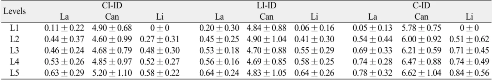
Table 2. Thickness (in millimeters; mean ± SD) of cortical and cancellous bones at each level in the interdental area of the tooth on the maxilla
CI-ID LI-ID C-ID
Levels
La Can Li La Can Li La Can Li
L1 0.11±0.22 4.90±0.68 0±0 0.20±0.30 4.84±0.88 0.06±0.16 0.05±0.13 5.78±0.75 0±0 L2 0.44±0.37 4.60±0.99 0.27±0.31 0.45±0.25 4.90±1.04 0.41±0.30 0.54±0.44 6.00±0.92 0.51±0.62 L3 0.46±0.24 4.68±0.79 0.48±0.30 0.53±0.18 4.70±0.88 0.55±0.29 0.69±0.33 6.21±0.59 0.71±0.45 L4 0.53±0.26 4.85±0.97 0.52±0.27 0.56±0.16 4.69±0.85 0.58±0.25 0.74±0.28 6.47±0.88 0.74±0.49 L5 0.63±0.29 5.20±1.10 0.58±0.22 0.64±0.24 4.83±1.05 0.64±0.26 0.78±0.32 6.62±1.04 0.84±0.56 Abbreviations; CI-ID: interdental area between CI and lateral incisor, LI-ID: interdental area between LI and canine, C-ID: interdental area between C and the first premolar.
2. The thickness of cortical and cancellous bone on mandible
1) The thickness of midline area
The cortical bone thickness of the midline area was thinnest in the CI on both labial and lingual sides, and thickest in the LI on the labial side and in the C on the lingual side among the tooth sites. As in the midline area in the maxilla, the cortical bone thickness increased from L1 to L5 in both labial and lin- gual sides, and the labial cortical bone thickness in L1 and L2 near the alveolar crest was thicker than the lingual side and the lingual side in L3, L4, and L5 under the middle level of the root was thicker than the labial side. The cancellous bone thickness of the midline area was thinnest in the CI, and thickest in the C among the tooth sites, and it became thinner at the middle level of the root (L3, L4) in all tooth sites (Table 3).
2) The thickness of the interdental area
The cortical bone thickness of the interdental area was thinnest in the CI-ID on both labial and lingual sides, and thick- est in the C-ID among the tooth sites. As like the midline area in the mandible, the cortical bone thickness increased from L1 to L5 in both the labial and lingual sides, and the lingual side under L2 was thicker than the labial side except for the L2 of LI-ID. The cancellous bone thickness of the interdental area was thinnest in the CI-ID, and thickest in the C-ID among the tooth sites. In addition, it became thinner at the middle level of the root (L3, L4) in all tooth sites (Table 4).
3. The average thickness of cortical and cancellous bone on the maxilla and mandible
1) Compared in the labial and lingual cortical bone thickness at each level in the anterior region
The average thickness of labial cortical bone according to the measurement levels on the maxilla was 0.12± 0.22 mm (L1), 0.40±0.33 mm (L2), 0.47±0.28 mm (L3), 0.54±0.26 mm (L4), and 0.63±0.27 mm (L5). The average thickness of lingual cortical bone according to the measurement levels on the maxilla was 0.02±0.09 mm, 0.30±0.37 mm, 0.55±0.33 mm, 0.62± 0.31 mm, and 0.66 ± 0.32 mm, respectively (Fig. 3). The average thickness of labial cortical bone accord- ing to the measurement levels on the mandible was 0.13±0.29 mm, 0.48±0.32 mm, 0.56±0.29 mm, 0.69±0.31 mm, and 0.88±0.31 mm, respectively. The average thickness of lingual cortical bone according to the measurement levels on the mandible was 0.05± 0.21 mm, 0.49 ± 0.43 mm, 0.89 ± 0.47 mm, 1.04± 0.41 mm, and 1.21 ± 0.42 mm, respec- tively (Fig. 4). On both the maxilla and mandible, the cortical bone thickness increased from L1 to L5 both the labial and lin- gual sides, and the labial cortical bone thickness in L1 and L2 was thicker than the lingual side, and the lingual side in L3, L4, and L5 was thicker than the labial side (Figs. 3, 4). On the max- illa, the labial and lingual cortical bone thickness showed- significant difference at all levels (Fig. 3), but on the mandible, there was significant difference only in L3, L4, and L5 under the middle level of the root (Fig. 4).
Cortical and cancellous bone thickness on the anterior region of alveolar bone in Korean: a study of dentate human cadavers Kim HJ et al.
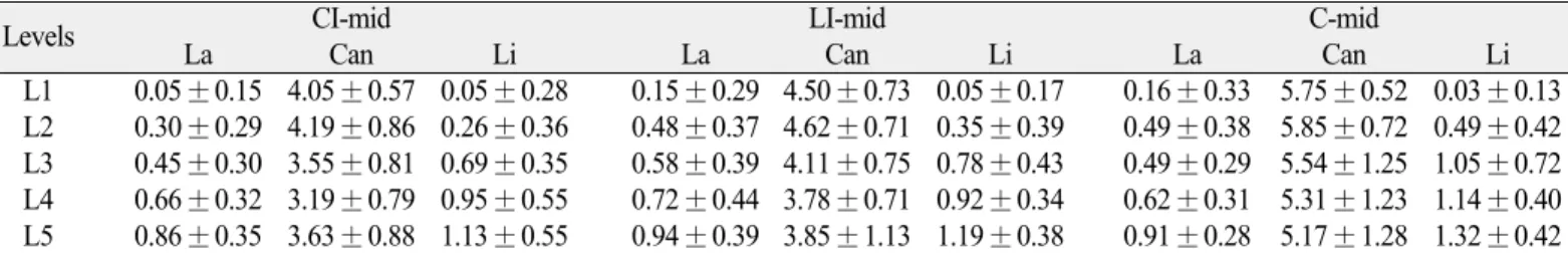
Table 3. Thickness (in millimeters; mean ± SD) of cortical and cancellous bones at each level in the middle area of the tooth on the mandible
CI-mid LI-mid C-mid
Levels
La Can Li La Can Li La Can Li
L1 0.05±0.15 4.05±0.57 0.05±0.28 0.15±0.29 4.50±0.73 0.05±0.17 0.16±0.33 5.75±0.52 0.03±0.13 L2 0.30±0.29 4.19±0.86 0.26±0.36 0.48±0.37 4.62±0.71 0.35±0.39 0.49±0.38 5.85±0.72 0.49±0.42 L3 0.45±0.30 3.55±0.81 0.69±0.35 0.58±0.39 4.11±0.75 0.78±0.43 0.49±0.29 5.54±1.25 1.05±0.72 L4 0.66±0.32 3.19±0.79 0.95±0.55 0.72±0.44 3.78±0.71 0.92±0.34 0.62±0.31 5.31±1.23 1.14±0.40 L5 0.86±0.35 3.63±0.88 1.13±0.55 0.94±0.39 3.85±1.13 1.19±0.38 0.91±0.28 5.17±1.28 1.32±0.42
Table 4. Thickness (in millimeters; mean ± SD) of cortical and cancellous bones at each level in the interdental area of the tooth on the mandible
CI-mid LI-mid C-mid
Levels
La Can Li La Can Li La Can Li
L1 0.10±0.29 3.88±0.52 0.05±0.28 0.15±0.34 4.27±0.78 0.07±0.25 0.15±0.29 5.26±0.84 0.12±0.28 L2 0.44±0.27 4.27±0.82 0.46±0.43 0.60±0.26 4.43±0.57 0.57±0.39 0.56±0.28 5.00±1.19 0.79±0.43 L3 0.51±0.24 3.86±0.89 0.77±0.41 0.67±0.21 4.24±0.71 0.90±0.36 0.63±0.25 5.19±1.52 1.12±0.29 L4 0.61±0.21 3.45±0.82 0.97±0.43 0.74±0.25 4.06±0.95 1.00±0.37 0.79±0.26 5.29±1.61 1.22±0.33 L5 0.77±0.26 3.53±1.04 1.15±0.42 0.84±0.27 4.34±1.13 1.13±0.38 0.93±0.28 5.43±1.66 1.30±0.37
2) The average thickness of cortical and cancellous bones in each level
Using the average value of the midline and interdental area mentioned in the materials and methods, this study evaluated a diagram of the overall shape of the cortical and cancellous bone at each level on both maxilla and mandible (Fig. 5). As a result, the cancellous bone thickness increased from L1 to L5 on the maxilla, and was thickest at L2 and thinnest at L4 on the mandible (Fig. 5).
DISCUSSION
The sufficient thickness of the cortical bone is a primary fac- tor in an initial fixation of implant placement.4,12Although the thickness of the alveolar bone normally increases from the alve- olar crest to the root apex, in the anterior region, the thickness of cancellous bone between the labial and lingual cortical bone is important because the width of the anterior alveolar process is narrower than that of molar.3After tooth extraction, the alve- olar bone undergoes the reparative resorption sequentially and, in particular, the labial cortical bone resorbs the first in the labi- al and alveolar crest directions.5Therefore, the cortical and can- cellous bone thickness in the anterior region should be an accu- rate assessment preoperatively for initial and successive implant stability.
In previous studies on the cortical bone thickness, Flanagan5 reported that the lingual cortical bone thickness was 2.33 mm, which was thicker than that of the labial side by 1.79 mm in edentulism, and the lingual side was always thicker than the labial side when bone absorption had already progressed.
Katranji et al.12reported that the cortical bone was thicker in the maxilla than the mandible, and the labial cortical bone was thicker than that of the lingual side in the dentate anterior region.
In the maxilla the labial cortical bone thickness was 1.59 mm and the lingual cortical bone thickness was 1.95 mm, on the other hand, in the mandible they were 0.99 mm and 1.24 mm, respectively.
In the present study using dentate cadavers, the lingual cortical bone was thicker than the labial one below L3 in both the maxilla and mandible. In particular, the lingual cortical bone in the mandible was thick enough to support the bone by L3 (0.89 mm), L4 (1.04 mm) and L5 (1.21 mm). The mandible was thicker than the maxilla at all levels in both the labial and lin- gual sides. In addition, the labial cortical bone thickness in the maxilla was thin by L2 (0.40 mm), L3 (0.47 mm), L4 (0.54 mm) and L5 (0.63 mm). These results constrained the functional and esthetic recovery because a change in alveolar socket volume was observed in a certain amount for the first 8 weeks after extraction when prosthetic restoration and the labial side bone of the extraction socket is absorbed more than the lingual side.13,14Therefore, for successful prosthetic restoration and implant placement after extraction, it is important to predict the initial absorption with consideration of the thinner labial cortical bone around the alveolar crest. At this time, in 6 months after implant placement the mean vertical bone resorption from the platform of implant was 1.32± 0.86 mm, and the residual labial bone thickness to implant fixture was 1.91±0.45 mm. Therefore, the labial bone thickness more than 1.91 mm could prevent a failure of implant placement that could result from severe labial bone resorption.15
In particular, to solve the problem caused by the resorption of paper-thin labial cortical bone after extraction, clinicians rec- ommended the placement of an immediate implant and the use of graft material as a remedy.16-18Yoo et al.19reported that the change of bone level in the alveolar crest was a recommend- able level at an immediate implant placement after extraction,
Fig. 5. Diagram showing the average thickness of cortical and cancellous bones at each level in the anterior region. A: maxilla, B: mandible.
A B
Co (Li) Can Co (La)
L1 L2 L3 L4 L5
0.12 0.02
0.40 0.30
0.47 0.55
0.54 0.62
0.63 0.66
0.13 0.05
0.48 0.49
0.56 0.89
0.69 1.04
0.88 1.21
and the alveolar crest was absorbed more in the mandible than in the maxilla after immediate placement. Although full recovery occurred without guided bone regeneration in an imme- diate implant placementwhere the bone loss around the implant was small, Nevins et al.18recommended the guided bone regeneration using graft materials because the thin labial alveolar bone on the anterior maxilla was absorbed easily. Covani et al.17,20 reported that the alveolar bone distance between the labial and the lingual side was 8.1 mm and 5.8 mm in imme- diate and delayed implant placement, respectively, as the secondary surgery. In addition, when immediate implant placement after extraction was performed on the anterior region, the vertical bone loss around the implant did not cause esthetic side effects given that it was restored completely 6 months after implant placement through a bone regeneration process.
On the other hand, Arau′jo et al.21,22and Cardaropoli23report- ed that the interspace between the implant and alveolar sock- et wall disappeared as a result of bone regeneration and absorption of the alveolar crest. At this time, the vertical bone absorption on the anterior region was more at the labi- al side than the lingual side, and when that was compared between the immediate implant placement and placement on edentulous bone, the result was similar to each other.
Therefore, an immediate implant placement did not guarantee better results. However, according to this study and previous reports, immediate implant placement after extraction and guid- ed bone regeneration are recommended for stability and aes- thetics because the labial cortical bone of the anterior maxil- la is particularly thin and more esthetically exposed than the mandible.17,19,24-26
Clinicians need to consider not only the cortical bone thick- ness but also the cancellous bone thickness for good blood sup- ply and, therefore, successful implant placement. Bra�nemark, who made dental implants popular, classified the remaining bone quality into 4 types. He said that class 2, which is thick cortical bone and a high density of cancellous bone, and class 3, which is thin cortical bone and a high density of cancellous bone, are suitable bone types for successful implant placement. In addition, Misch27 classified the bone quality into 5 types using the Houndsfield units (HU) for easy application by an objective standard.
In the present study, the cancellous bone thickness in the max- illa was thinnest in the LI-ID and thickest in the C-mid, and the average thickness increased from the alveolar crest (L1) to the root (L5). In the present study, the cancellous bone thickness in the maxilla was thinnest in the LI-ID and thickest in the C- mid, and the average thickness according to measurement lev- els increased from the alveolar crest (L1) to the root apex (L5).
The root width of maxillary incisors became suddenly narrower from 6 mm below the CEJ,9in the present study, the cancel- lous bone thickness was measured including the root width.
Therefore, the increasing thickness of cancellous bone toward the root apex (L5) could be reflected practical increasing of can- cellous bone.
The cancellous bone thickness in the mandible was thinnest in the CI-ID and thickest in the C-mid, and on each level, the average thickness was thickest at L2 (4.74 mm) and thinnest at L4 (4.21 mm). That is, unlike the cancellous bone of max- illa, the cancellous bone thickness on the mandible became nar- rower at the middle level of the root. Miyamoto et al.4report- ed that the cortical bone thickness is more important in the ear- ly stages of implant placement than the length of the implant fixture for stability, and the cancellous bone/cortical bone ratio is also important in the placement area. Therefore, the cancellous bone thickness of middle level of the root should be consid- ered at first to select the diameter of implant fixture on the ante- rior mandibular region. In addition, the cancellous bone thickness and bone quality are important for implant placement.
More studies using micro CT will be needed to measure the entire volume and cancellous bone density.
In the present study, used cadavers, whose age at death ranged from 40 to 90 years, were the elderly. However, Swasty et al.6 measured the cortical bone thickness in the mandible using CT according to age, and reported that it was thinnest in the first decade and thickest in the fifth decade with a decrease thereafter. So, this measured data related to the cortical and cancellous bone thickness could be smaller than that of the young generation. Therefore, fur- ther studies are needed to reveal the change of cortical bone thickness by age with supplementing a number of cadavers.
CONCLUSION
The cortical bone thickness was thicker in the lingual side than the labial side both on the maxilla and mandible except for L1, L2 around the alveolar crest. In particular, the labial cortical bone thickness in the maxilla was thinnest compared to the oth- er regions. In addition, the cancellous bone thickness in the max- illa increased to the root apex, and it was thinnest at the middle level of the root in the mandible. For implant placement on the anterior region, a careful evaluation and full knowledge on the thickness of cortical and cancellous bone are necessary, providing an anatomic guideline to clinicians.
REFERENCES
1. Ku JE, Yang HS, Yun KD. A morphometric analysis of maxillary central incisor on the basis of facial appearance in Korea. J Adv Prosthodont 2012;4:13-7.
2. Bernard JP, Schatz JP, Christou P, Belser U, Kiliaridis S.
Long-term vertical changes of the anterior maxillary teeth ad- jacent to single implants in young and mature adults. A retro- spective study. J Clin Periodontol 2004;31:1024-8.
3. Buser D, Martin W, Belser UC. Optimizing esthetics for implant restorations in the anterior maxilla: anatomic and surgical
Cortical and cancellous bone thickness on the anterior region of alveolar bone in Korean: a study of dentate human cadavers Kim HJ et al.
considerations. Int J Oral Maxillofac Implants 2004;19:43-61.
4. Miyamoto I, Tsuboi Y, Wada E, Suwa H, Iizuka T. Influence of cortical bone thickness and implant length on implant stability at the time of surgery-clinical, prospective, biomechanical, and imaging study. Bone 2005;37:776-80.
5. Flanagan D. A comparison of facial and lingual cortical thick- nesses in edentulous maxillary and mandibular sites measured on computerized tomograms. J Oral Implantol 2008;34:256-8.
6. Swasty D, Lee JS, Huang JC, Maki K, Gansky SA, Hatcher D, Miller AJ. Anthropometric analysis of the human mandibular cor- tical bone as assessed by cone-beam computed tomography. J Oral Maxillofac Surg 2009;67:491-500.
7. Deguchi T, Nasu M, Murakami K, Yabuuchi T, Kamioka H, Takano-Yamamoto T. Quantitative evaluation of cortical bone thickness with computed tomographic scanning for orthodon- tic implants. Am J Orthod Dentofacial Orthop 2006;129:721.
8. Lim WH, Lee SK, Wikesjo¨UM, Chun YS. A descriptive tissue evaluation at maxillary interradicular sites: implications for orthodontic mini-implant placement. Clin Anat. 2007;20:760- 5.
9. Kim JH, Lee JG, Han DH, Kim HJ. Morphometric analysis of the anterior region of the maxillary bone for immediate implant placement using micro-CT. Clin Anat 2011;24:462-8.
10. Park HD, Min CK, Kwak HH, Youn KH, Choi SH, Kim HJ.
Topography of the outer mandibular symphyseal region with ref- erence to the autogenous bone graft. Int J Oral Maxillofac Surg 2004;33:781-5.
11. Shin JW. Dental anatomy. 3rded. Seoul; DaehanNarae Publishing, Inc.; 2010. p. 61-99.
12. Katranji A, Misch K, Wang HL. Cortical bone thickness in den- tate and edentulous human cadavers. J Periodontol 2007;78:874- 8.
13. Cardaropoli G, Arau′jo M, Lindhe J. Dynamics of bone tissue for- mation in tooth extraction sites. An experimental study in dogs. J Clin Periodontol 2003;30:809-18.
14. Arau′jo MG, Lindhe J. Dimensional ridge alterations following tooth extraction. An experimental study in the dog. J Clin Periodontol 2005;32:212-8.
15. Cho YB, Moon SJ, Chung CH, Kim HJ. Resorption of labial bone in maxillary anterior implant. J Adv Prosthodont 2011;3:85-9.
16. Covani U, Cornelini R, Barone A. Bucco-lingual bone remod-
eling around implants placed into immediate extraction sockets:
a case series. J Periodontol 2003;74:268-73.
17. Covani U, Cornelini R, Barone A. Vertical crestal bone changes around implants placed into fresh extraction sockets. J Periodontol 2007;78:810-5.
18. Nevins M, Camelo M, De Paoli S, Friedland B, Schenk RK, Parma-Benfenati S, Simion M, Tinti C, Wagenberg B. A study of the fate of the buccal wall of extraction sockets of teeth with prominent roots. Int J Periodontics Restorative Dent 2006;26:19-29.
19. Yoo RH, Chuang SK, Erakat MS, Weed M, Dodson TB.
Changes in crestal bone levels for immediately loaded im- plants. Int J Oral Maxillofac Implants 2006;21:253-61.
20. Covani U, Bortolaia C, Barone A, Sbordone L. Bucco-lingual crestal bone changes after immediate and delayed implant placement. J Periodontol 2004;75:1605-12.
21. Arau′jo MG, Sukekava F, Wennstro¨m JL, Lindhe J. Ridge al- terations following implant placement in fresh extraction sock- ets: an experimental study in the dog. J Clin Periodontol 2005;32:645-52.
22. Arau′jo MG, Wennstro¨m JL, Lindhe J. Modeling of the buccal and lingual bone walls of fresh extraction sites following implant installation. Clin Oral Implants Res 2006;17:606-14.
23. Cardaropoli G, Lekholm U, Wennstro¨m JL. Tissue alterations at implant-supported single-tooth replacements: a 1-year prospective clinical study. Clin Oral Implants Res 2006;17:165- 71.
24. Kan JY, Rungcharassaeng K. Immediate placement and pro- visionalization of maxillary anterior single implants: a surgical and prosthodontic rationale. Pract Periodontics Aesthet Dent 2000;12:817-24.
25. Wagenberg BD, Ginsburg TR. Immediate implant placement on removal of the natural tooth: retrospective analysis of 1,081 im- plants. Compend Contin Educ Dent 2001;22:399-404, 406, 408.
26. Juodzbalys G, Wang HL. Soft and hard tissue assessment of im- mediate implant placement: a case series. Clin Oral Implants Res.
2007;18:237-43.
27. Misch CE. Density of bone: effect on treatment plans, surgical approach, healing, and progressive boen loading. Int J Oral Implantol 1990;6:23-31.