Necessity of Surgical Site Closed Suction Drain for Pterional Craniotomy
Su Yong Choi, Sung Min Yoon, Chan Jong Yoo, Cheol Wan Park, Young Bo Kim, Woo Kyung Kim Department of Neurosurgery, Gil Medical Center, Gachon University, Incheon, Korea
Objective : The aim of this study was to assess the benefit of using a prophylactic surgical site closed suction drain in pterional craniotomy.
Materials and Methods : A retrospective review was conducted on 607 consecutive patients who underwent a pterional craniotomy for treatment of intracranial anterior circulation aneurysms over a 5-year period. Between January 2000 and December 2004, 607 patients were divided into two groups, those who had a prophylactic suction drain during closure of the surgical site (drain group, DG) and those who did not (non-drain group, NDG). Head computed tomography (CT) was taken routinely on postoperative day (POD) 1, 7, and 14. Patients' demographics, incidence of surgical site complications, and courses of surgical site healing which were evaluated radiologically by the thickness of the surgical site myocutaneous layer, were analyzed between DG and NDG.
Results : Patients' demographics and characteristics did not differ significantly between the two groups. The head CT showed that the degree of changes in the postoperative surgical site thickness was 148% at POD 1, 209% at POD 7, and 198% at POD 14 in DG, and 118% at POD 1, 152% at POD 7, and 158% at POD 14 in NDG compared to the preoperative value. Postoperative surgical site hematoma was 7.9% (22/274) in DG and 2.4% (8/333) in NDG.
Conclusion : Prophylactic use of an epidural and/or subgaleal closed suc- tion drain does not appear to be necessary for prevention of postoperative surgical site hematoma as well as for promotion of surgical site healing in pterional craniotomy.
J Cerebrovasc Endovasc Neurosurg.
2015 September;17(3):194-202 Received : 25 May 2015
Revised : 8 July 2015 Accepted : 27 July 2015
Correspondence to Cheol Wan Park Department of Neurosurgery, Gachon University Gil Medical Center, 21 Namdong-daero 774 beon-gil, Namdong-gu, Incheon 21565, Korea Tel : 82-32-460-3304
Fax : 82-32-460-3899 E-mail : cwpark@gilhospital.com
ORCID : http://orcid.org/0000-0002-4682-9100
This is an Open Access article distributed under the terms of the Creative Commons Attribution Non- Commercial License (http://creativecommons.org/li- censes/by-nc/3.0) which permits unrestricted non- commercial use, distribution, and reproduction in any medium, provided the original work is properly cited.
Keywords Craniotomy, Drainage, Surgical wound infection
INTRODUCTION
After Hippocrates and recently, the introduction of a portable closed suction drain by Redon and Jost in 1954, the surgical drain has been forever changed in its form and utility.20)21) Several retrospective and pro- spective studies spanning various surgical disciplines have examined the efficacy and complications asso- ciated with prophylactic closed suction drainage and
have yielded inconsistent results as to whether or not it reduces the number of postoperative surgical site complications.1-3)5)15)16)21)26) In addition, there is increas- ing evidence that routine placement of a prophylactic closed suction drain does not decrease the rate of postoperative surgical site complications after various surgical procedures.2)3)5)6)14-16)21)
The necessity or utility of surgical site drains in a craniotomy is also controversial. Anecdotally, some
neurosurgeons drain all craniotomy sites, while others do not. Epidural or subgaleal drains are commonly placed and connected to a closed vacuum system at the end of the craniotomy to prevent accumulation of intracranial and extracranial blood, and to promote healing of the surgical site. Paradoxically, the use of drains in a craniotomy may potentially increase the incidence of surgical site complications. However, few studies have reported on the necessity or advantage of surgical site drainage in craniotomy. Our sporadic and anecdotal experiences on surgical site complica- tions in recent years have shown that a craniotomy without a surgical site drain had at least equal or bet- ter outcomes in terms of surgical site healing and complications, which encouraged us to conduct this study. One of the authors (CWP) had never used a routine surgical site drain for a pterional craniotomy since 2003. In this retrospective study, we compared the result of surgical site complications and healing processes in pterional craniotomy between a surgical site drain group (DG) and non-drain group (NDG) before and after 2003.
MATERIALS AND METHODS
Patient selection
From January 2000 to December 2004, a total of 652 pterional craniotomies were performed in 645 patients due to ruptured anterior circulation cerebral aneur- ysms by one of the surgeons who is part of this team of researchers (CWP). Of the 652 craniotomies, 45 cas- es (38 patients) involving patients who died before postoperative day 14, patients who had undergone a decompressive craniectomy, patients with a medical condition or on drug therapy likely influenced blood coagulation or immunosuppression, or the patient's data were incomplete were excluded. Patients were divided into 2 groups based on the use of a surgical site closed suction drain during closure of pterional craniotomy, the drain-group (DG) and the non-drain group (NDG). Patients' data including medical re-
cords and imaging study results were reviewed and analyzed in detail.
Operative methods and wound management
All pterional craniotomies were performed in the operating room using a laminar air flow system, as described by Yasargil et al.28) A compressive stocking was applied to every patient to prevent development of deep vein thrombosis, however subcutaneous hep- arin was not administered. Prophylactic antibiotics were administered to all patients: first generation cephalosporin (2 gm), was administered intravenously at the induction of general anesthesia followed by 1 gm every 8 hours for 3 days postoperatively. To pre- pare the scalp for incision, the whole scalp of each pa- tient was shaved, prepared by iodine derivatives, and the surgical site was covered with antimicrobial film.
At the end of the craniotomy, a drain was placed be- tween the bone flap and the temporalis muscle flap in the DG group, then brought out through a separate stab incision and attached to the commercially avail- able closed suction drain system (3.2 mm in outer di- ameter, round and transparent polyvinyl chloride tube with a 400 mL spring evacuator chamber; Hemovac, Zimmer, UK). After satisfactory hemostasis was ach- ieved and layer by layer closure performed, a light adhesive dressing was applied. Other than the use of a drain, the surgical sites for patients of both groups (DG and NDG) were closed in exactly the same man- ner using identical suture materials.
The surgical site and dressing was inspected daily in all patients. The closed suction drain system was emptied every 8 hours and the amount was measured in the DG. The drain was removed under sterile con- ditions, when the volume of drainage was less than 50 mL during the last 24 hours. Most drains were re- moved within 48 hours after insertion. During re- moval, the tip of the drain was cut off approximately 2 cm from the end, placed in culture medium and sent to the clinical laboratory. A culture was taken to identify the presence of any microorganisms, which were then reported using standard laboratory techniques.
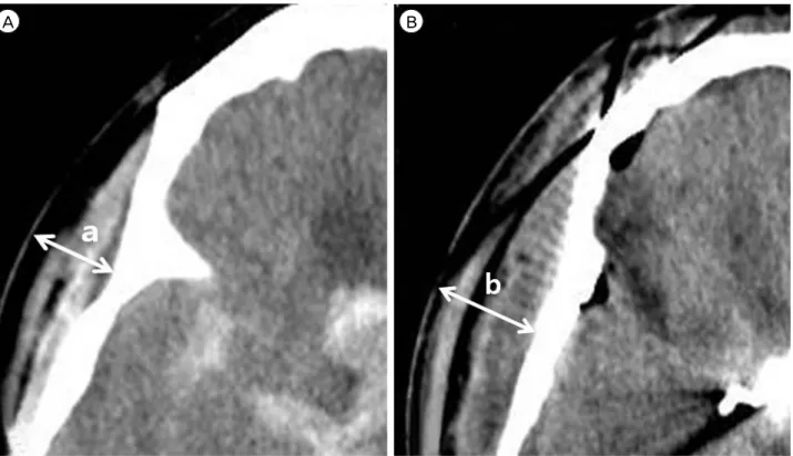
A B
Fig. 1. Thickness of myocutaneous layer (in mm) from the surface of the skull to that of the scalp on the pterional craniotomy site at or just above the level of zygomatic arch on the head CT slice. A: Preoperative thickness of myocutaneous layer (a). B:
Postoperative 7 day’s thickness of myocutaneous layer (b). Postoperative thickness of myocutaneous layer value ; (b/a) x 100 (%).
Evaluation of wound healing process
Computed tomography (CT) was checked routinely on postoperative day (POD) 1, 7, and 14 in all pa- tients for early detection of clinically silent lesions and development of complications such as intra- and extra-axial hematoma, hydrocephalus, and cerebral infarction. To assess healing of the surgical site with- out bias, two of the authors who were blinded to the patients' grouping measured the thickness of the my- ocutaneous layer (in mm) from the surface of the skull to that of the scalp on the surgical site at or above the level of the zygomatic arch on the post- operative serial head CT slices using calipers. The re- sults from the two neurosurgeons were then averaged.
The thickest myocutaneous layer slice on each post- operative head CT was selected, and that value was compared with the corresponding preoperative head CT slice. The postoperative thickness of the myocuta- neous layer was expressed as a % of the preoperative value (Fig. 1).
Extra-axial blood collection
In this study, a postoperative surgical site epidural hematoma was defined as extra-axial blood collection over 5 mm in maximum thickness underneath the pter- ional bone flap on head CT scan. The maximum thick- ness of hematoma was classified as small, medium, and large if less than 1 cm, between 1 and 2 cm, and more than 2 cm, respectively. However, extra-axial blood collection less than 0.5 cm in thickness was regarded as within the normal limit of postoperative changes.
Surgical site infection
A positive result in the culture of the drain tip with- out clinical findings of a surgical site infection in the DG was not considered a surgical site infection. The criteria for infection were limited to purulent or se- rous discharge from the surgical site with the clinical signs of inflammation including local heat, erythema, increasing tenderness, and swelling. Wound dehiscence was defined as spontaneous or iatrogenic separation of sutured edges requiring drainage of a small amount
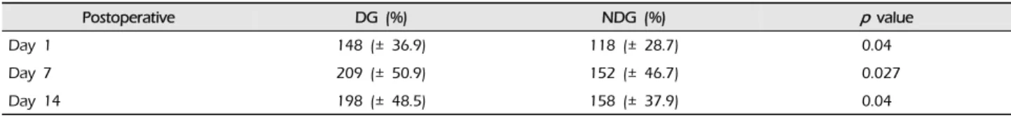
Postoperative DG (%) NDG (%) p value
Day 1 148 (± 36.9) 118 (± 28.7) 0.04
Day 7 209 (± 50.9) 152 (± 46.7) 0.027
Day 14 198 (± 48.5) 158 (± 37.9) 0.04
DG = drain group; NDG = non-drain group
Table 2. Comparison of rate of increase in postoperative surgical site myocutaneous layer thickness between DG and NDG
Variables DG NDG p value
Number of patient 274 333 0.285
Age (years) 49.2 (26-75) 51.5 (25-76) 0.374
Sex (M:F) 126 : 148 157 : 176 0.167
Immunosuppressive state 0 0
Uncontrolled DM 0 0
DG = drain group; NDG = non-drain group; DM = Diabetes mellitus
Table 1. Patients' demographic and clinical characteristics of DG and NDG
of serous discharge or pus.
Statistical analysis
Statistical analyses were performed using SPSS ver- sion 10.0 (SPSS Inc., Chicago, IL, USA). Values of con- tinuous variables were presented as mean with stand- ard deviation and categorical variables as counts with percentages. The Pearson chi-square test and Student's T test were used to compare the frequency distribution of categorical or continuous variables between DG and NDG. A p value of < 0.05 was considered statisti- cally significant.
RESULTS
A total of 607 patients (607 craniotomies) who un- derwent pterional craniotomy for clipping of ruptured cerebral aneurysms were enrolled int this study. DG included 274 patients; 126 patients were male, and the male to female ratio was 1:1.17. The mean age was 49.2 (range, 26.0-75.0) years. NDG included 333 pa- tients; 157 patients were male, and the male to female ratio was 1:1.2. The mean age was 51.5 (range, 25.0-76.0) years. There was no significant difference in the patients' basic demographics and clinical charac- teristics that might affect surgical site complications
between DG and NDG (p > 0.05) (Table 1).
Surgical site healing process
In the DG, postoperative thickness of the myocuta- neous layer was measured as 148% at POD 1, 209%
at POD 7, and 198% at POD 14. In the NDG, the re- sults were 118% at POD 1, 152% at POD 7, and 158%
at POD 14. In the NDG, the rate of increase in thick- ness was less remarkable compared with DG, partic- ularly at POD 7, and these differences were statisti- cally significant (p < 0.05) (Table 2).
Extra-axial blood collection
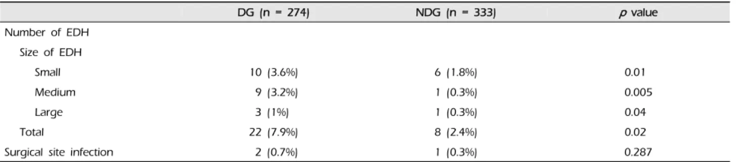
None of the patients in either group had a comorbidity related to blood coagulation disorders, which could influence the extra-axial blood collection. Based on our definition described in the Materials and method sec- tion, 22 (7.8%) of the 274 patients in the DG devel- oped epidural hematomas; they were small in 10 pa- tients, medium in nine, and large in three, while 8 (2.4%) of 333 patients in the NDG group presented with epidural hematomas.
They were small in size in six patients, and one was medium and the other was large. Statistically sig- nificant difference in the incidence of postoperative epidural hematoma was observed between the two
DG (n = 274) NDG (n = 333) p value Number of EDH
Size of EDH
Small 10 (3.6%) 6 (1.8%) 0.01
Medium 9 (3.2%) 1 (0.3%) 0.005
Large 3 (1%) 1 (0.3%) 0.04
Total 22 (7.9%) 8 (2.4%) 0.02
Surgical site infection 2 (0.7%) 1 (0.3%) 0.287
EDH = epidural hematoma; DG = drain group; NDG = non-drain group
Table 3. Comparison of postoperative surgical site EDH and infection between DG and NDG
groups (p < 0.05) (Table 3). All but two epidural hem- atomas were managed conservatively. Surgical evacu- ation was required for one epidural hematoma in the DG and one in the NDG.
Surgical site infection
None of the patients in either group had a comorbid condition increasing the risk of surgical site infection, such as immunodeficiency and/or suppression and uncontrolled diabetes mellitus (DM). A total of 102 patients in both groups had a medical history of type II DM (50 patients [18.2%] in the DG and 52 patients [15.6%] in the NDG), but during the perioperative pe- riod all patients' blood sugar levels were adequately controlled without any clinically evident complica- tions and/or sequelae from diabetes. Three (0.4%) of 607 patients showed signs of a surgical site infection throughout their hospital stay. In the DG, two (0.7%, 2/274) patients showed signs of a surgical site infection. Despite meticulous wound care, one of them required surgical wound debridement. In the NDG, only one (0.3%, 1/333) patient presented with a superficial infection which was resolved by anti- biotics and intensive surgical wound management without surgery. No statistically significant difference in the rate of surgical site infections was observed be- tween the two groups (p > 0.05) (Table 3).
DISCUSSION
An extensive review of the literature was performed in an attempt to define and compare the surgical site
healing process and the incidence of surgical site complications with or without use of a closed suction drain in various surgical procedures. However, few studies have reported on the surgical site healing process and complications including infection related to the use or non-use of a closed suction drain during craniotomy, thus the necessity or advantages of using a closed suction drain for a craniotomy remained elusive. Accordingly, we decided that it was neces- sary to conduct a comparative study between two pa- tient populations, DG and NDG, who have under- gone pterional craniotomies. To the best of the au- thors' knowledge, this is the first report in English to determine the necessity of a surgical site closed suc- tion drain for pterional craniotomy.
Results of this study concerning the rate of hema- toma formation and healing process at the surgical site might seem to contradict what was initially hy- pothesized, particularly in that the DG would have a lower incidence of hematoma formation at the surgi- cal site and would heal better than the NDG.
Although the use of a drain in a variety of surgical wounds has a long history, it very often falls into the realm of the surgeons' habit, rather than based in sci- ence or evidence-based medicine in surgical practice.
The utility of a prophylactic closed suction drain in surgical sites may be questionable in craniotomies, however, neurosurgeons have routinely used surgical drains to avoid surgical site hematomas and seromas, and/or to promote faster surgical site healing.
In a general surgery report, suction drains greatly improved the apposition of a large skin flap to surgi-
cal wounds to the underlying raw tissue surface while obliterating surgically created dead spaces, draining their exudates, and promoting rapid adher- ence and healing of the surgical sites.1) Waugh and Stinchfield, who published the first article describing the use of a closed suction drain in a prospective con- trolled survey examining orthopedic surgical wounds, reported that complete hemostasis was very difficult, and the use of a suction drain could improve surgical wound healing through reducing the formation of a hematoma in the dead space. They reported a 1% in- cidence of surgical site infections in patients with a closed drain system versus 3% in matched cases with- out a drain26). While this result did not reach stat- istical significance due to the sample size, the poten- tial to decrease the surgical site infection rate with a prophylactic suction drain was deemed encouraging.
An orthopedic surgeon also reported on the use of a suction drain that could reduce pain and edema at surgical sites in the postoperative period, promote surgical site healing, and decrease infection risk26). Royster reported that a suction drain was specifically helpful after head and neck procedures in which the incidence of seroma and hematoma formation was higher in non-drained surgical wounds.
In contrast, other investigators have found that the insertion of a drain at the end stage of surgery, partic- ularly in orthopedic surgery, did not significantly af- fect the incidence of surgical site complications.
Browett et al.,3) who studied the use of drains in or- thopedic surgery, found them to be of no use. Cobb, who evaluated the use of surgical site drain tubes in hip fracture patients, concluded that their use was rather detrimental.5) In 1998, Kim et al.15) reported that there was no benefit in placement of drains in or- thopedic surgical wounds and found that placement of drains required more frequent surgical wound care due to oozing from the surgical site, which resulted in broad petechiae. In vascular, general, and orthopedic surgeries, the results of prospective randomized trials did not show any benefit with regard to use of surgi-
cal wound drains.2)9)12)16)21) In a large prospective study of 23,949 general surgical wounds, 14,243 clean wounds without drains had a lower infection rate than 2,503 clean wounds with drains.6) Esler et al.,10) in a prospective, randomized study, reported no sig- nificant difference between closed suction drain and non-drain groups in postoperative surgical site swel- ling, petechiae, and fever occurrence. Guangming et al.11) also reported that an epidural drain did not pre- vent formation of epidural hematomas and subgaleal CSF collection after supratentorial craniotomy, and Walid et al.25) reported no increased risk of wound in- fection in patients who had a drain, whereas some re- ported its impact on the prevalence of postoperative fever.
In literature review, the main reason for use of a drain is the surgeon's fear of hematoma formation at the surgical site, and it has been a general belief that by reducing the formation of a hematoma, surgical site healing would be improved. Objective assessment of the surgical wound healing process is difficult and this would be one of the most complicated causes, making it difficult to prove the effect of a surgical wound drain. To date, there has been no defined method and/or generally accepted guideline for the objective and quantitative assessment of surgical wound healing in the English literature, particularly in neurosurgery. We measured the thickness of the myocutaneous layer (in mm) from the surface of the skull to that of the scalp at the surgical site as an ob- jective and quantitative indicator of surgical wound healing. The authors hypothesized that because there is always surgical site swelling due to the accumu- lation of exudates in the pterional craniotomy scalp flap, changes in the thickness of the myocutaneous layer might be a good indicator for the objective and quantitative assessment of surgical wound healing. In the current study, quantitative assessment of surgical site healing evaluated by the changes in the thickness of the myocutaneous layer during pterional craniot- omy tells us that the healing process in the NDG is
significantly superior to that of the DG.
Insertion of a closed suction drain into the space be- neath the bone flap and/or subgaleal layer in craniot- omies, including the pterional approach, is a common clinical practice among neurosurgeons. We placed a closed suction drain within the subgaleal space in the DG. This study also measured collection of the size of the hematoma in the epidural space, and compared DG to NDG. The result was also significantly superior in the NDG. Although the exact reasons remain un- clear, the authors assume that the major mechanism for our result is the negative pressure by the closed suction drain. Negative pressure created within a closed suction drain system causes oozing from the myocutaneous flap, which in turn can aggravate my- ocutaneous layer swelling and epidural blood collection.
Another reason for our results is the authors presume there might be more attention to the hemostasis in NDG during closure of the operative site.
Evidence in the literature has demonstrated that it is not the surgical wound hematoma itself, but the in- oculation of cutaneous bacteria that increases the postoperative morbidity and even mortality associated with surgical site drains. Magee, a general surgeon, showed how surgical wound drains caused a clinical infection with what would be a subinfective dose of bacteria if no foreign body such as a surgical drain might be present.17) In 1969, Jepsen et al.,14) a general surgeon and his team of researchers, who reported in- creased sepsis in drained surgical wounds, suggested that their result could be attributed to the foreign body effect of surgical drains, creating a direct route of infection into the surgical site by a drain, the de- creased inoculum requirements with drains, and the condition of the surgical wound during surgery which necessitated use of a surgical drain. Stevens published the results of a study comparing wound complications among orthopedic procedures.22) The foreign body effect and decreased host immune re- sponse with surgical drains were of concern in his report.22) This is particularly evident when consider- ing studies with surgical wound cultures positive for
organisms before closure. Overgaard et al. reported that 4 of 81 orthopedic surgical wounds had positive cultures before closure, along with five closed suction drain tips and six tracks that tested positive for organisms.19) The incidence of positive surgical site cultures increases steadily the longer the drain is left in place. Willemen et al.27) reported that 25% of cul- tures were positive 48 hours after knee arthroplasty, while Drinkwater and Neil reported culture positivity rates of 18% at 48 hours and 21% at 72 hours follow- ing orthopedic joint surgeries.8)
This study can be applied to craniotomies for these reports, because few studies have reported on surgical site healing or infection rates with or without closed suction drains in craniotomies. Even if there is not enough space between the scalp and surgical bone flap, when surgical site healing is delayed, an in- fection can develop within the swollen myocutaneous layer and travel directly to the meninges through the narrow gaps and holes rendered by the craniotomy.
Then surgical site infection after craniotomy can po- tentially spread into the subdural space and/or brain parenchyma. Although our results showed no statisti- cally significant difference in the rates of surgical site infection between DG and NDG, 2 of 274 in the DG showed overt infections at the surgical site with con- siderable pus discharge, and one of them required wide wound debridement while only 1 of 333 in the NDG presented with a superficial surgical site in- fection with a little serous oozing and tiny pus dis- charge from the wound margin which was managed and cured without surgery. In addition, if our method of measuring the thickness of the myocutaneous layer was reasonable, our result demonstrated that the sur- gical site healing process was significantly better in the NDG than the DG. Therefore, the findings of the current study may indicate that use of a drain for a pterional craniotomy is not necessary.
Beyond epidural hematoma formation and surgical site infection, various and numerous studies have reported fatal complications and potentialities such as sub- or epidural hematoma and blood loss involv-
ing a closed suction drain during craniotomy.4)13)18) Toshniwal et al.23) reported bradycardia following ap- plication of negative pressure to the subgaleal drain and Van Roost et al.24) reported harmful upward her- niation syndrome and pseudohypoxic brain swelling after uneventful brain surgery, likely related to suc- tion drainage. Other risks due to the use of drains are non-serious complications such as skin allergy, newly developed stab wound, and meshing of the drain line.
It should be noted that our study has several limi- tations as follows, and a few of them might sub- stantially influence the results of the current study: 1) This report has limitations inherent to a retrospective study where clinical data were exclusively dependent on medical records and radiography as well as being non-randomized. Then we might not consider im- portant clinical factors that could significantly influ- ence surgical site healing, such as peripheral artery disease, chronic inflammation, and sensory neuro- pathy7), injecting considerable bias into our results; 2) It was also difficult to make a completely fair compar- ison between the pre- and post-operative head CT scans due to difference in the level of the correspond- ing slice; 3) Although comparison of the thickness of myocutaneous layers seems objective, quantitative, and straightforward, there are more proper and valid clinical indicators for the pterional craniotomy site healing process besides the thickness of the myocuta- neous layer. 4) The authors assume that there may be several clinical conditions such as the total amount of traction exerted on the scalp and temporalis muscle during the pterional craniotomy, cerebral vasospasm, and activity status of the patient, likely influencing the changes in the thickness of the myocutaneous layer.
CONCLUSION
This study shows that there is no advantage in us- ing a closed suction drain for a pterional craniotomy.
Scientific evidence supporting the use of a closed suc- tion drain during pterional craniotomy appears to be limited. Real evidence on the usefulness or useless-
ness of a closed suction drain during pterional cra- niotomy remains to be evaluated by a prospective, randomized, and controlled multi-institutional study.
Disclosure
The authors declare that they have no vested inter- est that could be construed to have inappropriately influenced this study.
REFERENCES
1. Alexander JW, Korelitz J, Alexander NS. Prevention of wound infections. A case for closed suction drainage to remove wound fluids deficient in opsonic proteins. Am J Surg. 1976 Jul;132(1):59-63.
2. Beer KJ, Lombardi AV Jr, Mallory TH, Vaughn BK. The efficacy of suction drains after routine total joint arthroplasty. J Bone Joint Surg Am. 1991 Apr;73(4):584-7.
3. Browett JP, Gibbs AN, Copeland SA, Deliss LJ. The use of suction drainage in the operation of meniscectomy. J Bone Joint Surg Br. 1978 Nov;60-B(4):516-9.
4. Chan KW, Datta NN. Iatrogenic acute subdural hematoma due to drainage catheter. Surg Neurol. 2000 Dec;54(6):444-6.
5. Cobb JP. Why use drains? J Bone Joint Surg Br. 1990 Nov;72(6):993-5.
6. Cruse PJ, Foord R. A five-year prospective study of 23,649 surgical wounds. Arch Surg. 1973 Aug;107(2):206-10.
7. Diegelmann RF, Evans MC. Wound healing: an over- view of acute, fibrotic and delayed healing. Front Biosci.
2004 Jan;9:283-9.
8. Drinkwater CJ, Neil MJ. Optimal timing of wound drain removal following total joint arthroplasty. J Arthroplasty.
1995 Apr;10(2):185-9.
9. Dunlop MG, Fox JN, Stonebridge PA, Clason AE, Ruckley CV. Vacuum drainage of groin wounds after vascular sur- gery: a controlled trial. Br J Surg. 1990 May;77(5):562-3.
10. Esler CN, Blakeway C, Fiddian NJ. The use of a closed-suction drain in total knee arthroplasty. A pro- spective, randomised study. J Bone Joint Surg Br. 2003 Mar;85(2):215-7.
11. Guangming Z, Huancong Z, Wenjing Z, Guoqiang C, Xiaosong W. Should epidural drain be recommended af- ter supratentorial craniotomy for epileptic patients? Surg Neurol. 2009 Aug;72(2):138-41; discussion 141.
12. Healy DA, Keyser J 3rd, Holcomb GW 3rd, Dean RH, Smith BM. Prophylactic closed suction drainage of femoral wounds in patients undergoing vascular reconstruction. J Vasc Surg. 1989 Aug;10(2):166-8.
13. Higazi I. Epidural hematoma as complication of ventricular drainage. Report of a case and review of literature. J Neurosurg. 1963 Jun;20:527-8.
14. Jepsen OB, Larsen SO, Thomsen VF. Post-operative wound sepsis in general surgery. II. An assessment of factors influencing the frequency of wound sepsis. Acta Chir
Scand Suppl. 1969;396:80-90.
15. Kim YH, Cho SH, Kim RS. Drainage versus nondrainage in simultaneous bilateral total hip arthroplasties. J Arthroplasty. 1998 Feb;13(2):156-61.
16. Lewis RT, Goodall RG, Marien B, Park M, Lloyd-Smith W, Wiegand FM. Simple elective cholecystectomy: to drain or not. Am J Surg. 1990 Feb;159(2):241-5.
17. Magee C, Rodeheaver GT, Golden GT, Fox J, Edgerton MT, Edlich RF. Potentiation of wound infection by sur- gical drains. Am J Surg. 1976 May;131(5):547-9.
18. Meguro T, Terada K, Hirotsune N, Nishino S, Asano T.
Postoperative extradural hematoma after removal of a subgaleal drainage catheter--case report. Neurol Med Chir (Tokyo). 2007 Jul;47(7):314-6.
19. Overgaard S, Thomsen NO, Kulinski B, Mossing NB.
Closed suction drainage after hip arthroplasty. Prospective study of bacterial contamination in 81 cases. Acta Orthop Scand. 1993 Aug;64(4):417-20.
20. Redon H, Jost, Troques. Closure under reduced atmospheric pressure of extensive wounds. Mem Acad Chir (Paris). 1954 Mar-Apr;80(12-14):394-6.
21. Reilly TJ, Gradisar IA Jr, Pakan W, Reilly M. The use of postoperative suction drainage in total knee arthroplasty.
Clin Orthop Relat Res. 1986 Jul;(208):238-42.
22. Stevens DB. Postoperative orthopaedic infections. A study of etiological mechanisms. J Bone Joint Surg Am. 1964 Jan;46:96-102.
23. Toshniwal GR, Bhagat H, Rath GP. Bradycardia following negative pressure suction of subgaleal drain during cra- niotomy closure. Acta Neurochir (Wien). 2007 Oct;149(10):
1077-9; discussion 1079.
24. Van Roost D, Thees C, Brenke C, Oppel F, Winkler PA, Schramm J. Pseudohypoxic brain swelling: a newly de- fined complication after uneventful brain surgery, prob- ably related to suction drainage. Neurosurgery. 2003 Dec;53(6):1315-26; discussion 1326-7.
25. Walid MS, Abbara M, Tolaymat A, Davis JR, Waits KD, Robinson JS 3rd, et al. The role of drains in lumbar spine fusion. World Neurosurg. 2012 Mar-Apr;77(3-4):564-8.
26. Waugh TR, Stinchfield FE. Suction drainage of orthopae- dic wounds. J Bone Joint Surg Am. 1961 Oct;43-A:939-46.
27. Willemen D, Paul J, White SH, Crook DW. Closed suc- tion drainage following knee arthroplasty. Effectiveness and risks. Clin Orthop Relat Res. 1991 Mar;(264):232-4.
28. Yasargil MG, Reichman MV, Kubik S. Preservation of the frontotemporal branch of the facial nerve using the interfascial temporalis flap for pterional craniotomy.
Technical article. J Neurosurg. 1987 Sep;67(3):463-6.