J Cerebrovasc Endovasc Neurosurg.
2020 September;22(3):176-181
Received: 2 September 2019 Revised: 19 April 2020 Accepted: 30 April 2020
Correspondence to Emil Settarzade
Department of Radiology, Faculty of Medi- cine, Hacettepe University, Sıhhiye, 61000, Ankara, Turkey
Tel +90-312-305-1080 Fax +90-312-310-0580 E-mail [email protected] ORCID http://orcid.org/0000-0002-7417-595X
Case Report
Clinical challenges associated with the endovascular treatment of acute stroke in a patient with infective
endocarditis
Emil Settarzade1, Ahmet Peker1, M. Akif Topcuoglu2, E. Murat Arsava2, Metin Demircin3, Anıl Arat1
1Department of Radiology, Hacettepe University Faculty of Medicine, Ankara, Turkey
2Department of Neurology, Hacettepe University Faculty of Medicine, Ankara, Turkey
3Department of Cardiovascular Surgery, Hacettepe University Faculty of Medicine, Ankara, Turkey
Although stroke is common in infective endocarditis (IE), only 26 cases of thrombec- tomy have been reported to date for IE-related acute stroke. We report a 40-year-old man who presented with left middle cerebral artery occlusion of unknown cause.
Multiple attempts of mechanical aspiration thrombectomy and stentrievers failed to recanalize the artery. Effective revascularization was eventually achieved by placing a self-expanding intracranial stent. Post-procedurally the patient was diagnosed with IE with mitral valve insufficiency, mandating emergent valvular replacement while the patient was still on tirofiban infusion. On follow-up, the patient had a modified Rankin’s score of 0, had no recurrent stroke, and the intracranial stent remained patent yet stenosed. Based on the use of a self-expanding intracranial stent in the setting of IE, we discuss the consequences of the fibrotic and inflammatory content of the embolus and the associated high risk of intracranial hemorrhage which com- plicates clinical decision making.
Keywords Stroke, Infective endocarditis, Stent
This is an Open Access article distributed under the terms of the Creative Commons Attribution Non-Commercial License (http://creativecommons.org/licenses/
by-nc/3.0/) which permits unrestricted noncommercial use, distribution, and reproduction in any medium, provided the original work is properly cited.
INTRODUCTION
The initial treatment for infective endocarditis (IE) is antibiotic therapy to reduce mortality and morbidity related to heart failure and systemic embolism. Stroke occurs in about 20% of all IE cases9) and may result in a mortality rate of up to 56 percent.8) Intravenous or intraarterial thrombolysis for IE-related stroke is not well-defined in the literature.8)10) Notwithstanding the few successful cases of systemic thrombolytic therapy reported in the literature,6) an increased risk of intracerebral hemorrhage has repeatedly been documented.3) In addition, the overall thrombolysis results are not encouraging.13) Some authors claimed that intravenous thrombolysis is contra- indicated in acute stroke secondary to infective Endocarditis.1) There are 26 cases of
mechanical thrombectomy for IE-related acute stroke in the literature.1)2)5)8-12) We describe periprocedural and postprocedural management and clinical challenges of a case of acute IE associated with embolic stroke.
CASE REPORT
A 40-year-old male presented to our emergency de- partment with an acute onset of dysarthria and facial pa- ralysis. He had been investigated for fewer of unknown origin for the last several weeks. At the time of admis- sion, his temperature was 36°C, blood pressure was 140/80 mm Hg, respiratory rate was 18, his pulse was regular (107 bpm), and the Jump to search the National Institutes of Health Stroke Scale (NIHSS) score was 3.
Initial imaging work-up revealed no signs of ischemia on a non-contrast head computed tomography (CT) and occlusion of the superior trunk of the left middle cerebral artery on CT-angiography (Fig. 1). Three hours after the onset of stroke, IV tissue plasminogen activator (t-PA) was initiated. Despite the presence of low NIHSS score, he was transferred to the angiography suite for revascularization.
By the time he was taken the angiography suite, his NIHSS score had increased to 6. A cerebral angiogram under general endotracheal anesthesia confirmed total
occlusion of superior trunk of the left middle cerebral artery (MCA) (Fig. 2). After ten attempts of thrombec- tomy with thromboaspiration and/or stentrievers, the thrombus was seen to migrate more proximally, into the M1 segment. As the clot was very hard to traverse, we opted to deploy a closed-cell intracranial stent (En-
A
Fig. 1. In coronal (A) and axial (B) planes CT-angiography showing occlusion of the superior trunk of the left middle cerebral artery.
B
Fig. 2. The cerebral angiogram in the AP (A) and lateral views (B) confirming the total occlusion of the superior trunk of the left middle cerebral artery (MCA).
A B
terprise, Codman & Shurtleff, Raynham, MA, USA) through a 0.021-inch microcatheter. The stent was par- tially deployed over the hard clot. After verification of the patency of M1 segment of MCA by an angiogram, it was fully deployed. The patient was started on a bolus of GP IIb/IIIa antagonist (tirofiban, 33 mL first 3 min- utes, 12 mL/hr as maintenance infusion). Angiography
showed a Thrombolysis in Cerebral Infarction (TICI) grade 3 reperfusion of the left MCA territory (Fig. 3).
The post-procedural NIHSS score was 3. However a control brain CT without contrast obtained several hours after the procedure showed bilateral small in- traparenchymal hemorrhagic lesions and a focal sub- arachnoid hemorrhage (Fig. 4). The laboratory workup
Fig. 3. Just after replacement of stent, AP (A) and lateral view (B) angiography showing a Thrombolysis in Cerebral Infarction (TICI) score 3 reperfusion of the left middle cerebral artery territory.
A B
Fig. 4. After the procedure, non-enhanced brain computed tomography images passing through the level of thalamus (A) and sylvian fissures (B) revealing bilateral small intraparenchymal hemorrhagic lesions and a focal subarachnoid hemorrhage.
A B
revealed a white blood cell count of 13×10^3/μL, the erythrocyte sedimentation rate of 33 mm/hr and C-reactive protein level of 5.49 mg/dL. Transthoracic and transe- sophageal echocardiogram revealed a 17×5 mm vege- tation on the ventricular aspect of the mitral valve and a diagnosis of infective endocarditis was made. Strep- tococcus sanguinis was identified on multiple sets of blood cultures and treatment with sulbactam/ampicillin and gentamicin was initiated. The patient was kept on IV tirofiban during follow-up and underwent valve re- placement under tirofiban infusion on the fifth day of the endovascular procedure.
The patient was discharged with a NIHSS score of 0 af- ter two weeks on clopidogrel and warfarin therapy. Two control angiograms at 2 months and 6 months showed stent patency with persistent residual stenosis (Fig. 5).
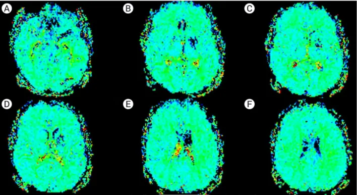
The modified Rankin Scale (mRS) and NIHSS scores were 0 at 4 months respectively. Magnetic resonance (MR) perfusion imaging at 10 months showed a chron- ic infarct of the putamen and no significant perfusion asymmetry between two hemispheres (Fig. 6). Based on the results of the perfusion study and due to sthe patients asymptomatic status, we decided to follow the patient medically.
Fig. 6. Color perfusion maps obtained at the level of basal ganglia (A-E) and corona radiata (F) show no significant asymmetry between the two hemispheres except sequelae changes of chronic infarct in the left putamen on MR perfusion imaging obtained after 10 months.
A B C
D E F
Fig. 5. The AP (A) and lateral views (B, C) of the control angiogram obtained at 6 months demonstrating stent patency with persistent residual stenosis.
A B C
DISCUSSION
A review of literature revealed 26 cases of mechanical thrombectomy in IE related stroke. Twentythree were successful and in 3 the authors had to revert to intra- cranial stenting due to the failure of mechanical throm- bectomy.1)2)5)8-12) Bain et al. used balloon expandable coronary stent, postulating that a self-expanding stent would not be sturdy enough to hold the firm clot against the arterial wall and maintain MCA patency.2) Hernan- dez-Fernandez et al. used a Solitaire (Medtronic, Dub- lin, Ireland) device for revascularization and stenting but they also had to perform an in-stent angioplasty to maintain patency during the same procedure.5) That is to say, a definitive stenting together with angioplasty was needed in more than 10% of the thrombectomy cases with acute IE.
Our review of the relevant literature also suggests that thrombectomy for embolic IE differs from rou- tine thrombectomy in several aspects: To begin with, these patients are frequently anticoagulated and may have acute/subacute infarctions in unrelated cerebral territories. Consequently, there is an increased risk of intracranial hemorrhage both during the thrombectomy procedure and after the thrombectomy procedure. The likelihood of observing distal mycotic aneurysms is not low, further increasing the perioperative risk of hem- orrhage. Last but not the least, previous reports have argued that the embolus in IE may be tighter due to a higher inflammatory content and compacted fibrotic nature, decreasing the likelihood of successful recanali- zation, especially when aspiration is not successful.2)5)
Our case is the first one in which only stenting was utilized for intracranial revascularization in IE. Con- sequently, residual stenosis occurred after intracranial stenting of the recalcitrant embolus.
However, during almost a year of follow-up, the pa- tient was asymptomatic and imaging was unremarkable except for residual stenosis. MR perfusion imaging was interpreted to be normal. Contrary to the allegation of Bain et al.2), it was possible to achieve sufficient recanal- ization with a closed-cell intracranial stent to keep the
patient asymptomatic. Arterial patency was sustained on the six-month digital subtraction angiography fol- low-up. Since it is not always possible to navigate a coronary stent intracranially and since acute post-stent angioplasty may be associated with a risk of arterial dissection or perforation, this method may be reserved as a rescue procedure for such intractable emboli. As we demonstrated, it is possible to test if the intracrani- al stent is strong enough to keep the artery patent by obtaining angiograms while the stent is half-deployed during the thrombectomy procedure. If the partial de- ployment fails to keep the artery patent, the stent can be resheated and a balloon-expandable coronary stent may then be considered.
Intracranial stenting is not risk-free. First of all, it al- ters the anticoagulation protocols in these patients. We used tirofiban during the procedure and for the next 5 days. As previously mentioned, both IE itself and a possible need for thoracic surgery impose the need for lifelong anticoagulation in these patients.9) In a single previously reported case, valve replacement was nec- essary due to the development of severe mitral insuffi- ciency but unlike our case, the timing of surgery was not mentioned by the authors.5) In our case, tirofiban use complicated the management of the patient. Tirofiban is known to increase the risk of intracranial hemorrhage, especially in acute stroke cases like our patient.7) As a matter of fact, tirofiban is contraindicated in patients with intracranial hemorrhage due to a risk of exacerba- tion of the bleeding.4) Although our patient had a post procedure intracranial bleeding, we were unable to dis- continue tirofiban (we took the risk of hematoma expan- sion) since he already had an intracranial stent in place.
Secondly, as a previously overlooked concern, there is a theoretical possibility that placement of a foreign body (stent) over a possibly infected embolus in a an acutely septic patient might be risky in terms of arterial patency and integrity. As the case by Bain et al.2) did not undergo follow-up vascular imaging, our case and a case report- ed by Hernandez-Fernandez et al. are the only cases that demonstrate the absence of arterial wall involvement by an infectious process on follow-up.5) Perioperative anti-
biotic coverage is critical for febrile patients presenting for intracranial thrombectomy to avoid a potentially fatal arterial damage on follow-up.
CONCLUSIONS
A recent history of febrile status or presentation with fever, as we noted in our patient, should raise a suspicion of IE as a cause of acute stroke. Due to time constraints, a workup (e.g. transesophageal ultrasound) may not be possible in this setting. The operator may consider cardiologic evaluation on the angiography suite, or as an alternative may choose to start the procedure with thromboaspiration, keeping in mind the condensed na- ture of the clot and reserve intracranial stent placement for resistant cases for reasons we described.
Disclosure
The authors report no conflict of interest concerning the materials or methods used in this study or the find- ings specified in this paper.
REFERENCES
1. Ambrosioni J, Urra X, Hernandez-Meneses M, Almela M, Falces C, Tellez A, et al. Mechanical Thrombectomy for Acute Ischemic Stroke Secondary to Infective Endocarditis. Clin Infect Dis. 2018 Apr;66(8):1286-9.
2. Bain MD, Hussain MS, Gonugunta V, Katzan I, Gupta R.
Successful recanalization of a septic embolus with a balloon mounted stent after failed mechanical thrombectomy. J Neu- roimaging. 2011 Apr;21(2):170-2.
3. Bhuva P, Kuo SH, Claude Hemphill J, Lopez GA. Intracranial hemorrhage following thrombolytic use for stroke caused by infective endocarditis. Neurocrit Care. 2010 Feb;12(1):79-82
4. Bukow SC, Daffertshofer M, Hennerici MG. Tirofiban for the treatment of ischaemic stroke. Expert Opin Pharmacother.
2006 Jan;7(1):73-9.
5. Hernandez-Fernandez F, Rojas-Bartolome L, Garcia-Garcia J, Ayo-Martin O, Molina-Nuevo JD, Barbella-Aponte RA, et al. Histopathological and bacteriological analysis of throm- bus material extracted during mechanical thrombectomy in acute stroke patients. Cardiovasc Intervent Radiol. 2017 Dec;40(12):1851-60.
6. Junna M, Lin CC, Espinosa RE, Rabinstein AA. Successful intravenous thrombolysis in ischemic stroke caused by infec- tive endocarditis. Neurocrit Care. 2007;6(2):117-20.
7. Kellert L, Hametner C, Rohde S, Bendszus M, Hacke W, Ringleb P, et al. Endovascu lar stroke therapy: tirofiban is as- sociated with risk of fatal intracerebral hemorrhage and poor outcome. Stroke. 2013 May;44(5):1453-5.
8. Kim JM, Jeon JS, Kim YW, Kang DH, Hwang YH, Kim YS.
Forced arterial suction thrombectomy of septic embolic mid- dle cerebral artery occlusion due to infective endocarditis: an illustrative case and review of the literature. Neurointerven- tion. 2014 Sep;9(2):101-5.
9. Ladner TR, Davis BJ, He L, Kirshner HS, Froehler MT, Moc- co J. Complex decision-making in stroke: preoperative me- chanical thrombectomy of septic embolus for emergency car- diac valve surgery. J Neurointerv Surg. 2015 Dec;7(12):e41.
10. Scharf EL, Chakraborty T, Rabinstein A, Miranpuri AS. En- dovascular management of cerebral septic embolism: three recent cases and review of the literature. J Neurointerv Surg.
2017 May;9(5):463-5.
11. Sloane KL, Raymond SB, Rabinov JD, Singhal AB. Mechan- ical thrombectomy in stroke from infective endocarditis:
case report and review. J Stroke Cerebrovasc Dis. 2019 Jan;29(1):104501.
12. Sveinsson O, Herrman L, Holmin S. Intra-arterial mechani- cal thrombectomy: an effective treatment for ischemic stroke caused by endocarditis. Case Rep Neurol. 2016 Oct;8(3):229-33.
13. Walker KA, Sampson JB, Skalabrin EJ, Majersik JJ. Clinical characteristics and thrombolytic outcomes of infective endo- carditis-associated stroke. Neurohospitalist. 2012 Jul;2(3):87-91.