submit.radiology.or.kr J Korean Soc Radiol 2011;65(6):603-605
603 INTRODUCTION
Osteogenic sarcoma of the ribs accounts for less than 3% of all osteosarcomas (1). Most rib osteosarcomas have been associated with pre-existing lesions such as Paget’s disease or irradiated bone. Few studies have been reported on the primary osteosar- coma of the rib (2). Even fewer studies have described CT or magnetic resonance (MR) findings of a primary osteosarcoma of the rib. We report both the CT and MR imaging (MRI) find- ings of primary osteoblastic osteosarcoma of the rib with histo- pathologic correlation.
CASE REPORT
A 31-year-old woman without previous medical history presented with a bony hard mass in the right lower back area.
Chest radiographs demonstrated a calcified mass involving the posterior arc of right 10th rib (Fig. 1A). A CT scan showed a densely calcified mass measuring about 5.5 × 5.0 × 2.5 cm in
the right lower back, with bone destruction of the right 10th rib (Fig. 1B). MR images revealed a heterogeneous high signal intensity lesion with multiple signal voids on T2-weighted image (T2WI) (Fig. 1C) and intermediate signal intensity mass with multiple signal voids on T1-weighted image (T1WI) (Fig. 1D) and well-enhancing portions of the tumor mass on fat-suppressed T1WI (Fig. 1E). The initial differential diagno- sis was chondrosarcoma, possibly low grade, due to high sig- nal intensity lobulated portions on T2WI. Mild uptake on a F-18 fluorodeoxyglucose positron emission tomography im- aging study corresponding to the mass was suggestive of a low grade tumor-like lesion with a max SUV of 1.4. The patient underwent resection of the right 9-10th ribs. The histopatholog- ic examination revealed the mass to be an osteoblastic type of osteosarcoma (Fig. 1F, G).
DISCUSSION
Osteogenic sarcoma of the ribs accounts for less than 3% of
Case Report
pISSN 1738-2637
J Korean Soc Radiol 2011;65(6):603-605
Received July 20, 2011; Accepted August 27, 2011 Corresponding author: Jung-Ah Choi, MD Department of Radiology, Seoul National University Bundang Hospital, 166 Gumi-ro, Bundang-gu, Seongnam 463-707, Korea.
Tel. 82-31-787-7609 Fax. 82-31-787-4011 E-mail: [email protected]
Copyrights © 2011 The Korean Society of Radiology
We report the CT and magnetic resonance (MR) imaging appearances in an adult case of primary osteoblastic osteosarcoma of the rib. Osteosarcoma of the rib pres- ents a diagnostic challenge because of the rarity of the lesion, especially with plain radiographs. The tumor should be suspected if CT and MR images demonstrate mineralization, suggestive of an osteoid matrix.
Index terms Rib
Osteosarcoma CT
Magnetic Resonance
Primary Osteoblastic Osteosarcoma of the Rib in an Adult:
A Case Report
1성인 늑골에 발생한 원발성 골모세포성 골육종: 증례 보고1
Inseon Ryoo, MD
2, Jung-Ah Choi, MD
1,2, Hyo Jin Park, MD
3, Jin-Haeng Chung, MD
3, Joo Han Oh, MD
4, Heung Sik Kang, MD
1,21Department of Radiology, Seoul National University Bundang Hospital, Seongnam, Korea
2Department of Radiology and Institute of Radiation Medicine, Seoul National University College of Medicine, Seoul, Korea Departments of 3Pathology, 4Orthopedic Surgery, Seoul National University Bundang Hospital, Seongnam, Korea
Primary Osteoblastic Osteosarcoma of the Rib in an Adult
submit.radiology.or.kr
J Korean Soc Radiol 2011;65(6):603-605
604
quite similar to the usual osteosarcomas in long bones, which have a slightly higher signal intensity than muscle on T1WI as well as mixed and high signal intensity on T2WI with multiple signal void areas at the mineralized matrix. Due to dense calci- fication of the majority of the mass, this case showed diffuse low signal intensity with multifocal high signal intensity within the mass on T2WI, which is consistent with previous studies.
Osteosarcoma of the rib presents a diagnostic challenge be- cause of the rarity of this lesion. This lesion is usually initially evaluated with plain radiographs. Suboptimal visualization of calcifications in plain radiographs may lead to misinterpreta- tion such as lung cancer, extraosseous chest wall tumors, or lytic rib metastases (3). Even with the visualization of calcifica- tions in plain radiographs, other lesions with calcifications, in- cluding chondrosarcoma, osteochondroma, fibrous dysplasia, extraosseous chest wall tumors, myositis occificans, infections and fractures should be included in the differential diagnoses (1, 3, 5-7). Therefore CT and MRI findings for osteosarcoma all osteosarcomas. The tumor most commonly occurs during
the 2nd decade of life. When it manifests in older patients, un- derlying pathology should be considered. Only a few reports exist on the CT appearance of primary osteosarcoma of the rib (2-6), most of which had a prior history of radiation or pre-existing pathology. Several cases described the CT find- ings of primary osteosarcoma of the rib to provide optimal assessment of the tumor matrix, dense central calcification within a large soft tissue mass with sparing of peripheral ar- eas, and localization of the epicenter of the soft tissue mass to the rib. Some authors suggested that it is possible to differen- tiate an osteosarcoma from a chondrosarcoma based on these findings (3, 6). CT also delineates the involvement of adjacent structures. This case also showed typical dense calcification with a relatively spared marginal peripheral area of the mass lesion.
Even fewer reports have described the MRI findings of pri- mary osteosarcoma of the rib (6, 7). MR findings of primary osteosarcoma of the rib described in previous studies were
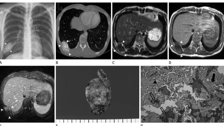
Fig. 1. Radiograph, CT, MRI, and histopathologic specimen images of 31-year-old woman with osteoblastic osteosarcoma of the rib.
A. Chest radiograph shows non homogenous calcification (white arrowheads) involving the right 10th rib.
B. CT scan of chest shows the tumor mass with central dense calcification (white arrowheads).
C. Axial T2-weighted image shows the tumor (white arrowheads) with extensive central signal loss due to dense calcification.
D. Axial T1-weighted image shows intermediate signal intensity lesion (white arrowheads) with multiple central signal void areas.
E. Axial T1-weighted contrast-enhanced image shows well enhancing portions of the soft tissue mass of the tumor (white arrowheads).
F. Gross specimen image shows lobulated mass with mineralized matrix.
G. Mineralized (open arrows) or unmineralized (arrow) osteoid is haphazardly distributed and neoplastic hyperchromatic cells (arrowhead) are noted between the osteoid (H&E, × 200).
A
E
B
F
C
G
D
Inseon Ryoo, et al
submit.radiology.or.kr J Korean Soc Radiol 2011;65(6):603-605
605
643: Osteosarcoma of ribs with giant rosettoid structures.
Skeletal Radiol 1990;19:609-612
3. Abdulrahman RE, White CS, Templeton PA, Romney B, Moore EH, Aisner SC. Primary osteosarcoma of the ribs: CT findings. Skeletal Radiol 1995;24:127-129
4. Chattopadhyay A, Nagendhar Y, Kumar V. Osteosarcoma of the rib. Indian J Pediatr 2004;71:543-544
5. Lawson JP, Barwick KW. Case report 162: Periosteal osteo- sarcoma of rib. Skeletal Radiol 1981;7:63-65
6. O’Sullivan P, O’Dwyer H, Flint J, Munk PL, Muller NL. Ma- lignant chest wall neoplasms of bone and cartilage: a pic- torial review of CT and MR findings. Br J Radiol 2007;
80:678-684
7. Lee TJ, Collins J. MR imaging evaluation of disorders of the chest wall. Magn Reson Imaging Clin N Am 2008;16:355- 379, x
of the rib can help to correctly diagnose the lesion. In this case, the correct diagnosis was not suggested from the chest radiographs. CT and MR scans provided valuable informa- tion about the tumor. CT provided the characteristic features of primary rib osteosarcoma, allowing for the differentiation from other more common lesions. MRI also depicted the full extent of osseous and soft tissue extension, which could not be seen on CT, especially in cases with dense calcification such as this case.
REFERENCES
1. Burt M, Fulton M, Wessner-Dunlap S, Karpeh M, Huvos AG, Bains MS, et al. Primary bony and cartilaginous sarco- mas of chest wall: results of therapy. Ann Thorac Surg 1992;54:226-232
2. Kim H, Park C, Lee YB, Jin SY, Ro JY, Ayala AG. Case report
성인 늑골에 발생한 원발성 골모세포성 골육종: 증례 보고1
유인선
2· 최정아
1,2· 박효진
3· 정진행
3· 오주한
4· 강흥식
1,2성인에서 생긴 늑골의 원발성 골모세포성 골육종(osteoblastic osteosarcoma)의 CT와 MRI 소견을 보고하고자 한다. 단 순 촬영상 늑골의 골육종은 그 희소성으로 인해 진단하기 매우 어렵다. 그러나 CT와 magnetic resonance상 특징적인 골 기질(osteoid matrix)을 시사하는 무기질 침착(mineralization)의 소견을 보인다면 반드시 의심해봐야 한다.
1분당서울대학교병원 영상의학과, 2서울대학교 의과대학 영상의학과학교실, 분당서울대학교병원 3병리과, 4정형외과