Sinusitis is a common condition; however, serious complications associated with sinusitis, such as os- teomyelitis, subperiosteal scalp abscess, orbital celluli- tis, and intracranial extension have become quite rare with the advent of antibiotics (1). Occasionally, serious complications may occur with mild symptomatic pre- sentation. In such cases, prompt medical and surgical therapy is required to minimize potentially serious se- quelae. Pott’s puffy tumor is an unusual but important complication of frontal sinusitis and may occur as a re- sult of the spread of sinusitis to the frontal bone.
Recognition of this entity is important, because of the risk of intracranial extension and its attendant complica- tions (2). We present a patient who had clinical signs of diplopia, blurred vision, and mild tenderness on glabel- lar area. A CT examination showed osteomyelitis of the frontal bone as well as frontal sinusitis.
Case Report
A 64-year-old male patient presented with complaints of diplopia, blurred vision, and mild tenderness on the glabellar area over the previous 25-days. The patient had no history of trauma, however did have a general history of hypertension and a 5-year history of stroke.
Upon physical examination, swelling and edema of the right periorbital was observed, which caused diplopia.
On the PNS series, a blurred margin of the right frontal sinus wall (Fig. 1A) and soft tissue swelling on prefrontal area were seen (Fig. 1B). A CT scan of the or- bits and sinuses revealed full opacification of the frontal sinus, erosive thinning of the anterior wall of the frontal sinus (Fig. 1C), sclerotic bone marrow of the frontal skull (Figs. 1C, D) as well as soft tissue swelling and en- hancement over the forehead (Fig. 1E).
The patient was diagnosed with frontal sinusitis, fore- head, periorbital cellulitis, and osteomyelitis of the frontal calvarium. The otolaryngology service was per- formed on the trephination of the frontal sinus and a large amount of pus in the frontal sinus was seen with erosion of the frontal bone. The microbiology broth iso-
J Korean Soc Radiol 2010;62:101-104
─ 101 ─
Pott’s Puffy Tumor Arising from Frontal Sinusitis
1Ji Yeon Lim, M.D., Hyun Koo Kang, M.D.
1Department of Diagnostic Radiology, Seoul Veterans Hospital Received June 9, 2009 ; Accepted August 25, 2009
Address reprint requests to : Hyun Koo Kang, M.D., Department of Diagnostic Radiology, Seoul Veterans Hospital, 6-2 Doonchon-dong, Gangdong-gu, Seoul 134-791, Korea.
Tel. 82-2-2225-1426 Fax. 82-2-2225-1488 E-mail: [email protected]
Pott’s puffy tumor is an extremely rare and potentially life-threatening complication of frontal sinusitis. We report a case of a 64-year-old man who presented at our emer- gency department with mild tenderness on the glabellar area and diplopia. Computed Tomography (CT) revealed frontal sinusitis and osteomyelitis of the frontal bone.
Following sinus trephination and long-term antibiotic therapy, the patient achieved a complete recovery.
Index words :Frontal bone Osteomyelitis
Tomography, X-Ray Computed
lated streptococcus viridans.
On contrast enhanced brain magnetic resonance imag- ing (MRI) performed four months later, enhancement of the frontal bone marrow was seen, however no intracra- nial abnormal findings were present (Fig. 1F).
Discussion
Pott’s puffy tumor was first described as “a puffy, cir- cumscribed, indolent tumor of the scalp and a sponta- neous separation of the pericranium from the skull un- der such a tumor” by Percivall Pott, in London in 1760 (3, 4). The disorder is an eponym for frontal os-
teomyelitis and is associated with a subperiosteal ab- scess. With the advent of antibiotics, this disorder is con- sidered to be extremely rare (4).
Sir Percival Pott originally described this condition as a complication of trauma; however, it is more common- ly observed as a complication of frontal sinusitis (5).
More rare causes of Pott’s puffy tumor include insect bites, a craniotomy, or hair transplantation (5). Pott’s puffy tumor can occur as a result of the spread of sinusi- tis to the frontal bone, along with the development of os- teomyelitis in the frontal bone and the extension of pu- rulent material (2). Trauma to the prefrontal region of the skull and surrounding soft tissues can also be a
Ji Yeon Lim, et al: Pott’s Puffy Tumor Arising from Frontal Sinusitis
─ 102 ─
A B C
D E F
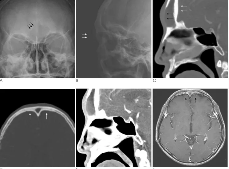
Fig. 1. 64-year-old man complaining of diplopia, blurred vision, and mild tenderness on the glabellar area.
A, B. On a PNS series, the blurred margin of the bony wall of the right frontal sinus (black arrows) (A), and soft tissue swelling on the prefrontal area (white arrows) are seen (B).
C. Sagittal PNS CT scan reveals full opacification of the frontal sinus, erosive thinning of the anterior wall of the frontal sinus (black arrows), and sclerotic bone marrow of the frontal skull (white arrows).
D. On an axial PNS CT scan, sclerotic bone marrow of the frontal skull (white arrows) is observed.
E. Soft tissue swelling and enhancement is observed over the forehead.
F. A post-operative brain MRI shows enhancement of the frontal bone marrow (black arrows). No abnormal intracranial findings are present.
cause of the disease (6). When forehead swelling after trauma is associated with fever and sinusitis, clinical suspicion should be raised for the occurrence of Pott’s puffy tumor.
Pott’s puffy tumor presents with symptoms including frontal scalp swelling, fever, headache, frontal sinus ten- derness, and photophobia (1). The frontal scalp swelling may be less tender or less erythematous than expected due to the depth of the infection (1). Headaches are of- ten relieved as the sinus drains through the frontal bone.
Occasionally, the patient’s symptoms are diminished as the forehead enlarges (6).
Pott’s puffy tumor can be associated with intracranial complications such as subdural empyema, an epidural abscess, and cortical vein thrombosis with or without di- rect erosion of the frontal bone. Because the mucosal ve- nous drainage of the frontal sinus occurs through the diploic veins, which communicate with the dural ve- nous plexus, septic thrombi can potentially evolve from foci within the frontal sinus and propagate through this venous system (2, 5). Also, because the frontal sinus shares a thin posterior plate with the frontal cranial fos- sa, when the frontal sinusitis spreads to the brain; it most commonly results in an infection of the frontal lobe (7). An orbital subperiosteal abscess, which is a known complication of adjacent sinusitis, can be associ- ated with frontal sinusitis and has been shown to place the patient with orbital cellulitis at a greater risk of in- tracranial involvement (7).
In previously reported cases, the cultured etiologic or- ganisms included microaerophilic streptococci such as alpha-hemolytic streptococcus, staphylococcus, peptostreptococcus, bacteroides species, and other anaerobes. Because of the relatively anaerobic condi- tions in the frontal sinus caused by compromised ostial patency, microaerophilic organisms may be more com- mon in the frontal sinusitis (2, 5).
Pott’s puffy tumor cannot be excluded without proper imaging studies. A contrast-enhanced head and sinus CT scan is the diagnostic modality of choice (1). Bone scanning will detect osteomyelitis.
Pott’s puffy tumor can be differentiated from a muco- cele and a malignancy. On a CT scan, a mucocele is ex- pansile and causes an erosive change of the surrounding bony structure; however, no osteomyelitis or soft tissue inflammatory changes are seen. In malignant paranasal sinus lesions, well enhancing soft tissue lesions, bizarre shaped surrounding bony structure destruction, and soft tissue infiltration are seen.
Antibiotic therapy alone is rarely adequate for the treatment of Pott’s puffy tumor. The definitive treat- ment consists of a combination of surgery, including de- bridement and removal of the sequestrum, as well as treatment with antibiotics to prevent further suppura- tive complications (1, 4).
Pott’s puffy tumor is a complicated infection that re- quires hospital admission and aggressive therapy.
Patients with Pott’s puffy tumor generally don’t appear acutely ill and have a subtle symptomatic presentation.
Therefore, proper imaging diagnosis, such as a contrast- enhanced CT scan, is critical to prevent aggravation of the complications associated with Pott’s puffy tumor (1).
This case report describes simple X-ray, CT, and MR imaging findings of Pott’s puffy tumor.
References
1. Bannon PD, McCormack RF. Pott’s puffy tumor and epidural ab- scess arising from pansinusitis. J Emerg Med 2008;18. [Epub ahead of print]
2. Blackman SC, Schleiss MR. Forehead swelling caused by Pott’s puffy tumor in a 9-year-old boy with sinusitis. Pediatr Int 2005;47:
704-707
3. Tattersall R, Tattersall R. Pott’s puffy tumor. Lancet 2002;359:
1060-1063
4. Martinez-Diaz GJ, Hsia R. Pott’s puffy tumor after minor head trauma. Am J Emerg Med 2008;26:739.e1-739.e3
5. Raja V, Low C, Sastry A, Moriarty B. Pott’s puffy tumor following an insect bite. J Postgrad Med 2007;53:114-116
6. Lamoreau KP, Fanciullo LM. Pott’s puffy tumour mimicking pre- septal cellulitis. Clin Exp Optom 2008;91:400-402
7. Reynolds DJ, Kodsi SR, Rubin SE, Rodgers IR. Intracranial infec- tion associated with preseptal and orbital cellulitis in the pediatric patient. J AAPOS 2003;7:413-417
J Korean Soc Radiol 2010;62:101-104
─ 103 ─
Ji Yeon Lim, et al: Pott’s Puffy Tumor Arising from Frontal Sinusitis
─ 104 ─
대한영상의학회지 2010;62:101-104
전두동염에 의한 Pott’s Puffy 종괴
1서울보훈병원 영상의학과 임 지 연∙강 현 구
Pott’s puffy 종괴는 매우 드물지만, 치명적인 전두동염의 합병증이다. 미간 동통과 복시를 주소로 한 64세 남자 환자가 본원 응급실에 내원하였다. 컴퓨터 단층 촬영상 전두동염과 전두골 골수염으로 진단되었고, 전두동 천자와 장기간 항생제 투여로 환자는 회복되었다.