Movement of the eye is a complex phenomenon regu- lated by a high level of coordination between sensory and motor functions which center stationary or moving objects in the central retina (fovea). The securing and stabilization of object images on the fovea during head movement are the basic functions of eye movements (1). Recent advances in the techniques of magnetic reso- nance (MR) imaging have opened a new horizon for the evaluation of ocular motility disorders; pathologies once considered beyond the scope of imaging are now fre- quently visualized. Knowledge of the basic pathway and physiology involved in eye movement provides a better understanding of the ocular motility disorders.
The supranuclear control of eye movement involves the pathway extending from the cerebral cortex to the ocu- lar motor nuclei located in the brain stem. In this paper,
in addition to describing the normal supranuclear path- way of eye movement, we demonstrate MR imaging findings of the typical ocular manifestations caused by disorders involving the supranuclear pathway, provid- ing the anatomic explanations for certain clinical signs.
Classification of Eye Movement
Many theories and assumptions have attempted to ex- plain the mechanism of eye movement. For the sake of simplicity, we assume that there are two independent major subsystems involved in the control of eye move- ments, namely version and vergence (1). The version subsystem controls all conjugate movements, while the vergence subsystem controls all those that are disconju- gate. The former encompasses two distinct eye move- ments, saccades (fast eye movements) and pursuit (slow eye movements), and the latter mediates vergence eye movements. While all three movements share a com- mon neural pathway from the ocular motor nuclei to the extraocular muscles, each is subject to a different supranuclear neural control.
Neuroradiology in the Ocular Motility Disorders :
I. Supranuclear Pathway
1H y u ng -Jin Kim, M.D.1 , 2, Byung Hoon Lim, M.D.3, Jae Bum Na, M.D.
Jae Hyoung Kim, M.D., Sung Hoon Chung, M.D.
The supranuclear control of eye movement invo l ves the pathway extending from the cerebral cortex to the ocular motor nuclei located in the brain stem. This paper de- scribes the normal supranuclear pathway, which controls eye movement. We also in- clude magnetic resonance imaging findings of the typical ocular manifestations caused by disorders involving the supranuclear pathway, providing the anatomic ex- planations for certain clinical signs.
Index words : Brain, anatomy Brain, abnormalities Brain, MR
1Department of Diagnostic Radiology, Gyeongsang National University H o s p i t a l
2Department of Radiology, College of Medicine, Inha University
3Department of Neurology, Gyeongsang National University Hospital Supported in part by the 1996 Joint Study Fund of the Gyeongsang National University Hospital
Received November 4, 1998 ; Accepted December 11, 1998
Address reprint requests to: Hyung-Jin Kim, M.D. Department of Radiology, College of Medicine, Inha University,
7-206, 3rd St., Shinheung-dong, Choong-ku, Inchon, 400-103, Korea.
Tel. 82-32-890-2761 Fax. 82-32-890-2743
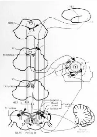
Normal Supranuclear Pathway (Fig. 1)
Fast Eye Movements(Saccades)
The visual stimulus for fast eye movements (saccades) is target displacement in space (1). Reticular formation in the pons and midbrain plays a critical role in the gen- eration of all types of saccades. The paramedian pontine reticular formation (PPRF) contains neurons essential for horizontal saccades, while the rostral interstitial nu- cleus of the medial longitudinal fasciculus (riMLF), lo- cated in the rostral midbrain, is essential for vertical sac- cades. The PPRF receives inputs from the contralateral frontal eye fields (FEF, Brodmann area 8) by way of the anterior limb of the internal capsule and the medial por- tion of the cerebral peduncle. This pathway decussates in the lower midbrain and upper pons. The riMLF prob- ably receives input from both the FEF and the PPRF.
The superior colliculi receive visual inputs from both the posterior cortical areas and the FEF via direct and indirect (basal ganglia) pathways, and probably play a role in both spontaneous and reflexive saccades.
Voluntary saccades are initiated in the FEF, which re- ceive inputs from the supplemental motor and posterior cortices (2). The pars reticulata of the substantia nigra inhibits the superior colliculi, suppressing excessive spontaneous saccades, while neurons in the caudate nu-
Fig. 2. Saccadic palsy caused by acute cerebral infarction. Axial T2-weighted MR image shows
large cerebral infarction in the right MCA territory. The patient’s eyes were deviated to the right side and the patient had leftward conjugate gaze palsy.
Fig. 3. Saccadic palsy caused by frontal lobe hemorrhage. Axial gradient-echo MR image shows multiple hemorrhagic foci in the brain with the largest being located just anterior to the right central sulcus in a 42-year-old man with seizure. His eyes were deviat- ed to the left side and he had rightward conjugate gaze palsy.
Fig. 4. Horizontal gaze palsy caused by a grade II pontine astrocytoma. Enhanced axial T1-weighted MR image shows a mass with peripheral rim-like enhancement in the right pontine tegmentum, and the patient had horizontal rightward gaze palsy.
2 3 4
Fig. 1. Diagram of overall supranuclear, nuclear, and infranuclear pathways of eye movement control.
DLPN=dorsolateral pontine nucleus ; IC=inferior colliculus ; MLF=medial longitudinal fasciculus ; OEF=occipital eye fields ; RF=reticular formation ; FEF=frontal eye fields ; riMLF=rostral interstitial nucleus of medial longitudinal fas- ciculus ; SC=superior colliculus.
cleus projecting to the pars reticulata of the substantia nigra inhibit substantia nigra cells.
Slow Eye Movement(Pursuit)
The major stimulus for slow eye movement (pursuit) is a fixated target that moves(1). Clinical and experi- mental observations have suggested that the occipital visual cortex (Brodmann area 17) contains neurons that respond to a moving visual stimulus and project to the middle temporal and medial superior temporal vi- sual areas with some influence from the adjacent pari- etal cortex. From these extrastriate visual areas neu- rons descend to the dorsolateral pontine nuclei and subsequently to the contralateral flocculus and dorsal vermis of the cerebellum, from which projections re- lay to the ipsilateral vestibular nuclei and finally to the ocular motor nuclei. If unilateral, pursuit abnormality usually occurs in the direction ipsilateral to the side of a lesion (2, 3).
Vergence Eye Movement
The stimulus for vergence eye movement is target dis- placement or motion along the visual z axis(toward or away from the observer) (1). Motor cells involved in the vergence system are intermixed in the midbrain reticu- lar formation, from where commands are transmitted to the ocular motor nuclei (2).
Supranuclear Pathology Resulting in Ocular Motility Disorders
Horizontal Version Abnormalities
Unilateral saccadic palsy usually results from an acute destructive frontal stroke, which causes deviation of the eyes toward the side of the lesion and impaired gaze to- ward the contralateral side (Fig. 2). It is usually associat- ed with contralateral hemiparesis or hemianopsia (2). If the opposite hemisphere is intact, saccadic palsy is tran- sient and normal function is usually regained in days to weeks. In contrast, irritative frontal lesions secondary to focal seizure activity or acute cerebral hemorrhage cause tonic deviation of the eyes to the contralateral side and gaze is impaired toward the same side of the lesion (Fig. 3). Bilateral lesions of the frontomesencephalic sys- tem cause saccadic palsy in both directions. In this situ- ation, patients rely on vestibulo-ocular reflex to change fixation (3). A similar manifestation can be observed in congenital or acquired diseases such as multiple sclero- sis, Huntington’s disease, Wilson’s disease, and spin- ocerebellar degeneration (2, 3). Unilateral pursuit pare- sis occurs with posterior hemispheric disease and mani- fests as saccadic pursuit in the direction ipsilateral to the side of the lesion; it is usually accompanied by contralat- eral homonymous hemianopsia (2). Bilateral saccadic pursuit may accompany the use of sedative drugs, inat- tention, fatigue, or impaired consciousness, and is also seen in cases involving diffuse cerebral, cerebellar or
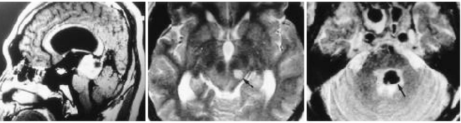
Fig. 5. Vertical gaze palsy caused by pineal germinoma (Parinaud’s syndrome) .Enhanced sagittal T1-weighted MR image shows a well enhanced pineal mass which causes compression of the midbrain resulting in patient’s upgaze palsy.
Fig. 6. Internuclear ophthalmoplegia caused by type 2 neurofibromatosis. Axial T2-weighted MR image shows an oviod mass with high signal intensity in the rostral midbrain (arrow) just posterior to the left red nucleus. After gadolinium infusion, the lesion was well enhanced (not shown). Presumptive diagnosis is glioma.
Fig. 7. One-and-a-half syndrome caused by pontine hemorrhage. Axial T2-weighted MR image shows acute hemorrhage with dark signal intensity (arrow) in the very area of left abducens nerve nucleus. The hemorrhage extended up to the ipsilateral midbrain (not shown).
5 6 7
brain stem disease (2). The saccadic and pursuit subsys- tems converge anatomically at subthalamic and upper brain stem levels (2). The PPRF is believed to be the fi- nal prenuclear anatomical pathway of ipsilateral sac- cades. All unilateral PPRF lesions are associated with ip- silateral gaze paralysis, which is usually persistent.
Excitatory projections from the PPRF mediating hori- zontal saccades are directed to the ipsilateral abducens nucleus. From synapses in intranuclear interneurons lo- cated in the abducens nucleus, they are transmitted to the contralateral MLF which, in turn, conveys impulses to the medial rectus subnucleus. Axons carrying hori- zontal vestibulo-ocular and pursuit signals also connect with internuclear interneurons (2). Lesions that are s- mall enough to be confined to the PPRF may, therefore, spare the vestibulo-ocular pathway, while those that in- volve the abducens nucleus frequently impair the vestibulo-ocular reflex (Fig. 4).
Vertical Gaze Abnormalities
Vertical gaze palsy typically occurs in diseases involv- ing the midbrain and thalamus. While projections for downward gaze are more or less directly dorsolateral from the riMLF, where they are primarily arranged lat- erally, to the oculomotor and trochlear nuclei, those for upward gaze are more sophisticated, reflecting the fact that upward gaze palsy result from lesions variously
placed in the midbrain. The critical structures involved are the riMLF, the interstitial nucleus of Cajal, the pos- terior commissure, the pretectal area and the dorsal pe- riaqueductal gray matter (2, 3). Dorsal midbrain ( P a r i n a u d’s) syndrome is the most well-known abnor- mality affecting vertical gaze with pineal region tumors and midbrain infarction as a leading cause (Fig. 5). In addition to upgaze palsy, the syndrome is frequently ac- companied by mydriasis, light-near dissociation, lid re- traction, lid lag and convergence-retraction nystagmus.
Internuclear Ophthalmoplegia (INO)
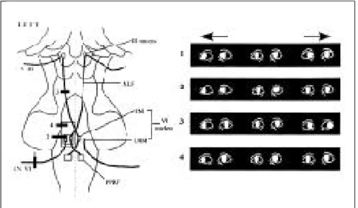
The MLF conveys saccadic, vestibulo-ocular, and pur- suit impulses to the ipsilateral medial rectus subnucleus (2). Unilateral disruption of the MLF prevents the ipsi- lateral medial rectus from adduction during horizontal versions; it is commonly accompanied by nystagmus of the abducting eye and is known as internuclear ophthal- moplegia (INO) (Fig. 6). Most cases of INO are exophor- ic in the primary position, without symptomatic diplop- ia. In bilateral INO, exotropia is occasionally seen. This is the so-called WEBINO (wall-eyed bilateral INO) syn- drome, which might be caused by simultaneous in- volvement of the medial rectus subnuclei. Lesions that are large enough to involve an MLF as well as the ipsi- lateral abducens nucleus or the PPRF result in so-called one-and-a-half syndrome, in which condition ipsilateral horizontal gaze palsy occurs in combination with INO during gaze towards the opposite side (3-6). Abduction of the opposite eye is the only spared movement in this condition (Fig. 7). Deficits of horizontal gaze following damage to the various sites of the abducens/MLF sys- tem are demonstrated in Figure 8.
R e f e r e n c e s
1. Dell’Osso LF, Daroff RB. Eye movement characteristics and recording techniques. In: Glaser GS, ed. N e u r o o p h t h a l m o l o g y, 2nd ed. New York: J. B. Lippincott Company, 1990;279-297 2. Daroff RB, Troost BT, Leigh RJ. Supranuclear disorders of eye
m o v e m e n t s. In: Glaser GS, ed. N e u r o o p h t h a l m o l o g y, 2nd ed.
New York: J. B. Lippincott Company, 1990;299-323
3. Digre KB, Osborn AG, Subbaratnam H. Neuro-radiologic evalu- ation of supranuclear and infranuclear disorders of eye move- ment. Ophthalmol Clin North Am 1994;7:459-486
4. Atlas SW, Grossman RI, Savino PJ, et al. Internuclear ophthal- moplegia: MR-anatomic correlation. AJNR 1987;8:243-247 5. Bronstein AM, Morris J, Boulay GD, Gresty MA, Rudge P.
Abnormalities of horizontal gaze. Clinical, oculographic and magnetic resonance imaging findings. I Abducens palsy. J Neurol Neurosurg Psychiatry1990;53:194-199
6. Bronstein AM, Rudge P, Gresty MA, Boulay GD, Morris J.
Fig. 8. Diagrams showing deficits of horizontal gaze following damage to the abducens/MLF system. Lesions labeled 1-4 on the left diagram can produce the characteristic ocular mani- festations shown on the right diagram: 1, abducens palsy caused by lesion in left abducens nerve; 2, lateral gaze palsy caused by lesion in left abducens nucleus; 3, internuclear oph- thalmoplegia caused by lesion in left MLF; and 4, one-and-a- half syndrome caused by lesion in left abducens nerve nucle- us and left MLF.INI=intranuclear interneuron; LRM=lateral rectus motor neuron; MLF=medial longitudinal fasciculus; P- PRF=paramedian pontine reticular formation.frequency en- coding pulse, PE - phase encoding pulse)
Abnormalities of horizontal gaze. Clinical, oculographic and magnetic resonance imaging findings. II Gaze palsy and inter-
nuclear ophthalmoplegia. J Neurol Neurosurg Psychiatry 1 9 9 0 ; 53:200-207
안구운동질환의 신경방사선학적 소견 : I. 핵상경로1
1경상대학교 의과대학 진단방사선과학교실
2인하대학교 의과대학 방사선과학교실
3경상대학교 의과대학 신경과학교실
김형진1 , 2・임병훈3・나재범・김재형・정성훈
안구운동에 관여하는 핵상경로는 대뇌 피질로부터 뇌간에 위치한 안구운동핵에 이르는 신경통로를 의미한다.
이 임상화보에서는 안구운동을 조절하는 정상 핵상경로와 이를 침범하는 질환에 의하여 야기되는 특징적인 안 구운동 장애의 자기공명영상 소견을 해부학적 근거와 더불어 기술하였다.
대한방사선의학회지 1 9 99;40: 4 29- 4 3 3