CASE REPORT
Copyright © 2011, the Korean Surgical Society J Korean Surg Soc 2011;81:427-430
http://dx.doi.org/10.4174/jkss.2011.81.6.427
JKSS
Journal of the Korean Surgical Society pISSN 2233-7903ㆍeISSN 2093-0488
Received November 25, 2010, Revised February 17, 2011, Accepted February 22, 2011 Correspondence to: Hyung-Il Seo
Department of Surgery, Pusan National University Hospital, Pusan National University School of Medicine, 10-1 Ami-dong 1-ga, Seo-gu, Busan 602-739, Korea
Tel: +82-51-240-7238, Fax: +82-51-247-1365, E-mail: [email protected]
cc Journal of the Korean Surgical Society is an Open Access Journal. All articles are distributed under the terms of the Creative Commons Attribution Non-Commercial License (http://creativecommons.org/licenses/by-nc/3.0/) which permits unrestricted non-commercial use, distribution, and reproduction in any medium, provided the original work is properly cited.
Histologic confirmation of huge pancreatic lipoma: a case report and review of literatures
Jee Yeon Lee, Hyung-Il Seo, Eun Young Park, Gwang Ha Kim
1, Do Youn Park
2, Suk Kim
3Departments of Surgery, 1Internal Medicine, 2Pathology and 3Diagnostic Radiology, Pusan National University School of Medicine, Yangsan, Korea
Pancreatic lipomas are commonly diagnosed based on radiologic images, although the prevalence of lipomas has not been established. Histologic confirmation of pancreatic lipomas is extremely rare because surgical treatment is unnecessary in most cases. Endoscopic ultrasound-guided fine-needle aspiration cytology has been suggested to avoid unnecessary surgery to distinguish between a lipoma and a well-differentiated liposarcoma; however, surgery would be needed when the tumor is associated with symptoms or difficult to distinguish from a liposarcoma. We present a case of a pancreatic lipoma in a 54-year-old male patient that was histologically-confirmed by subtotal pancreatectomy.
Key Words: Lipoma, Pancreas
INTRODUCTION
Most pancreatic tumors arise from epithelial cells. In fact, only 1 to 2% of these tumors originate from the mesenchyme. Mesenchymal tumors are classified accord- ing to histologic origin and include benign and malignant neoplasms. Because pancreatic lipomas can be observed if there is no specific clinical symptom or change in tumor size, the histologic confirmation of pancreatic lipoma is rare. However, when the pancreatic tumor is symptomatic and difficult to distinguish from well-differentiated lip- osarcoma, surgical management would be needed. We re- port a large pancreatic lipoma with histologic confir- mation which was difficult to distinguish from a lipo-
sarcoma.
CASE REPORT
A 54-year-old male patient with type 2 diabetes mellitus was evaluated for assessment of an incidental pancreatic tumor. There were no specific findings based on labo- ratory testing (amylase, 84 IU/L; lipase, 23 IU/L; carci- noembryonic antigen, 1.86 ng/mL; carbohydrate antigen 19-9, 12.78 U/mL), with the exception of an elevated glu- cose level (172 mg/dL). Computed tomography (CT) dem- onstrated a 9-cm, well-circumscribed, non-invasive tumor in the body of the pancreas with a -100 HU density which
Jee Yeon Lee, et al.
428 thesurgery.or.kr
Fig. 1. A 54-year-old male patient with a pancreatic lipogenic mass. (A) Contrast-enhanced abdominal computed tomography shows a 9-cm lipoma in the body of the pancreas (arrow), which had a density of -100 HU (consistent with subcutaneous adipose tissue). (B) After 3 months, the pancreatic tumor increased from 9 cm to 10.5 cm (arrow).
Fig. 2. (A) Lipogenic tumor of the pancreas. The axial T1WI shows a 10.5 cm well-defined hyperintense mass lesion in the body and tail of the pancreas. (B) The axial 3-dimensional fat suppression T1WI shows a well-defined hypointense mass containing mural nodules (arrows).
Fig. 3. (A) The 10.4 × 6.9 cm well-circumscribed mass in the body of the pancreas. The cut surface of the mass consists of yellow focal fibrous tissue and a hemorrhagic area. This hemorrhagic area (arrow) was noted on the magnetic resonance imaging as a mural nodule. (B) Mature adipocytes were noted adjacent to the pancreatic parenchyma (H&E, original magnification, ×40).
was homogeneous in appearance with the fat composition of the abdominal wall (Fig. 1A). Although the initial im- pression was a lipoma, a liposarcoma could not be com-
pletely ruled out because of a focal enhancing soft tissue component in the mass. On magnetic resonance imaging, there was a 10.5 cm well-defined, hyperintense mass con-
Histologic confirmation of pancreatic lipoma
thesurgery.or.kr 429
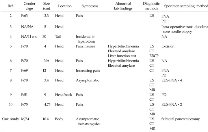
Table 1. Histologic confirmation of pancreatic lipomas
Ref. Gender
/age
Size
(cm) Location Symptoms Abnormal
lab findings
Diagnostic
methods Specimen sampling methods
2 F/63 3.3 Head Pain US FNA
PD
3 NA/NA 5 Head Intra-operative trans-duodenal
core needle biopsy 4 NA/11 mo 30 Tail Incidental in
laparotomy
NA
5 F/70 4 Head Pain, nausea Hyperbilirubinemia
Elevated amylase Liver function test
US CT ERCP
Excision
6 F/70 NA Head Pain Hyperbilirubinemia
Elevated amylase
US CT
NA
7 F/69 12 Head Increasing pain CT FNA
PD
8 F/70 3.4 Head Asymptomatic US
CT MR
EUS-FNA × 4
9 F/51 9 Head/neck Pain US
CT PD
10 F/75 4.75 Head Pain US
CT MR
EUS-FNA × 2
Our study M/54 10.4 Body Asymptomatic, increasing size
US CT MR
Subtotal pancreatectomy
NA, not available; US, ultrasound; CT, computed tomography; ERCP, endoscopic retrograde cholangiopancreatography; MR, magnetic resonance; FNA, fine needle aspiration cytology; PD, pancreaticoduodenectomy; EUS-FNA, endoscopic ultrasound-guided fine needle aspiration biopsy.
taining mural nodules in the body and tail of the pancreas on axial T1-weighted images (Fig. 2). After 3 months, a subtotal pancreatectomy with a splenectomy was per- formed to distinguish the mass from a liposarcoma be- cause the pancreatic tumor increased from 9 to 10.5 cm in size on contrast-enhanced abdominal CT (Fig. 1). In patho- logic finding, a 10.4 × 6.9 cm pancreatic lipoma containing yellow focal fibrous tissue and hemorrhagic areas was identified which was suggested as a mural nodule on im- age findings. Based on the microscopic findings, only ma- ture adipocytes were noted adjacent to the pancreatic pa- renchyma (Fig. 3). Post-operatively, the patient was treat- ed to control a worsening glucose level and discharged without specific complications in 14 days.
DISCUSSION
Since the first pancreatic lipoma was documented by Bigard et al. [1] in 1989, only 45 cases of pancreatic lipomas have been reported. Although the tumor size could vary from <1 to 30 cm in size, the observation would be possi- ble because they are nearly stable. For this reason, the his- tologic confirmation of pancreatic lipoma is rare [2-9].
While lipomas located with the head of the pancreas may originated from trapped retroperitoneal or mesen- teric fat between the dorsal and ventral pancreatic buds during embryonic fusion, the etiopathogenesis of lipomas within the body and tail of the pancreas are unclear [10].
Lipomas within the head of the pancreas exhibit clinical symptoms, such as abdominal pain, whereas lipomas within the body and tail of the pancreas are usually silent, even when >10 cm in size (Table 1) [1-9].
Jee Yeon Lee, et al.
430 thesurgery.or.kr
A CT scan is a useful imaging modality for detecting pancreatic lipomas. The diagnostic criteria for pancreatic lipomas include well-circumscribed lesions without any extravisceral continuation with the peri-pancreatic adi- pose tissue, densitometric measurement between -30 and -150 HU, well-delineated thin-homogenous capsule, and no sign of invasion of adjacent organs. The thick septa within the tumor, calcifications, rapid growth, and focal fatty infiltration within the peri-pancreatic adipose tissue, in contrast to pancreatic lipomas, and the absence of dis- tinct capsules are significant indicators of malignancy [10].
A surgical procedure is needed for well-differentiated lip- osarcomas which resemble benign lipomas [1]. The differ- ential diagnosis of pancreatic lipomas should include lip- omatosis, cystic teratomas, fibrolipomas, lipoblastomas, and liposarcomas. In the current case, liposarcomas could not be ruled out because of the increasing size and enhanc- ing soft tissue component. An endoscopic ultrasound- guided fine-needle aspiration (EUS-FNA) was not per- formed because EUS-FNA sometimes leads to needle-track seeding metastasis when the mass is malignant.
There are 10 histologically-confirmed lipomas based on surgical resection or EUS-FNA, including the current case (Table 1). The mean size of 8 pancreatic lipomas confirmed with surgery was 10.5 cm and the locations of the tumor were as follows: 6 cases in the head of the pancreas; 1 case in the body of the pancreas; and 1 case in the tail of the pancreas. Abdominal pain was associated with most pan- creatic lipomas and pancreatitis occurred in two cases, but not in the current case.
Although pancreatic lipomas are not common, the diag- nosis with imaging scans is quite accurate and EUS-FNA is a useful procedure for histologic confirmation of the tumor. Surgical excision should be considered when the tumor is symptomatic or difficult to distinguish from a liposarcoma.
CONFLICTS OF INTEREST
No potential conflict of interest relevant to this article was reported.
ACKNOWLEDGEMENTS
This work supported by clinical reasearch grant from Pusan National University Hospital 2010.
REFERENCES
1. Bigard MA, Boissel P, Regent D, Froment N. Intrapancre- atic lipoma. First case in the literature. Gastroenterol Clin Biol 1989;13:505-7.
2. De Jong SA, Pickleman J, Rainsford K. Nonductal tumors of the pancreas. The importance of laparotomy. Arch Surg 1993;128:730-4.
3. Boglino C, Inserra A, Silvano A, Ciprandi G, Boldrini R, Caione P. Intrapancreatic lipoma: a case report. Pediatr Med Chir 1993;15:397-9.
4. Merli M, Fossati GS, Alessiani M, Spada M, Gambini D, Viezzoli A, et al. A rare case of pancreatic lipoma. Hepato- gastroenterology 1996;43:734-6.
5. Di Maggio EM, Solcia M, Dore R, Preda L, La Fianza A, Rodino C, et al. Intrapancreatic lipoma: first case diag- nosed with CT. AJR Am J Roentgenol 1996;167:56-7.
6. Raut CP, Fernandez-del Castillo C. Giant lipoma of the pancreas: case report and review of lipomatous lesions of the pancreas. Pancreas 2003;26:97-9.
7. Di Matteo FM, Shimpi L, Pandolfi M, Rabitti C, Fabio C, Gabbrielli A, et al. EUS diagnosis of pancreatic lipoma: a case report. Gastrointest Endosc 2006;64:146-8.
8. Celis Zapata J, Berrospi Espinoza F, Valencia Mariñas HD, Sánchez Lihón J, Abad Licham M, Farías Mejía I. Pancre- atic lipoma: presentation of a case and review of literature.
Rev Gastroenterol Peru 2008;28:56-9.
9. Suzuki R, Irisawa A, Hikichi T, Shibukawa G, Takagi T, Wakatsuki T, et al. Pancreatic lipoma diagnosed using EUS-FNA. A case report. JOP 2009;10:200-3.
10. Karaosmanoglu D, Karcaaltincaba M, Akata D, Ozmen M, Akhan O. Pancreatic lipoma computed tomography diag- nosis of 17 patients and follow-up. Pancreas 2008;36:434-6.