Primary pancreatic lymphoma (PPL) is a rare extra- lymphatic lymphoma. It is usually found as a large pan- creatic mass with no dilatation of the pancreatic duct.
Severe dilatations of the pancreatic duct and common bile duct in PPL have not been described. Moreover, in- ternal necrosis in PPL is extremely rare in the literature.
Here, we report CT and MRI findings of PPL with se- vere dilatation of the pancreatic duct, as well as internal necrosis in a 27-year-old man.
Case Report
A 27-year-old man presented with a history of fatigue, jaundice, and weight loss for 20 days. His past medical history revealed that he was a hepatitis B carrier.
However, his physical examination was normal without evidence of abdominal tenderness or masses. His serum total albumin, direct albumin, aspartate amino trans- ferase (AST), and alanine amino transferase (ALT) were elevated; however, his serum amylase and lipase levels were normal, as were other laboratory tests including leukocyte counts. Contrast enhanced multi-detector row CT (MDCT) showed a 5 cm long diameter and poorly enhancing mass containing a low attenuated area in the pancreas head, with moderate to severe dilatation of pancreatic duct, common bile duct, and both intrahepat- ic bile ducts (Fig. 1A). Lymphadenopathy was not demonstrated; however, displacement of portal vein, IVC, and duodenum was depicted. Abdominal MR imaging was performed for the purpose of planning surgery on a 3T MRI unit using a phased array coil. T1- weighted MR images showed a hypointense homoge- neous mass with an internal hyperintense area (Fig. 1B).
T2-weighted MR images demonstrated a hyperintense mass with a hyperintense area, as well as severe dilata- tion of the pancreatic duct (Fig. 1C). A hyperintense area on T1- and T2-weighted MR images was considered as hemorrhage within the mass. However, gadolinium-en- hanced T1-weighted MR images showed homogeneous
Primary Pancreatic Lymphoma with Severe Dilatation of Pancreatic Duct: A Case Report1
Tae Wook Heo, M.D., Jin Woong Kim, M.D.2, Suk Hee Heo, M.D.2, Sang Soo Shin, M.D., Yong Yeon Jeong, M.D.2, Heoung Keun Kang, M.D.2, Yoo Duk Choi, M.D.3
1Department of Diagnostic Radiology, 3Department of Pathology, Chonnam National University Medical School, Chonnam National University Hospital
2Department of Diagnostic Radiology, Chonnam National University Medical School, Chonnam National University Hwasun Hospital Received February 2, 2010 ; Accepted April 21, 2010
Address reprint requests to : Jin Woong Kim, M.D., Department of Diagnostic Radiology, Chonnam National University Medical School, Chonnam National University Hwasun Hospital, 160 Ilsim-ri, Hwasun- eup, Hwasun-gun, Jeollanam-do 519-763, Korea.
Tel. 82-61-379-7104 Fax. 82-61-379-7133 E-mail: [email protected]
Primary pancreatic lymphoma is extremely rare and usually consists of a bulky tu- mor without internal necrosis and severe dilatation of the pancreatic duct. We report the unusual CT and MRI findings of a primary pancreatic lymphoma in a 27-years-old man, associated with severe dilatation of the pancreatic duct, common bile duct, and internal necrosis.
Index words :Pancreatic Neoplasms
Tomography, X-Ray Computed Magnetic Resonance Imaging Lymphoma, Non-Hodgkin
A
C D
E
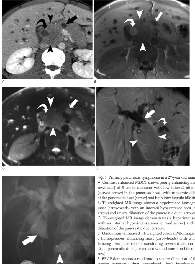
Fig. 1. Primary pancreatic lymphoma in a 27-year-old man.
A. Contrast enhanced MDCT shows poorly enhancing mass (ar- rowheads) of 5 cm in diameter with low internal attenuation (curved arrow) in the pancreas head, with moderate dilatation of the pancreatic duct (arrow) and both intrahepatic bile ducts.
B. T1-weighted MR image shows a hypointense homogeneous mass (arrowheads) with an internal hyperintense area (curved arrow) and severe dilatation of the pancreatic duct (arrow).
C. T2-weighted MR image demonstrates a hyperintense mass with an internal hyperintense area (curved arrow) and severe dilatation of the pancreatic duct (arrow).
D. Gadolinium-enhanced T1-weighted coronal MR image shows a homogeneous enhancing mass (arrowheads) with a non-en- hancing area (asterisk) demonstrating severe dilatation of the distal pancreatic duct (curved arrow) and common bile duct (ar- row).
E. ERCP demonstrates moderate to severe dilatation of the up- stream pancreatic duct (arrowhead), both intrahepatic bile ducts, and the common bile duct (arrow).
B
perform surgery because of the patient’s young age and high probability of malignancy on the imaging study. A pylorus preserving pancreaticoduodenectomy was per- formed, without preoperative chemotherapy.
Upon gross inspection of the surgical specimen, a well circumscribed whitish mass was identified with the nar- rowing of the downstream pancreatic duct and common bile duct. The tumor invaded the pancreatic duct and duodenal wall (Fig. 2A). A microscopic examination showed focal, well-circumscribed coagulative necrosis within the tumor, considered as hemorrhage on a preop- erative MRI (Fig. 2B). The tumor cells displayed a large, round to oval shaped nucleus, scanty cytoplasm, and prominent nucleoli. Immunostaining results indicated that the tumor cells showed positive immunoreactivity for CD20 and CD79a, and negative immunoreactivity for cytokeratin, CD45RO, and CD3 (Fig. 2C). The tumor
was pathologically confirmed as malignant diffuse large B-cell lymphoma. The patient had received postopera- tive adjuvant CHOP (cyclophosphamide, doxorubicin, vincristine, and prednisone) chemotherapy and has been living without evidence of tumor recurrence over 17 months after surgery.
Discussion
PPL is an extremely rare disease which occurs in pan- creatic situ, with or without involvement of the peripan- creatic lymph nodes (1). The diagnostic criteria for PPL include: 1) no palpable superficial lymphadenopathy, no mediastinal lymphadenopathy on chest radiography, 2) a normal leukocyte count in peripheral blood, 3) a main mass in the pancreas with lymph nodal involvement confined to the peripancreatic region, and 4) no hepatic A
C
B
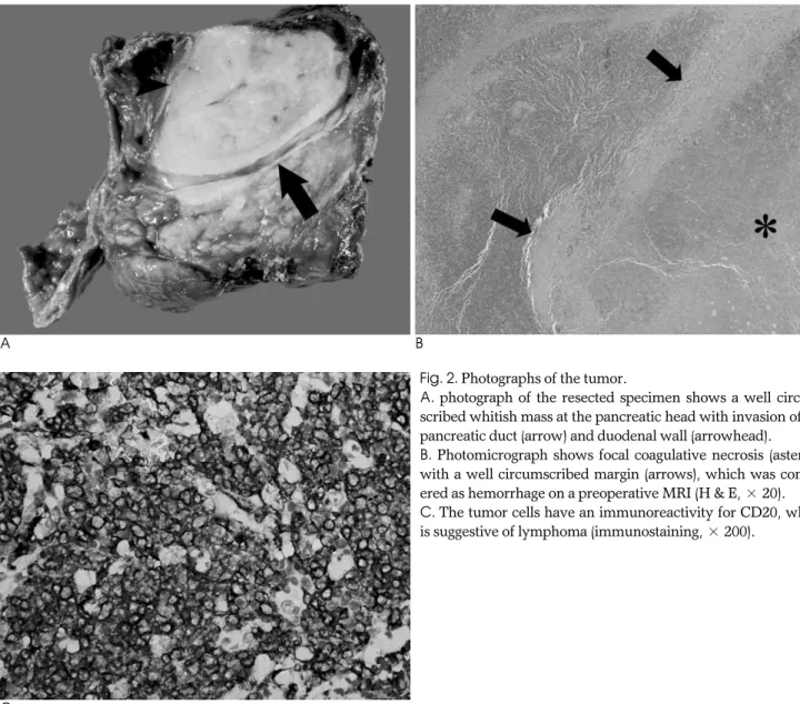
Fig. 2. Photographs of the tumor.
A. photograph of the resected specimen shows a well circum- scribed whitish mass at the pancreatic head with invasion of the pancreatic duct (arrow) and duodenal wall (arrowhead).
B. Photomicrograph shows focal coagulative necrosis (asterisk) with a well circumscribed margin (arrows), which was consid- ered as hemorrhage on a preoperative MRI (H & E, × 20).
C. The tumor cells have an immunoreactivity for CD20, which is suggestive of lymphoma (immunostaining, × 200).
the latter is a diffusely enlarged infiltrating or replacing form occupying most of the pancreatic gland (3). In a well circumscribed tumoral type, an MRI shows a ho- mogeneous hypointense mass within the pancreas on T1-weighted images with subtle enhancement after gadolinium enhancement, as well as a heterogeneous mass with low to intermediate signal intensity on T2- weighted images. As for a diffuse infiltrating tumor type, T1- and T2-weighted images showed hypointense enlarged pancreas, with mild to moderate enhancement after gadolinium enhancement (4). In our case, the mass was also homogeneous on CT and MRI with mild homo- geneous enhancement, but the mass contained a necrot- ic area as a hypoattenuation on CT and a hyperintense area on T1- and T2-weighted MR images.
Internal hypoattenuation on CT and a hyperintense area on T1- and T2-weighted MR images were consid- ered as hemorrhage within the mass. The presences of calcification or necrosis are reliable findings for ruling out non-Hodgkin’s lymphoma (5). However, a small het- erogeneous area which was necrosis within a tumor can be seen in an isolated case (6). In our case, focal necrosis within the mass was shown on CT and MRI. This find- ing was very unusual in untreated non-Hodgkin’s lym- phoma and made the diagnosis of PPL difficult.
Unlike pancreatic ductal adenocarcinoma, moderate to severe dilatation of the pancreatic duct is apparently extremely rare in PPL because the pancreatic duct is ei- ther normal, displaced, or simply narrowed in patients with PPL (4). Van Beers B et al. (3) reported that dilata- tion of the pancreatic duct can be usually mild, with a ratio of duct diameter to distal gland width invariably less than 0.5 in PPL. Severe dilatation of the pancreatic duct and common bile duct in our case was demonstrat- ed on CT and MRI. A dilated pancreatic duct and com- mon bile duct on CT and MRI led us to preoperatively diagnose a neuroendocrine tumor or pancreatic ductal adenocarcinoma.
During the surgical procedure, the hard mass with in- ternal necrosis was seen at the pancreatic head along with moderate to severe dilatation of common bile duct and pancreatic duct. The hardness of the tumor and the
differential diagnosis of PPL from the pancreatic ductal adenocarcinoma is very important. For the pancreatic ductal adenocarcinoma, the primary treatment is con- sidered to be surgical excision; but in PPL, the primary treatment is nonsurgical, based on chemotherapy alone or a combination of chemotherapy and radiation thera- py (1). Ductal adenocarcinoma commonly infiltrates in- to pancreatic parenchyma, peripancreatic fat, as well as adjacent structures, and dilates the more distal pancreat- ic duct when the more proximal ductal invasion has tak- en place. Also CT findings of ductal adenocarcinoma typically manifest themselves as a lesion that is some- what inhomogeneous and hypoattenuating relative to the normally enhancing pancreatic parenchyma.
Neuroendocrine tumors such as the large non-function- ing islet cell tumor demonstrate hypervascular solid components, usually in the periphery, and central non- enhancing areas on CT, which may represent necrosis, fibrosis, or cystic degeneration (7). Also, islet cell tumors usually show early strong enhancement on early-dy- namic contrast CT or MRI as well as frequent hepatic metastasis (8). However PPL usually demonstrates poor but homogeneous enhancement (9).
In summary, we present a rare case of PPL with un- usual imaging findings. Although a bulky homogeneous mass with moderate to severe dilatation of the pancreat- ic duct and focal necrosis within tumor is more com- monly seen in pancreatic ductal adenocarcinoma or a neuroendocrine tumor, PPL must be included in the dif- ferential diagnosis.
References
1. Lin H, Li SD, Hu XG, Li ZS. Primary pancreatic lymphoma:
Report of six cases. World J Gastroenterol 2006;12:5064-5067 2. Dawson IM, Cornes JS, Morson BC. Primary malignant lymphoid
tumours of the intestinal tract. Report of 37 cases with a study of factors influencing prognosis. Br J Surg 1961;49:80-89
3. Van Beers B, Lalonde L, Soyer P, Grandin C, Trigaux JP, De Ronde T, et al. Dynamic CT in pancreatic lymphoma. J Comput Assist Tomogr 1993;17:94-97
4. Merkle EM, Bender GN, Brambs HJ. Imaging findings in pancreat- ic lymphoma: differential aspects. AJR Am J Roentgenol 2000;174:
671-675
5. Prayer L, Schurawitzki H, Mallek R, Mostbeck G. CT in pancreat- ic involvement of non-hodgkin lymphoma. Acta Radiol 1992;33:123-127
6. Shirkhoda A, Ros PR, Farah J, Staab EV. Lymphoma of the solid abdominal viscera. Radiol Clin North Am 1990;28:785-799 7. Buetow PC, Miller DL, Parrino TV, Buck JL. Islet cell tumors of
the pancreas: clinical, radiologic, and pathologic correlation in di- agnosis and localization. Radiographics 1997;17:453-472
8. Semelka RC, Ascher SM. MR imaging of the pancreas. Radiology 1993;188:593-602
9. Saif MW. Primary pancreatic lymphomas. JOP 2006;7:262-273
대한영상의학회지 2010;63:167-171
심한 췌관확장을 동반한 원발췌장림프암: 증례 보고1
1전남대학교 의과대학 영상의학교실, 전남대학교병원 영상의학과
2전남대학교 의과대학 영상의학교실, 화순전남대학교병원 영상의학과
3전남대학교 의과대학 병리학교실, 전남대학교병원 병리과 허태욱∙김진웅2∙허숙희2∙신상수∙정용연2∙강형근2∙최유덕3
췌장의 원발림프암은 매우 드물어 특징적인 영상소견은 잘 알려져 있지 않으나 췌관선암종과는 달리 췌관의 확장 이 드물고 종괴 내부에 괴사를 잘 동반하지 않는다고 알려져 있다. 우리는 심한 췌관 및 총담관의 확장과 종괴 내 괴 사를 동반하였던 27세 남자의 원발췌장림프암의 전산화단층촬영(CT) 및 자기공명영상(MR)소견을 보고하고자 한 다.