Vol. 21, No. 3, 2009 315
Received November 11, 2008, Accepted for publication March 1, 2009
*This work was supported by the research grant of the Chungbuk Na- tional University in 2008.
Reprint request to: Tae Young Yoon, M.D., Department of Dermatology, School of Medicine & Medical Research Institute, Chungbuk National University, 410, Seongbong-ro, Heungdeok-gu, Cheongju 361-763, Korea. Tel: 82-43-269-6369, Fax: 82-43-266-1698, E-mail: tyyoon@
chungbuk.ac.kr
Ann Dermatol Vol. 21, No. 3, 2009
CASE REPORT
A Case of Neonatal Lupus Erythematosus Showing Transient Anemia and Hepatitis
Kyu Ri Kim, M.D., Tae Young Yoon, M.D.
Department of Dermatology, School of Medicine and Medical Research Institute, Chungbuk National University, Cheongju, Korea
Neonatal lupus erythematosus (NLE) is an autoimmune disease that is associated with transplacental passage of maternal autoantibodies that are reactive to SSA/Ro and SSB/La antigens. Cardiac involvement, hematologic abnormality and hepatic disease may occur in the infants suffering with NLE, in addition to the characteristic skin lesions. We report here on a case of NLE in a 4-week-old female infant who was born to an asymptomatic mother, and the baby displayed the characteristic clinical and histological features of cutaneous NLE with transient anemia and hepatitis. Both the infant and mother were positive for anti-SSA/Ro and anti-SSB/La. There have been 18 case reports of NLE in the Korean literature, including 7 case reports in the dermatological field. We describe herein another case of NLE that showed transient anemia and hepatitis, and we also review the case reports of NLE in the Korean literature. (Ann Dermatol 21(3) 315∼318, 2009) -Keywords-
Anemia, Anti-SSA/Ro, Anti-SSB/La, Hepatitis, Neonatal lu- pus erythematosus
INTRODUCTION
Neonatal lupus erythematosus (NLE) is an uncommon immune-mediated disease that is associated with trans- placental transfer of maternal IgG autoantibodies. Appro- ximately half of the affected neonates have congenital
heart block (CHB). Cutaneous lesions occur in approxi- mately 50% of these patients and they typically consist of transient, erythematous, annular, and scaling plaques in sun-exposed areas, and these lesions resemble subacute cutaneous lupus erythematosus both clinically and his- tologically. Skin lesions and heart block are seen simul- taneously in less than 10% of the patients with NLE1. Transient hepatitis, anemia, and thrombocytopenia can also occur2-10.
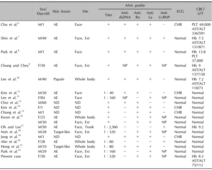
There have been 18 case reports of NLE in the Korean literature, including 7 case reports in the dermatological field of which 3 cases showed anemia and hepatitis (Table 1)6-22. We describe herein another case of NLE that displayed transient anemia and hepatitis. In addition, we review the cases of NLE in the Korean literature.
CASE REPORT
A 4-week-old, full-term, female infant, whose birth and life had been otherwise gone well, was brought to the dermatological clinic with a 3-week history of erythema- tous and annular plaques on the face and lower extre- mities (Fig. 1). The physical examination revealed no other abnormalities.
A biopsy from the face showed slight keratin plugging of the hair follicles, focal hydropic degeneration of the basal layer in the epidermis and a patchy perivascular, peri- adnexal, and interstitial infiltrate of lymphohistiocytes in the whole dermis (Fig. 2).
The patient’s antinuclear antibody (ANA) titer was 1:320 with a homogeneous pattern. The tests for anti- SSA/Ro antibody and anti-SSB/La antibody were positive. The results of the electrocardiogram (ECG) were normal, but mild anemia (hemoglobin 8.3 g/dl) and hepatitis (AST/
ALT 75/112 IU/L) were present.
The 25-year-old mother had no history of connective ti- ssue disease and she was asymptomatic. The serological
KR Kim and TY Yoon
316 Ann Dermatol
Table 1. The clinical and serologic features of the reported cases of neonatal lupus erythematosus in the Korean literature Sex/
Day-old Skin lesion Site
ANA profile
ECG CBC/
Titer Anti- LFT dsDNA Anti-
Ro Anti- La Anti-
U1RNP
Chu et al.6 M/1 AE Face + + + + − CHB PLT 69,000
AST/ALT 336/591
Shin et al.7 M/40 AE Face, Ext + + + + + Normal Hb 7.5
AST/ALT 131/871
Park et al.8 M/1 AE Face + + + + - Normal Hb 13.8
PLT 37,000
Chung and Chey9 F/30 AE Face, Ext + NP + + NP Normal Hb 9
AST/ALT 137/150
Lee et al.10 M/40 Papule Whole body + + + + - Normal Hb 7.2
AST/ALT 110/73
Kim et al.11 M/30 AE Face 1:40 + + - - CHB Normal
Lee et al.12 F/84 AE Face 1:160 NP - + NP Normal Normal
Choi et al.13 M/60 ND ND + + + - - Normal Normal
Kim et al.14 F/1 ND ND + - + + - CHB Normal
Chung et al.15 M/1 ND ND + - + + - CHB Normal
Kwon et al.16 F/25 AE Whole body + - + + NP Normal Normal
M/30 AE Face, Ext + - + + NP Normal Normal
Oh and Lee17 M/50 AE Face, Trunk 1:2,560 - - - + Normal Hb 9.2
Noh et al.18 M/28 Target-like Face, Ext 1:320 - + + NP Normal Normal
Jung et al.19 M/1 ND ND + + + + - CHB Normal
Ahn et al.20 F/28 AE Whole body 1:80 - - - + Normal Normal
Hong et al.21 M/10 Target-like Whole body 1:80 + + + - Normal Normal
Park et al.22 M/29 AE Face, Ext 1:160 - - + NP Normal Normal
Present case F/30 AE Face, Ext 1:320 - + + NP Normal Hb 8.3
AST/ALT 75/112 AE: annular erythema, ALT: alanine aminotransferase, ANA: antinuclear antibody, AST: aspartate aminotransferase, CBC: complete blood count, CHB: congenital heart block, ECG: electrocardiogram, Ext: extremity, Hb: hemoglobin, LFT: liver function test, ND:
not described, NP: not performed, PLT: platelet
Fig. 1. Erythematous and annular plaques on the face.
studies of the mother showed the presence of anti-SSA/Ro antibody and anti-SSB/La antibody. The mother’s ANA titer was 1:640 with a homogeneous pattern.
The patient received topical corticosteroid and the skin lesions completely disappeared 6 months later without any residual atrophy or hyperpigmentation. At that time, the infant had an ANA titer less than 1:40; both anti- SSA/Ro antibody and anti-SSB/La antibody became nega- tive and the complete blood cell count and liver function tests were normal.
DISCUSSION
NLE is an uncommon autoimmune disease that is asso- ciated with the transplacental transfer of maternal IgG
A Case of Neonatal Lupus Erythematosus Showing Transient Anemia and Hepatitis
Vol. 21, No. 3, 2009 317 Fig. 2. Slight keratin plugging of the hair follicles, focal hydropic
degeneration of the basal layer in the epidermis and a patchy perivascular, periadnexal, and interstitial infiltrate of lympho- histiocytes in the whole dermis (H&E, ×100).
autoantibodies. Only 1% of neonates develop NLE if the mother has autoantibodies. If an anti-SSA/Ro positive mo- ther has one child with NLE, then 25% of the future si- blings will also be affected. In 95% of the cases, the auto- antibody is anti-SSA/Ro, but it can be anti-SSB/La or anti- U1RNP1. Any of these autoantibodies can be found alone or in combinations. Only cutaneous disease has been reported for the cases where only anti-U1RNP antibodies are found20,23. There are even reports of histological do- cumented cases of NLE where none of the above-listed antibodies were found24. This finding suggests that anti- bodies other than anti-SSA/Ro, anti-SSB/La, or anti-U1RNP can be involved in some cases, and perhaps by some factor or cofactor that has yet to be determined.
The major clinical manifestations of NLE are cardiac problems, including CHB, and cutaneous lesions1. CHB can result in congestive heart failure and the subsequent placement of a pacemaker. In one investigation, 57% of these patients eventually required a pacemaker25. CHB is associated with a 20∼30% mortality rate during the neo- natal period. Deaths may also occur later in life as a result of the failure of the pacemaker. There have been five CHB patients reported in the Korean literature. Among them, three showed a spontaneous remission, one needed pace- maker insertion and one passed away. On the other hand, the cutaneous lesions of NLE consist of transient nonscar- ring erythematous annular plaques with a predilection for the periorbital and photodistributed areas. These lesions generally appear within the first 2 months of life and they resolve within 4 to 6 months when the maternal anti- bodies disappear. In rare cases, remnant telangiectasias can occur at the previously affected sites26.
Hepatic and hematologic abnormalities are observed in approximately 10% of the infants with NLE10. Hepatitis, hyperbilirubinemia, liver failure, thrombocytopenia, and anemia can occur2-10. The prognosis of a NLE patient with liver failure is poor, whereas for the NLE patients with other manifestations, the prognosis is good with transient involvement and spontaneous resolution. In the Korean literature, anemia occurred in 5 cases, thrombocytopenia occurred in 2 cases, and hepatitis occurred in 4 cases.
Only 3 cases showed anemia and hepatitis simultaneou- sly. Our patient showed typical cutaneous lesions, tran- sient hepatitis, and anemia without cardiac involvement.
The association of NLE with maternal autoantibodies is a strong indicator of the importance of autoantibodies for determining the pathogenesis of NLE27. In patients with NLE, the autoantibodies are maternal in origin and they disappear by 6 months of age. These autoantibodies have the capacity to cause direct injury to the skin and the cardiac, hepatobiliary and hematologic system28. The dis- appearance of autoantibodies parallels the disappearance of the cutaneous, hematologic, and hepatic abnormalities.
However, in patients suffering with CHB, immunodeposits of autoantibodies may result in fibrosis and calcification in and around the artrioventricular node, which can lead to permanent conduction defects.
The diagnosis of NLE is generally based on the clinical findings when there are maternal and/or neonatal autoan- tibodies present. Determining the titers of ANA and anti- SSA/Ro, anti-SSB/La, and anti-U1RNP antibodies is recom- mended for making the diagnosis. Liver function tests and a complete blood cell count should also be done. All the patients suspected of having NLE should undergo a tho- rough cardiac examination.
Spontaneous resolution is the natural course of the cu- taneous lesions. However, management of the skin lesions of NLE requires avoiding sun light and using sunscreen and low-potency topical corticosteroids to hasten resolu- tion. Treatment of the heart block is not necessary unless cardiac failure is evident.
Many (40%) of the affected infants’ mothers are asymp- tomatic. They might have Sjögren's syndrome, systemic lupus erythematosus, rheumatoid arthritis, overlap synd- rome or even leukocytoclastic vasculitis. Although the outlook of these mothers appears to be generally good, there is the possibility of developing serious autoimmune disease29. Thus, close observation is necessary for the mothers of infants with NLE.
In conclusion, we describe here an additional case of NLE that displayed transient anemia and hepatitis.
KR Kim and TY Yoon
318 Ann Dermatol
REFERENCES
1. Lee LA. Neonatal lupus erythematosus. J Invest Dermatol 1993;100:9S-13S.
2. Esterly NB. Neonatal lupus erythematosus. Pediatr Dermatol 1986;3:417-424.
3. Dickerson PA, Prendiville JS. Thrombocytopenia and hepato- splenomegaly in a newborn. Pediatr Dermatol 1989;6:346- 348.
4. Watson R, Kang JE, May M, Hudak M, Kickler T, Provost TT.
Thrombocytopenia in the neonatal lupus syndrome. Arch Dermatol 1988;124:560-563.
5. Wolach B, Choc L, Pomeranz A, Ben Ari Y, Douer D, Metz- ker A. Aplastic anemia in neonatal lupus erythematosus. Am J Dis Child 1993;147:941-944.
6. Chu TG, Back YW, Huh JW, Lee CY, Chung HK, Park JS. A case of neonatal lupus syndrome with congenital heart block. J Korean Soc Neonatol 1997;4:260-266.
7. Shin DY, Seok SM, Koo DW. Neonatal lupus erythematosus.
Korean J Dermatol 1999;37:1649-1654.
8. Park SH, Kim HJ, Yoo KH, Hong YS, Lee JW, Kim SK. A case of neonatal lupus erythematosus with pancytopenia and bradycardia. J Korean Soc Neonatol 2001;8:150-155.
9. Chung JY, Chey MJ. A case of neonatal lupus with abnormal liver function test and skin lesion. Korean J Pediatr 2005;
48:85-87.
10. Lee SM, Ham SS, Jeon IS, Son DW. Neonatal lupus ery- thematosus manifests as pancytopenia and mildly abnormal liver functions. Korean J Perinatol 2005;16:317-321.
11. Kim CR, Chang YP, Kim HS, Kim MJ, Kim BI, Choi JH, et al.
Outcome in infants of mothers with systemic lupus erythe- matosus. J Korean Pediatr Soc 1993;36:791-804.
12. Lee HP, Lee HN, Houh D, Byun DG, Baek SC. Neonatal lupus erythematosus. Ann Dermatol 1998;10:185-189.
13. Choi JW, Kim MH, Kim ST, Park HJ. A case of neonatal lupus syndrome with acute myocarditis. J Korean Pediatr Soc 1999;42:1298-1303.
14. Kim GS, Choi SM, Lee GH. Two cases of neonatal arrhy- thmia observed by fetal echocardiography. Korean J Perina- tol 1999;10:71-79.
15. Chung SH, Lee Y, Cheon YH, Jung IC, Yoon WS, Lee JS, et al. A case of neonatal lupus syndrome with congenital com-
plete heart block. Korean J Obstet Gynecol 2002;45:723- 727.
16. Kwon IH, Yoo JY, Lee JH, Cho KH. Two cases of neonatal lupus erythematosus. Korean J Dermatol 2003;41:956-959.
17. Oh DH, Lee CW. Neonatal lupus erythematosus. Korean J Dermatol 2003;41:1108-1110.
18. Noh Y, Lee GC, Eo SH, Ha TS, Kim MK, Kim CY, et al.
Neonatal lupus erythematosus: showing target-like skin lesions. Korean J Dermatol 2003;41:1228-1231.
19. Jung KS, Shin SH, Choi CW, Lee JH, Kim SI. A case of complete atrioventricular block in neonatal lupus erythema- tosus. Korean J Med 2004;67:s887-s891.
20. Ahn BH, Lee GC, Yoon TY, Kim MJ. A case of neonatal lupus erythematosus associated with anti-U1RNP antibodies.
Korean J Pediatr 2005;48:342-345.
21. Hong SJ, Hwang IJ, Kim DH. Neonatal lupus syndrome. J Korean Rheum Assoc 2005;12:245-246.
22. Park G, Jang HC, Chung H. A case of neonatal lupus erythe- matosus. Korean J Dermatol 2006;44:124-126.
23. Dugan EM, Tunnessen WW, Honig PJ, Watson RM. U1RNP antibody-positive neonatal lupus. A report of two cases with immunogenetic studies. Arch Dermatol 1992;128:1490-1494.
24. Crowley E, Frieden IJ. Neonatal lupus erythematosus: an unusual congenital presentation with cutaneous atrophy, ero- sions, alopecia, and pancytopenia. Pediatr Dermatol 1998;
15:38-42.
25. Neonatal lupus erythematosus. In: Hurwitz S, editor. Clinical pediatric dermatology: a textbook of skin disorders of child- hood and adolescence. 2nd ed. Philadelphia: W B Saun- ders, 1993:567-569.
26. Thornton CM, Eichenfield LF, Shinall EA, Siegfried E, Rabi- nowitz LG, Esterly NB, et al. Cutaneous telangiectases in neonatal lupus erythematosus. J Am Acad Dermatol 1995;
33:19-25.
27. Lee LA. Transient autoimmunity related to maternal autoanti- bodies: neonatal lupus. Autoimmun Rev 2005;4:207-213.
28. Sontheimer RD, McCauliffe DP. Pathogenesis of anti-Ro/SS- A autoantibody-associated cutaneous lupus erythematosus.
Dermatol Clin 1990;8:751-758.
29. McCune AB, Weston WL, Lee LA. Maternal and fetal out- come in neonatal lupus erythematosus. Ann Intern Med 1987;106:518-523.