Ann Hepatobiliary Pancreat Surg 2020;24:518-521
https://doi.org/10.14701/ahbps.2020.24.4.518
Case Report
Hepatocellular carcinoma with atrial tumor thrombus presenting as myxoma: Resection under cardiopulmonary bypass
Manoj K. Singh1, Rahul Roy1, Varun Shetty2, and Sanjay Goja1
1Liver Transplant/HPB Surgery, Narayana Health,
2Cardiothoracic Surgery, Narayana Health, Bengaluru, India
Surgical resection for Hepatocellular carcinoma (HCC) with atrial tumor thrombus is a rare life saving procedure. A case of left lateral segment liver tumor (HCC) with atrial tumor thrombus resected with use of cardio-pulmonary bypass is presented. (Ann Hepatobiliary Pancreat Surg 2020;24:518-521)
Key Words: Atrial tumor; Thrombus; Hepatocellular carcinoma; Cardiopulmonary bypass; Myxoma
Received: May 4, 2020; Revised: June 9, 2020; Accepted: June 10, 2020 Corresponding author: Manoj K. Singh
Liver Transplant/HPB Surgery, Mazumdar Shaw Medical Center, Narayana Health City, 258/A, Bommasandra Industrial Area, Hosur Road, Bengaluru 560099, Karnataka, India
Tel: +91-80-71222182, Fax: +91-80-27832648, E-mail: [email protected]
Copyright Ⓒ 2020 by The Korean Association of Hepato-Biliary-Pancreatic Surgery
This is an Open Access article distributed under the terms of the Creative Commons Attribution Non-Commercial License (http://creativecommons.org/
licenses/by-nc/4.0) which permits unrestricted non-commercial use, distribution, and reproduction in any medium, provided the original work is properly cited.
Annals of Hepato-Biliary-Pancreatic Surgery ∙ pISSN: 2508-5778ㆍeISSN: 2508-5859
INTRODUCTION
Surgical resection for advanced HCC is an uncommon operation and most society recommendations do not have clear guidelines for these tumors due to lack of class I data. We present a case of advanced left lateral segment HCC with atrial tumor thrombus extension, treated surgi- cally with simultaneous resection of liver tumor and tu- mor thrombus extending to right atrium using cardiopul- monary bypass (CPB). Most of the reported cases of liver tumor with atrial extension have historically been right lobe tumor where alternate resection strategies have also been successful.
CASE
A 49 year old male with type II diabetes mellitus and chronic obstructive pulmonary disease, was incidentally found to have left lateral segment liver tumor (Hepatocel- lular Carcinoma) with right atrial involvement during a screening echocardiography which showed an atrial mass with probable diagnosis of myxoma. On further evaluation he was found to have Hepatitis B, was Child A cirrhotic with normal liver function, platelet count of 219×109/L,
prothrombin time of 13 seconds, and alpha-fetoprotein value of 12103.8 ng/ml. Contrast enhanced computed to- mography showed mild hepatomegaly with normal paren- chymal echotexture along with a well defined 7.7×7.6×5.6 cm hypervascular lesion in segment II, III of liver show- ing early arterial enhancement and washout in portal ve- nous phase characteristic of HCC (Fig. 1A). The mass had a contiguous tumor thrombus extending into the left hep- atic vein (LHV), suprahepatic inferior vena cava (IVC) and right atrium (RA) (Fig. 1B). There was no ascites or any other evidence of distant metastases. Trans-thoracic echocardiography showed a large mass in right atrium ex- tending from the IVC. Upper gastrointestinal tract endos- copy showed no features of portal hypertension.
Surgical resection was planned with a team of hepato- biliary and cardiothoracic surgeons. Abdominal explora- tion was done first through an inverse L-shaped incision.
Upon palpation, the tumor invaded the suprahepatic IVC, and extended into the right atrium. A left lateral hep- atectomy, starting with parenchymal transection was per- formed using an ultrasonic dissector and harmonic scalpel after isolation and division of segment II/III pedicles sepa- rately lateral to the falciform ligament, leaving only the left and middle hepatic veins attached to the IVC (Fig.
Manoj K. Singh, et al. HCC with atrial tumor thrombus 519
Fig. 1. (A) Computed tomo- graphy image of segment II/III hepatocellular carcinoma with early arterial enhancement. (B) Computed tomography image of hepatocellular carcinoma tumor thrombus extending from left he- patic vein to right atrium.
Fig. 2. Intraoperative image after left lateral parenchymal transection and placement of cardiopulmonary bypass cannulas.
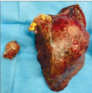
Fig. 3. Resected specimen; left lateral segment liver tumor with tumor thrombus in left hepatic vein and detached atrial tumor thrombus.
2). Next, a median sternotomy was performed, the cava was snared, liver vascular inflow was controlled with a vascular clamp. The patient was placed on CPB with the inferior vena cava cannula inserted distal to the hepatic veins, insertion of the superior vena cava cannula, and the arterial cannula in the ascending aorta. The cava was then opened caudal to the entrance of the left hepatic vein. The tumor was palpated superiorly past the atriocaval junction.
The entire en bloc specimen was then removed from the operative field, which included the left lateral liver seg- ment along with tumor thrombus extending into the IVC;
right atrial tumor thrombus was removed superiorly from the atrial incision (Fig. 3). Total hepatic vascular occlu- sion time was 14 minutes and CPB time was 43 minutes;
patient received four units of packed red cells transfusion during the procedure.
The patient’s postoperative course was uneventful and
he was discharged on POD#12. Histopathological exami- nation of the resected specimen showed 9 cm moderately differentiated HCC with tumor thrombus (pT4 Nx Mx).
Patient is alive and doing well at six month follow-up with no evidence of local HCC recurrence or systemic disease.
DISCUSSION
Liver cancer is the seventh most common and the third leading cause of cancer related mortality in the world;
HCC being the most common liver cancer.1 Among the various factors associated with poor prognosis in HCC, major vascular invasion i.e. tumor thrombus (TT) of por- tal vein (PVTT) or hepatic vein (HVTT)/ IVC (IVCTT) is one of the most important.2-4 The incidence of HVTT varies between 1.4-4.9%5 and in rare circumstances it can
520 Ann Hepatobiliary Pancreat Surg Vol. 24, No. 4, November 2020 www.ahbps.org
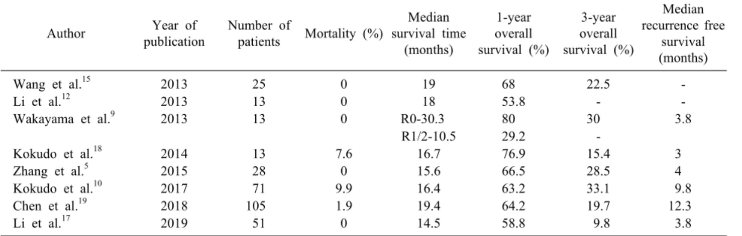
Table 1. Studies with Surgical resection outcomes for HCC with IVC TT/RA TT
Author Year of
publication
Number of
patients Mortality (%)
Median survival time
(months)
1-year overall survival (%)
3-year overall survival (%)
Median recurrence free
survival (months)
Wang et al.15 2013 25 0 19 68 22.5 -
Li et al.12 2013 13 0 18 53.8 - -
Wakayama et al.9 2013 13 0 R0-30.3 80 30 3.8
R1/2-10.5 29.2 -
Kokudo et al.18 2014 13 7.6 16.7 76.9 15.4 3
Zhang et al.5 2015 28 0 15.6 66.5 28.5 4
Kokudo et al.10 2017 71 9.9 16.4 63.2 33.1 9.8
Chen et al.19 2018 105 1.9 19.4 64.2 19.7 12.3
Li et al.17 2019 51 0 14.5 58.8 9.8 3.8
manifest as an intracardiac mass lesion. Treatment guide- lines issued by American Association for the study of the liver disease and Barcelona Clinic for Liver cancer Staging System consider PVTT and HVTT as advanced stage of the disease with Sorafenib as the only treatment recommended. However survival with Sorafenib is at best modest with low local tumor control with a reported sur- vival of 6-8 months.6,7 As a result, many centres have been advocating alternate treatment modalities including Hepatic resection (HR), Transcatheter arterial chemoembo- lization (TACE), conformal radiotherapy (RT) to improve tumor control and patient survival.
TT involving the IVC/RA is seen in 1.5-4.5% of pa- tients with HCC undergoing radiological imaging.8-10 Depending on its anatomic location with respect to heart, TT is classified into the following types: Type I (inferior hepatic IVCTT) involves IVC below diaphragm; Type II (superior hepatic IVCTT) involves IVC above the dia- phragm but outside the RA; Type III (Intracardiac IVCTT) involving the RA.11,12 If hepatic resection is being con- templated for HCC with HVTT then type I TT can be treated by radical hepatectomy, type II TT can be man- aged by abdominal exploration and total hepatic vascular occlusion and type III TT will require CPB.11,13
Prognosis is usually very poor without surgical treat- ment for HCC with right atrial tumor thrombus extension due to risk of pulmonary embolism or cardiac outflow tract obstruction, pulmonary and distant metastasis with median survival of 1.9 months.14 It is prudent to opt for simultaneous resection of HCC with atrial thrombectomy to prevent acute pulmonary embolism and lung metastasis.
Hepatic resection with concomitant thrombectomy is a technically challenging procedure particularly for the type III TT variant which requires CPB. Surgical outcomes have improved with time and the surgery can now be per- formed with acceptable post operative morbidity and mor- tality as is evident from Table 1.
As the incidence of HVTT is low very few comparative studies that assess the effect of different treatment modal- ities are available. Wang et al. showed a median survival time of 4.5 months with TACE compared with 19 months for surgical resection.15 When TACE is combined with three dimensional conformal radiotherapy the median sur- vival time increased to 11.7 months.12,16 Li et al.17 have compared radiotherapy versus surgery for HCC with TT of IVC/ right atrium and found comparable results be- tween the two with median survival time of 12.8 months and 14.5 months respectively.
Despite the survival benefit achieved by surgical re- section, it is evident from Table 1 that recurrence rate is high and therefore the poor recurrence free survival. The selection of patients for curative resection should not be made by anatomical criteria alone; tumor biology should also be taken into consideration. The factors associated with poor prognosis following surgery for HCC with HVTT/IVCTT include multiple tumors, tumor size >10 cm, co-existing PVTT, type III IVCTT, high preoperative AFP level, poor response to preoperative therapy (TACE, hepatic arterial infusion chemotherapy).15,17-20
Manoj K. Singh, et al. HCC with atrial tumor thrombus 521
CONCLUSION
Selected advanced HCC patients with HVTT/IVCTT can undergo surgical resection with acceptable post-oper- ative morbidity and mortality with a survival benefit better than currently advocated non-surgical therapy (Sorafenib/
TACE).
CONFLICT OF INTEREST
There is no conflict of interest.
ORCID
Manoj K. Singh: https://orcid.org/0000-0002-1909-1745 Rahul Roy: https://orcid.org/0000-0003-3487-8228 Varun Shetty: https://orcid.org/0000-0003-3120-5888 Sanjay Goja: https://orcid.org/0000-0002-9090-9087
AUTHOR CONTRIBUTIONS
MKS: Conception and design, drafting the article, re- vising it critically for important intellectual content and final approval of the version to be published. RR: Drafting the article and final approval of the version published.
VS: Data collection and final approval of the version published. SG: Conception and design, revision for in- tellectual content and final approval of the version to be published.
REFERENCES
1. Global Burden of Disease Cancer Collaboration, Fitzmaurice C, Abate D, Abbasi N, Abbastabar H, Abd-Allah F, et al. Global, regional, and national cancer incidence, mortality, years of life lost, years lived with disability, and disability-adjusted life-years for 29 cancer groups, 1990 to 2017: a systematic analysis for the global burden of disease study. JAMA Oncol 2019;5:1749-1768.
2. Vauthey JN, Lauwers GY, Esnaola NF, Do KA, Belghiti J, Mirza N, et al. Simplified staging for hepatocellular carcinoma.
J Clin Oncol 2002;20:1527-1536.
3. Llovet JM, Brú C, Bruix J. Prognosis of hepatocellular carcino- ma: the BCLC staging classification. Semin Liver Dis 1999;19:
329-338.
4. Ikai I, Yamaoka Y, Yamamoto Y, Ozaki N, Sakai Y, Satoh S, et al. Surgical intervention for patients with stage IV-A hep- atocellular carcinoma without lymph node metastasis: proposal as a standard therapy. Ann Surg 1998;227:433-439.
5. Zhang YF, Wei W, Guo ZX, Wang JH, Shi M, Guo RP. Hepatic resection versus transcatheter arterial chemoembolization for the
treatment of hepatocellular carcinoma with hepatic vein tumor thrombus. Jpn J Clin Oncol 2015;45:837-843.
6. Marrero JA, Kulik LM, Sirlin CB, Zhu AX, Finn RS, Abecassis MM, et al. Diagnosis, staging, and management of Hepatocellu- lar Carcinoma: 2018 practice guidance by the American Associa- tion for the Study of Liver Diseases. Hepatology 2018;68:723- 750.
7. Llovet JM, Ricci S, Mazzaferro V, Hilgard P, Gane E, Blanc JF, et al. Sorafenib in advanced hepatocellular carcinoma. N Engl J Med 2008;359:378-390.
8. Kojiro M, Nakahara H, Sugihara S, Murakami T, Nakashima T, Kawasaki H. Hepatocellular carcinoma with intra-atrial tumor growth. A clinicopathologic study of 18 autopsy cases. Arch Pathol Lab Med 1984;108:989-992.
9. Wakayama K, Kamiyama T, Yokoo H, Kakisaka T, Kamachi H, Tsuruga Y, et al. Surgical management of hepatocellular carcino- ma with tumor thrombi in the inferior vena cava or right atrium.
World J Surg Oncol 2013;11:259.
10. Kokudo T, Hasegawa K, Matsuyama Y, Takayama T, Izumi N, Kadoya M, et al. Liver resection for hepatocellular carcinoma associated with hepatic vein invasion: a Japanese nationwide survey. Hepatology 2017;66:510-517.
11. Sakamoto K, Nagano H. Outcomes of surgery for hepatocellular carcinoma with tumor thrombus in the inferior vena cava or right atrium. Surg Today 2018;48:819-824.
12. Li AJ, Zhou WP, Lin C, Lang XL, Wang ZG, Yang XY, et al.
Surgical treatment of hepatocellular carcinoma with inferior vena cava tumor thrombus: a new classification for surgical guidance.
Hepatobiliary Pancreat Dis Int 2013;12:263-269.
13. Asahara T, Itamoto T, Katayama K, Nakahara H, Hino H, Yano M, et al. Hepatic resection with tumor thrombectomy for hep- atocellular carcinoma with tumor thrombi in the major vasculatures. Hepatogastroenterology 1999;46:1862-1869.
14. Okuda K, Ohtsuki T, Obata H, Tomimatsu M, Okazaki N, Hasegawa H, et al. Natural history of hepatocellular carcinoma and prognosis in relation to treatment. Study of 850 patients.
Cancer 1985;56:918-928.
15. Wang Y, Yuan L, Ge RL, Sun Y, Wei G. Survival benefit of surgical treatment for hepatocellular carcinoma with inferior vena cava/right atrium tumor thrombus: results of a retrospective cohort study. Ann Surg Oncol 2013;20:914-922.
16. Koo JE, Kim JH, Lim YS, Park SJ, Won HJ, Sung KB, et al.
Combination of transarterial chemoembolization and three-di- mensional conformal radiotherapy for hepatocellular carcinoma with inferior vena cava tumor thrombus. Int J Radiat Oncol Biol Phys 2010;78:180-187.
17. Li Y, Liu F, Yang L, Meng Y, Li A, Pan M. External-beam radiation therapy versus surgery in the treatment of hepatocel- lular carcinoma with inferior vena cava/right atrium tumor thrombi.
Asia Pac J Clin Oncol 2019;15:316-322.
18. Kokudo T, Hasegawa K, Yamamoto S, Shindoh J, Takemura N, Aoki T, et al. Surgical treatment of hepatocellular carcinoma as- sociated with hepatic vein tumor thrombosis. J Hepatol 2014;61:
583-588.
19. Chen ZH, Zhang XP, Wang K, Sun JX, Chai ZT, Yang Y, et al. Liver resection versus transcatheter arterial chemoemboliza- tion for the treatment of patients with hepatocellular carcinoma and hepatic vein or inferior vena cava tumor thrombus: a propen- sity score matching analysis. Hepatol Res 2019;49:441-452.
20. Kasai Y, Hatano E, Seo S, Taura K, Yasuchika K, Okajima H, et al. Proposal of selection criteria for operative resection of hep- atocellular carcinoma with inferior vena cava tumor thrombus in- corporating hepatic arterial infusion chemotherapy. Surgery 2017;
162:742-751.