Intrathecal Gabapentin Increases Interleukin-10 Expression and Inhibits Pro-Inflammatory Cytokine in a Rat Model of Neuropathic Pain
We examined the possible anti-inflammatory mechanisms of gabapentin in the
attenuation of neuropathic pain and the interaction between the anti-allodynic effects of gabapentin and interleukin-10 (IL-10) expression in a rat model of neuropathic pain. The anti-allodynic effect of intrathecal gabapentin was examined over a 7-day period. The anti-allodynic effects of IL-10 was measured, and the effects of anti-IL-10 antibody on the gabapentin were assessed. On day 7, the concentrations of pro-inflammatory cytokines and IL-10 were measured. Gabapentin produced an anti-allodynic effect over the 7-day period, reducing the expression of pro-inflammatory cytokines but increasing the expression of IL-10 (TNF-α, 316.0 ± 69.7 pg/mL vs 88.8 ± 24.4 pg/mL; IL-1β,
1,212.9 ± 104.5 vs 577.4 ± 97.1 pg/mL; IL-6, 254.0 ± 64.8 pg/mL vs 125.5 ± 44.1 pg/
mL; IL-10, 532.1 ± 78.7 pg/mL vs 918.9 ± 63.1 pg/mL). The suppressive effect of gabapentin on pro-inflammatory cytokine expression was partially blocked by the anti- IL-10 antibody. Expression of pro-inflammatory cytokines was significantly attenuated by daily injections of IL-10. The anti-allodynic effects of gabapentin may be caused by upregulation of IL-10 expression in the spinal cord, which leads to inhibition of the expression of pro-inflammatory cytokines in the spinal cords.
Key Words: Allodynia; Gabapentin; Interleukin-10; Intrathecal; Pro-inflammatory Cytokines
Byung-Sang Lee,1 In-Gu Jun,2 Sung-Hoon Kim,2 and Jong Yeon Park2
1Department of Anesthesiology and Pain Medicine, CHA Medical Center, CHA University, Gumi;
2Department of Anesthesiology and Pain Medicine, Asan Medical Center, University of Ulsan College of Medicine, Seoul, Korea
Received: 6 September 2012 Accepted: 26 December 2012 Address for Correspondence:
Jong-Yeon Park, MD
Department of Anesthesiology and Pain Medicine, Asan Medical Center, University of Ulsan College of Medicine, 88 Olympic-ro 43-gil, Songpa-gu, Seoul 138-736, Korea
Tel: +82.2-3010-3868, Fax: +82.2-470-1363 E-mail: [email protected]
http://dx.doi.org/10.3346/jkms.2013.28.2.308 • J Korean Med Sci 2013; 28: 308-314 Anesthesiology & Pain
INTRODUCTION
Peripheral nerve injury may lead to a chronic pain condition called neuropathic pain, which is characterized by dysesthesia (unpleasant, abnormal sensations), hyperalgesia (increased sensitivity to a normally painful stimulus) and allodynia (pain due to a normally painless stimulus) (1). Because the underly- ing mechanisms of neuropathic pain are poorly understood, treatments are frequently unsatisfactory. In a rat model, unilat- eral ligation of the L5 and L6 spinal nerves has been shown to produce some signs that are indicative of neuropathic pain (2, 3). Of the several experimental animal models available, signs of mechanical allodynia are most evident in the nerve ligation model (4).
The intrathecal administration of cytokine antagonists or the interleukin-1 (IL-1) receptor antagonist (IL-1ra) have been found to decrease central cytokine production and glial activation, and to inhibit allodynia in neuropathic pain situations (5). Neuroin- flammation and the expression of pro-inflammatory cytokines are related to pain transmission and the development of inflam-
matory and neuropathic pain (6). It has been shown that pro- inflammatory cytokines and chemokines modulate, either di- rectly or indirectly, mechanical allodynia and thermal hyperal- gesia (7). Additionally, activated glial cells in the spinal cord can release pro-inflammatory cytokines, which initiate a signal trans- duction cascade and cause the release of excitatory amino acids and promote pain transmission (8).
IL-10 is a potent anti-inflammatory cytokine. It is produced by monocytes and macrophages and inhibits the synthesis of pro-inflammatory cytokines by activated macrophages (9). It has already been reported that IL-10 possesses anti-allodynic or anti-hyperalgesic effects by inhibiting the release of the pro- inflammatory cytokines TNF-α and IL-1β by peritoneal macro- phages (10).
Meanwhile, gabapentin is a 3-alkylated analogue of gamma- amino butyric acid with anticonvulsant effects (11). These ef- fects are due to modulation of α2δ calcium-channel subunits, which inhibits glutamate release and increases the threshold for neuronal excitation (12). Gabapentin also has a well estab- lished role in the treatment of chronic pain (13, 14), especially
neuropathic pain (15), such as diabetic neuropathy (13) and postherpetic neuralgia (14, 16). Analgesic effects of gabapentin are not affected by opioid antagonists and repeated administra- tion of gabapentin does not lead to analgesic tolerance (17). How- ever, the mechanisms underlying the analgesic effects of gaba- pentin are not fully understood and possible interactions be- tween gabapentin and IL-10 have not yet been reported.
In our present study, we examined the possible anti-inflam- matory mechanisms of gabapentin in the attenuation of neuro- pathic pain. This is the first direct investigation of the interac- tion between IL-10 expression and the anti-allodynic effect of gabapentin in a rat model of neuropathic pain.
MATERIALS AND METHODS Animals
Male Sprague-Dawley rats, weighing 200-250 g, were housed individually in plastic cages with soft bedding at room tempera- ture and maintained in a 12:12 hr light:dark cycle with free ac- cess to food and water. All animal experiments conformed to the guidelines of, and were approved by, the institutional ani- mal care and use committee at Asan Institute for Life Science, Seoul, Korea (IACUC No. 2012-13-233).
Surgical procedure: L5/6 spinal nerve ligation
All surgical procedures were performed under inhalational an- esthesia with sevoflurane in 100% oxygen, which was induced at 6% and maintained at 3%. Neuropathic pain was induced as described previously (2). Briefly, rats were anesthetized and placed under a microsurgical apparatus in a prone position. A midline incision was made in the back, and the left paraspinal muscles were separated from the spinous processes at the L4- S2 levels. The left L6 transverse process was carefully removed and the L4/5 spinal nerves were identified. The left L5 nerve was tightly ligated with a 6-0 silk thread. The left L6 spinal nerve, lo- cated just caudal and medial to the sacroiliac junction, was tight- ly ligated with a silk thread.
Intrathecal catheter implantation
The rats were chronically implanted with catheters for intrathe- cal drug administration as previously described (18). Briefly, un- der sevoflurane anesthesia, the rats were placed in a stereotaxic head holder and the occipital muscles were separated from their attachment points and retracted caudally to expose the cister- nal membrane at the base of the skull. Intrathecal catheters (PE- 10 polyethylene tubing, Becton Dickinson, Sparks, MD, USA) were passed caudally from the cisterna magna to the spinal cord level of the lumbar enlargement and then externalized through the skin. Proper intrathecal location was confirmed by a tem- porary motor block of both hind limbs after the injection of 10 μL 2% lidocaine, followed by saline. Only animals with no evidence
of neurological deficit after the operation were included in the study. At least a 5-day recovery period was allowed before the animals were used in experiments. All drugs were injected through the intrathecal catheter using a microinjection syringe (MicroliterTM #702, Hamilton, Reno, NV, USA) over a 60-s inter- val in a volume of 10 μL, followed by a 10 μL flush. The drugs were given in a blinded fashion.
Drugs
Gabapentin (molecular weight 171.24, lot number G154), rat IL-10 (I9154), and goat anti-IL-10 antibody (I5020) were pur- chased from Sigma, (St Louis, MO, USA). Gabapentin was dis- solved in 0.9% saline. The drug dosages were selected accord- ing to our previous study (19) in which the ED50 of intrathecal gabapentin was measured at 10.4 μg (95% confidence interval, 5.3-20.4) in spinal nerve ligation (SNL) rats. Hence, in our pres- ent study we selected a 30 μg gabapentin dose, which produces about 60% of the maximum possible effect (19). IL-10 (0.1, 0.3, and 1.0 μg in 10 μL of saline) was injected daily 1 hr before the behavior test to measure the anti-allodynic effect of IL-10 in the neuropathic pain rat model. Anti-IL-10 antibody (10 μg in 10 μL of saline) was intrathecally injected daily at 1 h before the intra- thecal administration of gabapentin and behavioral testing to investigate its effects on the anti-allodynic effects of gabapen- tin. The doses for IL-10 or anti-IL-10 antibody were determined based on a previous study (20). Neither IL-10 nor anti-IL-10 an- tibody produced any observable abnormal neurological behav- ior at these doses (data not shown).
Behavioral assessments (mechanical allodynia threshold) For each rat, the left hind paw withdrawal threshold to the von Frey filament test was determined before surgery (baseline, day 0) and once daily 30 min after intrathecal drug administration for 7 days post-surgery. All the behavioral tests were performed between 9 a.m. and 2 p.m. by an examiner blinded to the treat- ment groups. To quantify the mechanical sensitivity of the foot, the threshold of foot withdrawal in response to normally innoc- uous mechanical stimuli was determined using the von Frey filaments (North Coast Medical, Morgan Hill, CA, USA) and the
“up and down” method (21). A series of eight calibrated von Frey filaments (0.4, 0.6, 1.0, 2.0, 4.0, 6.0, 8.0, and 15.0 g) were applied serially to the paw in ascending order of strength. Each rat was placed in a transparent plastic dome with a metal mesh floor, which allowed access to the plantar surface of the hind paw.
Following habituation for 30 min to this environment, the von Frey filament was pressed perpendicular to the plantar surface of the left hind paw with sufficient force to cause slight buckling and was held for approximately 6 sec. A positive response was noted if the left hind paw was sharply withdrawn. Flinching im- mediately on removal of the filament was also considered a pos- itive response. Each trial was repeated twice at approximately 2
min intervals, and the mean value was used as the force required to produce withdrawal responses.
Spinal cord sample preparation and measurement of cytokine proteins
On day 7, at the end of all experiments, all rats were sacrificed by exsanguination under sevoflurane anesthesia. A laminecto- my was then performed at the lower edge of the 12th thoracic vertebra and the left L5-S3 segment of the spinal cord was re- moved immediately and stored at -80ºC until used for cytokine measurement. The frozen spinal cord segments were homoge- nized in ice-cold RIPA buffer (150 mM NaCl, 1.0% IGEPAL® CA- 630, 0.5% sodium deoxycholate, 0.1% sodium dodecyl sulfate, 50 mM Tris, pH 8.0; lot number R0278, Sigma) with a protease in- hibitor cocktail (104 mM AEBSF, 80 μM aprotinin, 4 mM bestatin, 1.4 mM E-64, 2 mM leupeptin, 1.5 mM pepstatin A; lot number P8340, Sigma) and centrifuged at 13,000 rpm for 20 min at 4ºC.
The supernatants were used for analysis of TNF-α, IL-1β, IL-6, and IL-10. Commercially available enzyme-linked-immunosor- bent assay (ELISA) kits were used for assessing cytokine proteins (R&D Systems, Minneapolis, MN, USA; sensitivity: 5 pg/mL).
Bradford protein assay was used to measure total protein con- centration in the tissue supernatant. ELISA microplates were analyzed using a Victor3 V multilabel counter (1420, PerkinEl- mer, Boston, MA, USA) and data were standardized as picograms of TNF-α, IL-1β, IL-6, and IL-10 per 200 μg of total supernatant protein. The concentration of each target cytokine was deter- mined based on an appropriate set of internal standard curves using recombinant rat cytokines.
Experimental groups
Experimental groups were established as follows:
1. Sham (n = 6) × 4 (for behavioral assessments and ELISA of TNFα, IL-1β, IL-6, and IL-10)
2. SNL group (n = 6) × 4 (for behavioral assessments and ELISA of TNFα, IL-1β, IL-6, and IL-10)
3. SNL + gabapentin 30 μg daily intrathecal injections (n = 6)
× 4 (for behavioral assessments and ELISA of TNFα, IL-1β, IL-6, and IL-10)
4. SNL + IL-10 (0.1 μg) daily intrathecal injections (n = 6) × 3 (for behavioral assessments and ELISA of TNFα, IL-1β, and IL-6)
5. SNL + IL-10 (0.3 μg) daily intrathecal injections (n = 6) × 3 (for behavioral assessments and ELISA of TNFα, IL-1β, and IL-6)
6. SNL + IL-10 (1.0 μg) daily intrathecal injections (n = 6) × 3 (for behavioral assessments and ELISA of TNFα, IL-1β, and IL-6)
7. Sham + anti-IL-10 antibody (10 μg) daily intrathecal injec- tions (n = 6) for behavioral assessments
8. SNL + gabapentin 30 μg + anti-IL-10 (10 μg) daily intrathe-
cal injections (n = 6) × 3 (for behavioral assessments and ELISA of TNFα, IL-1β, and IL-6)
9. SNL + IL-10 (single large dose, 7.0 μg) on day 7 (n = 6) for behavioral assessments
Statistical analysis
All behavioral data were expressed as the mean ± standard er- ror of the mean (SEM). Statistical analysis was carried out using one-way analysis of variance (ANOVA) and the Kruskal–Wallis one-way ANOVA on ranks in comparison to the control group at the same time point between groups and followed by a Tukey’s test for multiple comparisons. The significance of the differenc- es in the responses of treatment groups in comparison to the preoperative baseline value of each group was determined us- ing one-way repeated-measures ANOVA followed by a Tukey’s test for multiple comparisons. Statistical evaluation was per- formed with SigmaPlot® software version 11 (Systat Software, San Jose, CA, USA). P values of < 0.05 were considered statisti- cally significant.
RESULTS
Overview of rat usage
In our present study, 180 rats were prepared in total. However, 24 rats were excluded due to no allodynic production, neuro- logical deficit after intrathecal catheter implantation, obstruct- ed catheters, or catheter loss. Among the 156 rats, 30 rats were used for sham group, and 126 rats were prepared for SNL and drug administration. The mechanical allodynia threshold was measured for all animals and 144 rats were subsequently sacri- ficed for the tissue cytokine protein assay. All rats remained healthy and continued to gain weight throughout the experi- mental period. No motor dysfunction was observed in the rats that received experimental drugs.
Effects of intrathecally administered gabapentin on the mechanical allodynia threshold
As shown in Fig. 1, the mechanical allodynia threshold in the SNL group, as measured by von Frey filaments, was decreased to just under 4.0 g by the 2nd day after SNL. However, the intra- thecal administration of gabapentin (30 μg) produced a signifi- cant anti-allodynic effect over the 7 day period.
Effects of intrathecally administered gabapentin on pro-inflammatory cytokine expression and the IL-10 protein levels
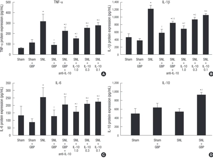
As shown in Fig. 2A-C, protein levels of the pro-inflammatory cytokines TNF-α, IL-1β, and IL-6 on day 7 were significantly in- creased after SNL, and this effect was significantly attenuated by intrathecal administration of gabapentin (TNF-α, 316.0 ± 69.7 pg/mL vs 88.8 ± 24.4 pg/mL, P < 0.05; IL-1β, 1,212.9 ± 104.5 vs
577.4 ± 97.1 pg/mL, P < 0.05; IL-6, 254.0 ± 64.8 pg/mL vs 125.5 ± 44.1 pg/mL, P < 0.05). In the sham group, no significant changes were seen on day 7 in the concentrations of TNF-α, IL-1β, and IL-6 with administration of gabapentin. As shown in Fig. 2D, IL- 10 protein was significantly increased with administration of gabapentin (918.9 ± 63.1 pg/mL) compared to the SNL alone group (532.1 ± 78.7 pg/mL, P < 0.05). In the sham group, admin- istration of gabapentin did not cause significant changes in the concentration of IL-10 protein.
The role of IL-10 in the anti-allodynic effect in SNL rats As shown in Fig. 3, daily intrathecal injections of IL-10 (0.1, 0.3, and 1.0 μg) for 7 days after SNL produced an anti-allodynic ef- fect in a dose-dependent manner throughout the 7 day treat- ment period. A single large dose injection of IL-10 (7 μg) on day 7 did not produce an anti-allodynic effect.
Fig. 1. Time course of paw withdrawal thresholds after spinal nerve ligation and with or without daily intrathecal injections of gabapentin (30 µg) over a 7-day period (6 rats per group). All data points are expressed as the mean ± SEM. *P < 0.05 vs SNL group, †P < 0.05 vs Sham group.
Paw withdrawal threshold (g)
Day
* *
* *
* *
*
*,†
*,†
*,†
*,†
*,†
*,†
*,†
0 1 2 3 4 5 6 7
16 14 12 10 8 6 4 2 0
Sham SNL SNL + Gabapentin
Fig. 2. Intrathecally injected gabapentin reduces the expression of TNF-α (A), IL-1β (B), and IL-6 (C) but increases the expression of IL-10 (D) in the dorsal horn in SNL rats (6 rats per group). The anti-allodynic effect of gabapentin on the expression of the pro-inflammatory cytokines TNF-α, IL-1β, and IL-6 are inhibited by daily injections of the anti- IL-10 antibody (10 µg). Protein expression of the pro-inflammatory cytokines TNF-α, IL-1β, and IL-6 on day 7 are significantly attenuated by IL-10 (1 µg) daily injections com- pared with the SNL alone group. All data points are expressed as the mean ± SEM. *P < 0.05 vs sham group; †P < 0.05 vs SNL group; ‡P < 0.05 vs SNL + gabapentin group.
SNL, spinal nerve ligation; Ab, antibody.
TNF-α protein expression (pg/mL) IL-1β protein expression (pg/mL)
TNF-α IL-1β
Sham Sham SNL SNL SNL SNL SNL SNL
+ + + + + +
GBP GBP GBP IL-10 IL-10 IL-10
+ 1.0 0.3 0.1
anti-IL-10
Sham Sham SNL SNL SNL SNL SNL SNL
+ + + + + +
GBP GBP GBP IL-10 IL-10 IL-10
+ 1.0 0.3 0.1
anti-IL-10 500
400
300
200
100
0
1,400 1,200 1,000 800 600 400 200 0
*
*
*,‡
*,‡
*,‡
*,‡
*,†
*,†
*,‡
*,†,‡
†
†
A B
IL-6 protein expression (pg/mL) IL-10 protein expression (pg/mL)
IL-6 IL-10
Sham Sham SNL SNL SNL SNL SNL SNL
+ + + + + +
GBP GBP GBP IL-10 IL-10 IL-10
+ 1.0 0.3 0.1
anti-IL-10
Sham Sham SNL SNL
+ +
GBP GBP
350 300 250 200 150 100 50 0
1,200 1,000 800 600 400 200 0
*
*,‡
*,‡
*,†
*,†
*,‡
†
C D
The role of anti-IL-10 antibodies and gabapentin in anti-allodynia in SNL rats
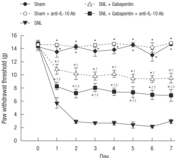
Daily intrathecal injections of anti-IL-10 antibody (10 μg) had an inhibitory effect on the anti-allodynic effects of gabapentin while intrathecal injections of anti-IL-10 antibody (10 μg) alone had neither allodynic nor anti-allodynic effects in the sham group of rats (Fig. 4). These data demonstrate that gabapentin beneficially modulates IL-10 expression, which not only coun- teracts the pro-inflammatory response but also facilitates anti- allodynia.
Effects of IL-10 and anti-IL-10 antibodies on pro- inflammatory cytokine expression
As shown in Fig. 2A-C, the protein expression of the pro-inflam- matory cytokines TNF-α, IL-1β, and IL-6 on day 7 were signifi- cantly reduced with daily IL-10 (1.0 μg) injections compared with the SNL alone group (TNF-α, 316.0 ± 69.7 pg/mL vs 151.6 ± 23.1 pg/mL, P < 0.05; IL-1β, 1,212.9 ± 104.5 vs 685.2 ± 85.2 pg/mL, P < 0.05; IL-6, 254.0 ± 64.8 pg/mL vs 155.3 ± 38.7 pg/mL, P <
0.05). Daily administration of low dose IL-10 (0.1 and 0.3 μg) pro- duced no significant differences compared with the SNL alone group. In contrast, daily anti-IL-10 antibody injections blocked the intrathecal gabapentin-induced decrease in the levels of the pro-inflammatory cytokines TNF-α, IL-1β, and IL-6 (TNF-α, 88.8 ± 24.4 pg/mL vs 225.2 ± 36.2 pg/mL, P < 0.05; IL-1β, 577.4 ± 97.1 vs 841.7 ± 51.5 pg/mL, P < 0.05; IL-6, 125.5 ± 44.1 pg/mL vs 205.4 ± 51.9 pg/mL, P < 0.05).
DISCUSSION
In the present study, intrathecally administered gabapentin showed an anti-allodynic effect in an SNL rat model of neuro- pathic pain. This effect could be explained, in part, by a gabap- entin-induced reduction in the expression of the SNL-induced spinal pro-inflammatory cytokines TNF-α, IL-1β, and IL-6. We observed increased expression of IL-10 protein with the intra- thecal administration of gabapentin in a rat model of SNL. In- trathecal IL-10 administration not only had an anti-allodynic effect but also inhibited pro-inflammatory cytokine production in the SNL rat spinal cord, both of which were blocked by daily intrathecal injections of anti-IL-10 antibody. It has previously been reported that both mRNA and protein expression of the pro-inflammatory cytokines TNF-α, IL-1β, and IL-6 are increased during neuropathic pain (7, 22, 23). Pro-inflammatory cytokines are released as a consequence of spinal glial cell activation in neuropathic pain (24). Taken together, we suggest from our cur- rent data and previous findings that intrathecally administered gabapentin promotes an advantageous control of neuroinflam- mation.
IL-10 was first reported to be a cytokine synthesis inhibitory factor (25). Later it was also described to attenuate nociception in many animal models through the inhibition of pro-inflam- matory cytokines and spinal glial cell activation (26). Intrathe- cal IL-10 gene therapy has been shown to have a therapeutic effect in a rat model of neuropathic pain (27). Our present re- sults support the idea that an increase in IL-10 expression in the spinal cord may be a mechanism by which intrathecally admin-
Fig. 3. Time course of paw withdrawal thresholds over the 7 day period of daily injec- tions of various doses (0.1, 0.3, or 1 µg) of IL-10 in SNL rats (6 rats per group). A sin- gle large dose injection of IL-10 (7 µg) on day 7 does not produce an anti-allodynic effect. *P < 0.05 vs SNL group, †P < 0.05 vs sham group. SNL, spinal nerve ligation.
Paw withdrawal threshold (g)
Day
* *
* *
* *
*
*,†
*,†
*,†
*,†
*,†
*,†
*,†
*,†
*,†
*,†
*,†
*,†
*,†
*,†
*,†
*,†
*,†
*,†
*,†
*,†
*,†
0 1 2 3 4 5 6 7
16 14 12 10 8 6 4 2 0
SNL + IL-10 1.0 µg
SNL + IL-10 7.0 µg single dose on day 7 SNL + IL-10 0.3 µg
Sham
SNL + IL-10 0.1 µg SNL
Fig. 4. Time course of paw withdrawal thresholds over the 7 day period of daily injec- tions of anti-IL-10 antibody (10 µg) with intrathecally administered gabapentin (30 µg).
*P < 0.05 vs SNL group, †P < 0.05 vs sham group. ‡P < 0.05 vs SNL + gabapentin group. SNL, spinal nerve ligation; Ab, antibody.
Paw withdrawal threshold (g)
Day
*
*
*
*
*
*
*
*
*
*
*
*
*
*
*,†
*,†
*,†,‡
*,†
*,†
*,†,‡
*,†,‡
*,†
*,†,‡
*,†,‡
*,†
*,†
*,†,‡
*,†,‡
0 1 2 3 4 5 6 7
16 14 12 10 8 6 4 2 0
SNL + Gabapentin + anti-IL-10 Ab SNL + Gabapentin
Sham SNL
Sham + anti-IL-10 Ab
istered gabapentin inhibits pro-inflammatory cytokine produc- tion and attenuates the mechanical allodynia induced by SNL.
In our present analyses, daily IL-10 intrathecal injections (0.1, 0.3 and 1.0 μg) attenuated mechanical allodynia in a dose-de- pendent manner. However, a single large dose injection of IL-10 (7 μg, equivalent to seven daily doses of 1 μg) on day 7 failed to produce anti-allodynic effect, suggesting that both the timing and dose of IL-10 are important factors in inhibiting inflamma- tory responses. The anti-allodynic effect produced by daily injec- tions of 1 μg IL-10 instead of a single injection of 7 μg IL-10 on day 7 in SNL rats strongly imply that it is the inhibiting effect of IL-10 on microglia activation and cytokine production that is responsible for the early phase of neuroinflammation induced by SNL. Accordingly, a neutralizing anti-IL-10 antibody was found to counteract the anti-allodynic effect of gabapentin in SNL rats.
Intracerebroventricular IL-10 administration has been shown to inhibit the lipopolysaccharide-induced production of TNF-α in the brain (28). It has been demonstrated that the degree of neuroinflammation induced by neuropathy in conjunction with chronic morphine administration in nerve-injured rats is higher than that induced by neuropathy alone (5). This suggests that neuroinflammation is the common mechanism in both neu- ropathy-induced and chronic morphine-induced glial activa- tion. In our present study, daily anti-IL-10 antibody injections produced significant pro-inflammatory cytokine expression and attenuated the anti-allodynic effect of gabapentin in the SNL rats on day 7. Conversely, on day 7, daily IL-10 (1 μg) injec- tions produced a significant reduction in pro-inflammatory cy- tokine expression compared with SNL alone rats. Taken together, the increased pro-inflammatory cytokine expression observed in SNL rats contributes to the development of allodynia induced by SNL (29) and inhibition of pro-inflammatory cytokine ex- pression can potentiate the anti-allodynic effects of gabapentin.
Based on current data, intrathecally administered gabapentin enhances IL-10 expression and counteracts SNL-induced neu- roinflammation, and may be responsible for preventing neuro- pathic pain and inhibiting SNL-induced pro-inflammatory cy- tokine expression in the rat spinal cord. Further studies will be needed to elucidate the underlying mechanism of action of in- trathecal gabapentin and the precise mechanism by which ga- bapentin induces the upregulation of IL-10 expression.
In conclusion, our present results demonstrate that intrathe- cally administered gabapentin attenuates mechanical allodynia in SNL rats. The effects of gabapentin may be due, at least in part, to the upregulation of anti-inflammatory cytokine IL-10 expres- sion in the spinal cord, which leads to inhibition of the expres- sion of pro-inflammatory cytokines TNF-α, IL-1β, and IL-6 in SNL rat spinal cords. Consequently, our present report provides an explanation for the manner by which intrathecally adminis- tered gabapentin beneficially modulates IL-10 expression to
counter a pro-inflammatory response triggered by SNL.
ACKNOWLEDGMENTS
The authors declare no conflict of interest.
REFERENCES
1. Woolf CJ, Mannion RJ. Neuropathic pain: aetiology, symptoms, mecha- nisms and management. Lancet 1999; 353: 1959-64.
2. Kim SH, Chung JM. An experimental model for peripheral neuropathy produced by segmental spinal nerve ligation in the rat. Pain 1992; 50:
355-63.
3. Sheen K, Chung JM. Signs of neuropathic pain depend on signals from injured nerve fibers in a rat model. Brain Res 1993; 610: 62-8.
4. Kim KJ, Yoon YW, Chung JM. Comparison of three rodent neuropathic pain models. Exp Brain Res 1997; 113: 200-6.
5. Raghavendra V, Rutkowski MD, DeLeo JA. The role of spinal neuroim- mune activation in morphine tolerance/hyperalgesia in neuropathic and sham-operated rats. J Neurosci 2002; 22: 9980-9.
6. Tsai RY, Jang FL, Tai YH, Lin SL, Shen CH, Wong CS. Ultra-low-dose naloxone restores the antinociceptive effect of morphine and suppresses spinal neuroinflammation in PTX-treated rats. Neuropsychopharma- cology 2008; 33: 2772-82.
7. del Rey A, Apkarian AV, Martina M, Besedovsky HO. Chronic neuropath- ic pain-like behavior and brain-borne IL-1β. Ann N Y Acad Sci 2012;
1262: 101-7.
8. Kawasaki Y, Xu ZZ, Wang X, Park JY, Zhuang ZY, Tan PH, Gao YJ, Roy K, Corfas G, Lo EH, et al. Distinct roles of matrix metalloproteases in the early- and late-phase development of neuropathic pain. Nat Med 2008;
14: 331-6.
9. Zhang JM, An J. Cytokines, inflammation, and pain. Int Anesthesiol Clin 2007; 45: 27-37.
10. Vale ML, Marques JB, Moreira CA, Rocha FA, Ferreira SH, Poole S, Cunha FQ, Ribeiro RA. Antinociceptive effects of interleukin-4, -10, and -13 on the writhing response in mice and zymosan-induced knee joint incapacitation in rats. J Pharmacol Exp Ther 2003; 304: 102-8.
11. Luo ZD, Calcutt NA, Higuera ES, Valder CR, Song YH, Svensson CI, My- ers RR. Injury type-specific calcium channel α2δ-1 subunit up-regulation in rat neuropathic pain models correlates with antiallodynic effects of gabapentin. J Pharmacol Exp Ther 2002; 303: 1199-205.
12. Maneuf YP, Luo ZD, Lee K. α2δ and the mechanism of action of gabap- entin in the treatment of pain. Semin Cell Dev Biol 2006; 17: 565-70.
13. Backonja M, Beydoun A, Edwards KR, Schwartz SL, Fonseca V, Hes M, LaMoreaux L, Garofalo E. Gabapentin for the symptomatic treatment of painful neuropathy in patients with diabetes mellitus: a randomized controlled trial. JAMA 1998; 280: 1831-6.
14. Rowbotham M, Harden N, Stacey B, Bernstein P, Magnus-Miller L. Ga- bapentin for the treatment of postherpetic neuralgia: a randomized con- trolled trial. JAMA 1998; 280: 1837-42.
15. Bennett MI, Simpson KH. Gabapentin in the treatment of neuropathic pain. Palliat Med 2004; 18: 5-11.
16. Chen SR, Pan HL. Effect of systemic and intrathecal gabapentin on allo- dynia in a new rat model of postherpetic neuralgia. Brain Res 2005;
1042: 108-13.
17. Field MJ, Oles RJ, Lewis AS, McCleary S, Hughes J, Singh L. Gabapentin (neurontin) and S-(+)-3-isobutylgaba represent a novel class of selective antihyperalgesic agents. Br J Pharmacol 1997; 121: 1513-22.
18. Yaksh TL, Rudy TA. Chronic catheterization of the spinal subarachnoid space. Physiol Behav 1976; 17: 1031-6.
19. Park JY, Jun IG. The interaction of gabapentin and N6-(2-phenylisopropyl)- adenosine R-(-)isomer (R-PIA) on mechanical allodynia in rats with a spinal nerve ligation. J Korean Med Sci 2008; 23: 678-84.
20. Lin SL, Tsai RY, Tai YH, Cherng CH, Wu CT, Yeh CC, Wong CS. Ultra- low dose naloxone upregulates interleukin-10 expression and suppresses neuroinflammation in morphine-tolerant rat spinal cords. Behav Brain Res 2010; 207: 30-6.
21. Chaplan SR, Bach FW, Pogrel JW, Chung JM, Yaksh TL. Quantitative assessment of tactile allodynia in the rat paw. J Neurosci Methods 1994;
53: 55-63.
22. Ma W, St-Jacques B, Duarte PC. Targeting pain mediators induced by injured nerve-derived COX2 and PGE2 to treat neuropathic pain. Expert Opin Ther Targets 2012; 16: 527-40.
23. Chen YW, Li YT, Chen YC, Li ZY, Hung CH. Exercise training attenuates neuropathic pain and cytokine expression after chronic constriction in-
jury of rat sciatic nerve. Anesth Analg 2012; 114: 1330-7.
24. Moalem G, Tracey DJ. Immune and inflammatory mechanisms in neu- ropathic pain. Brain Res Rev 2006; 51: 240-64.
25. Fiorentino DF, Bond MW, Mosmann TR. Two types of mouse T helper cell. IV. Th2 clones secrete a factor that inhibits cytokine production by Th1 clones. J Exp Med 1989; 170: 2081-95.
26. Watkins LR, Hutchinson MR, Ledeboer A, Wieseler-Frank J, Milligan ED, Maier SF. Norman Cousins Lecture, Glia as the “bad guys”: implica- tions for improving clinical pain control and the clinical utility of opioids.
Brain Behav Immun 2007; 21: 131-46.
27. Milligan ED, Sloane EM, Langer SJ, Hughes TS, Jekich BM, Frank MG, Mahoney JH, Levkoff LH, Maier SF, Cruz PE, et al. Repeated intrathecal injections of plasmid DNA encoding interleukin-10 produce prolonged reversal of neuropathic pain. Pain 2006; 126: 294-308.
28. Di Santo E, Sironi M, Pozzi P, Gnocchi P, Isetta AM, Delvaux A, Gold- man M, Marchant A, Ghezzi P. Interleukin-10 inhibits lipopolysaccha- ride-induced tumor necrosis factor and interleukin-1 beta production in the brain without affecting the activation of the hypothalamus-pituitary- adrenal axis. Neuroimmunomodulation 1995; 2: 149-54.
29. Inoue K. The function of microglia through purinergic receptors: neuro- pathic pain and cytokine release. Pharmacol Ther 2006; 109: 210-26.