In vitro study of the fracture resistance of monolithic lithium disilicate, monolithic zirconia, and lithium disilicate pressed on zirconia for three-unit fixed dental prostheses
Jae-Won Choi1,2, So-Yeun Kim3, Ji-Hyeon Bae1,2, Eun-Bin Bae1,2, Jung-Bo Huh1*
1Department of Prosthodontics, Dental Research Institute, Institute of Translation Dental Science, School of Dentistry, Pusan National University, Yangsan, Republic of Korea
2BK21 PLUS Project, School of Dentistry, Pusan National University, Yangsan, Republic of Korea
3Department of Prosthodontics, Pusan National University Hospital, Pusan, Republic of Korea
PURPOSE. The purpose of this study was to determine fracture resistance and failure modes of three-unit fixed dental prostheses (FDPs) made of lithium disilicate pressed on zirconia (LZ), monolithic lithium disilicate (ML), and monolithic zirconia (MZ). MATERIALS AND METHODS. Co-Cr alloy three-unit metal FDPs model with maxillary first premolar and first molar abutments was fabricated. Three different FDPs groups, LZ, ML, and MZ, were prepared (n = 5 per group). The three-unit FDPs designs were identical for all specimens and cemented with resin cement on the prepared metal model. The region of pontic in FDPs was given 50,000 times of cyclic preloading at 2 Hz via dental chewing simulator and received a static load until fracture with universal testing machine fixed at 10°. The fracture resistance and mode of failure were recorded. Statistical analyses were performed using the Kruskal-Wallis test and Mann-Whitney U test with Bonferroni’s correction (α=0.05/3=0.017).
RESULTS. A significant difference in fracture resistance was found between LZ (4943.87 ± 1243.70 N) and ML (2872.61 ± 658.78 N) groups, as well as between ML and MZ (4948.02 ± 974.51 N) groups (P<.05), but no significant difference was found between LZ and MZ groups (P>.05). With regard to fracture pattern, there were three cases of veneer chipping and two interfacial fractures in LZ group, and complete fracture was observed in all the specimens of ML and MZ groups. CONCLUSION. Compared to monolithic lithium disilicate FDPs, monolithic zirconia FDPs and lithium disilicate glass ceramics pressed on zirconia-based FDPs showed superior fracture resistance while they manifested comparable fracture resistances. [J Adv Prosthodont 2017;9:244-51]
KEYWORDS: Fixed dental prostheses; Lithium disilicate; Zirconia; Lithium disilicate pressed on zirconia;
Fracture resistance
INTRODUCTION
Porcelain-fused-to-metal (PFM) restorations have been widely
used for fixed dental prostheses (FDPs).1 However, PFM restorations have problems, such as metal exposure at the cervical margin, discoloration of porcelain, potential disad- vantage of the alloy, and possible bonding problem between the framework and veneering material.2 Also, they may cause allergy and staining along with the release of metal ions at the gingival tissue.3 These problems of PFM were resolved through the development of all-ceramic restora- tions without a metal. The usage of all-ceramic crowns in restorative dentistry has been increased due to its natural color, as well as its translucency as in feldspathic porcelain, high biocompatibility, relatively affordable price compared to precious metals, and increasing demand for metal-free FDPs.4,5
Corresponding author:
Jung-Bo Huh
Department of Prosthodontics, Institute for Dental Research, Institute of Translation Dental Science, School of Dentistry, Pusan National University, 20 Geumo-ro, Mulgeum-eup, Yangsan 50612, Republic of Korea Tel. +82553605146: e-mail, [email protected]
Received October 4, 2016 / Last Revision February 17, 2017 / Accepted March 15, 2017
© 2017 The Korean Academy of Prosthodontics
This is an Open Access article distributed under the terms of the Creative Commons Attribution Non-Commercial License (http://creativecommons.
org/licenses/by-nc/3.0) which permits unrestricted non-commercial use, distribution, and reproduction in any medium, provided the original work is properly cited.
As high-strength ceramic materials and systems have been developed due to the introduction of the computer- aided design/computer-aided manufacturing (CAD/CAM) technologies, the scope of the anterior and posterior FDPs usage has been expanded.6 Among the currently available all-ceramic materials, yttria-stabilized tetragonal zirconia polycrystalline (Y-TZP) shows the greatest fracture resis- tance, and it is considered as the gold standard in dental crown restorations.7 However, Y-TZP has limitations, including difficult control of its opacity and translucency, chipping of the veneered porcelain, and limited optical defects.8 As so, a lot of efforts, including coloring, have been made to overcome these problems, but the problems still remain. After all, porcelain-fused-to-zirconia (PFZ) has been chosen as suitable zirconia prosthesis.9
The method of veneering the zirconia core using the hand-layering technique with feldspathic porcelain has mainly been used, but as this method is technique sensitive and as veneer chipping often occurs,10 reducing the fracture rate of porcelain veneers has become a major concern in zirconia restorations.11 Due to the better mechanical proper- ties of lithium disilicate ceramic, a transition from feld- spathic porcelain to lithium disilicate ceramic for veneer fabrication has been suggested in order to improve the frac- ture resistance.12 CAD-on technique was used to mill frame and veneer from ceramic blanks. Either by using resin cement or by fusing ceramic, lithium disilicate veneer can be attached to zirconia framework after sintering the frame- work.13 Recently, heat-pressed method that presses lithium disilicate glass ceramic veneer has been introduced.14 As the lost wax press system can produce ceramic prostheses with or without CAD/CAM system, it is economical and simple, and it can effectively reconstruct the customized anatomical shape.15
A study utilized lithium disilicate glass ceramic as a heat- pressed veneered ceramic in fabrication of zirconia crown, and it showed a higher fracture resistance than fluroaptite- pressed zirconia crown and monolithic lithium disilicate
crown.14 However, there are almost no studies on lithium disilicate pressed zirconia FDPs. Hence, the aim of the present study was to investigate relative fracture resistance and failure mode of three-unit FDPs made of lithium disili- cate pressed on zirconia, monolithic lithium disilicate, and monolithic zirconia.
MATERIALS AND METHODS
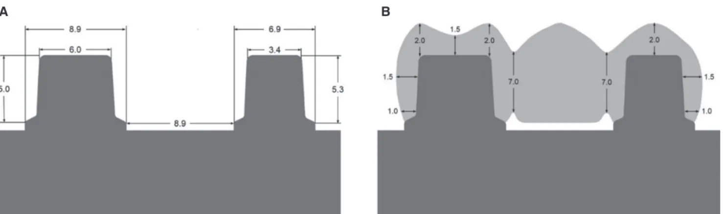
A master model with two abutments in the maxillary first premolar and first molar was fabricated. To fabricate the master model, each abutment was first designed using CAD software (Exocad, Exocad GmbH, Darmstadt, Germany).
Each abutment was fabricated with chamfer preparation of 120° and convergence angle of 15° (Fig. 1A).16 The STL file of the designed master model was transmitted to the five- axis milling machine (Ceramill Motion 2, AmannGirrbach, Koblach, Austria), and dry milling was performed with Co-Cr blanks (Ceramill Sintron, Amann Girrbach, Koblach, Austria). After that, a total of 15 master models were fabri- cated through sintering at a high-temperature sintering fur- nace (Ceramill Argotherm, AmannGirrbach, Koblach, Austria) (Fig. 2).
The experiment was performed by dividing the speci- men into three groups: the lithium disilicate glass ceramics pressed on zirconia-based FDPs (LZ group), the monolithic lithium disilicate FDPs (ML group), and the monolithic zir- conia FDPs (MZ group). All the ceramic materials are pre- sented in Table 1. Three-unit FDPs from the first premolar to the first molar were fabricated, with the same shape as the one used in clinical practice (Fig. 1B). A total of 15 three-unit FDPs, five for each group, were fabricated (Fig.
3). The master models were scanned using the AutoScan 3D Dental Scanner (Hangzhou Shining 3D Tech Co., Ltd., Hangzhou, China), and same-shaped crowns were designed using the Exocad software (Exocad GmbH, Darmstadt, Germany). The connectors included mesiodistal cross-sec- tional areas of 31.3 mm², a buccolingual width of 5.7 mm,
Fig. 1. Schematic drawing and dimensions. (A) Master model, (B) Fixed dental prostheses.
A B
and an occlusogingival height of 7 mm.17
To fabricate lithium disilicate glass ceramics pressed on zirconia-based FDPs, a 0.5 mm-thick zirconia coping (Zirtooth Fulluster, HASS, Gangneung, Korea) was pre- pared first. After making the coping, liner powder (Rosetta Ceram Liner, HASS, Gangneung, Korea) was applied to the surface of the zirconia to improve the bond strength and wettability between the zirconia and the veneer glass ceram- ic, and heat treatment was done according to the instruction of the manufacturer. Using the Exocad software, a wax veneer structure was fabricated by milling a wax block
(TOTEM, Qingdao Totem Candle Industry, Shandong, China). The veneer was fixed to the coping by applying heat to its margin, and after investing using a dedicated invest- ment ring, it was burned out at 880°C for 30 minutes (Burnout Furnace L 1/12, Nabertherm, Bremen, Germany).
After that, glass ingot (Rosetta UltraPress, HASS, Gangneung, Korea) was put in the investment ring, and the latter was pressed with the pressing furnace (Horizon Press, Shenpaz Dental Ltd., Migdal HaEmek, Israel) to join the zirconia and the lithium disilicate glass ceramic. According to the instruction of the manufacturer, sandblasting (50 μm glass Table 1. Ceramic materials used in this study, and properties of each group
Group Specifications Materials Manufacturer Translucency /
Shade
CTE (×10-6/°C)
Biaxial flexure strength (MPa)
LZ Heat-pressed lithium disilicate Rosetta UltraPress HASS LT /A2 9.7 450
Liner Rosetta Ceram Liner, prototype HASS - - -
CAD/CAM zirconia coping Zirtooth Fulluster HASS LT /A2 10.8 1250
ML Heat-pressed lithium disilicate FDPs Rosetta SuperPress HASS LT /A2 10.8 460
MZ CAD/CAM zirconia FDPs Zirtooth Fulluster HASS LT /A2 10.8 1250
LZ: lithium disilicate glass ceramic pressed on zirconia, ML: monolithic lithium disilicate, MZ: monolithic zirconia, LT: low translucency.
Fig. 3. Fabrication of three-unit FDPs. (A) LZ group: lithium disilicate glass ceramics pressed on zirconia-based FDPs, (B) ML group: monolithic lithium disilicate FDPs, (C) MZ group: monolithic zirconia FDPs.
A B C
Fig. 2. Preparation of the master model. (A) STL file of the master model, (B) Front view of the fabricated alloy master model, (C) Occlusal view of the fabricated alloy master model.
A B C
beads at 1 bar pressure) and glazing (IPS e.max Ceram glaze paste, Ivoclar Vivadent, Schaan, Leichtenstein) were per- formed. The monolithic lithium disilicate FDPs were fabri- cated by obtaining same-shaped wax patterns by milling the wax block, pressing the lithium disilicate ingots (Rosetta SuperPress, HASS, Gangneung, Korea) using the heat- pressing technique, and glazing (IPS e.max Ceram glaze paste, Ivoclar Vivadent, Schaan, Leichtenstein). Then, the monolithic zirconia FDPs were fabricated through milling with a milling machine (Roland DWX-50, Roland DGA, Irvine, CA, USA), final-sintering, and glazing (IPS e.max Ceram glaze paste, Ivoclar Vivadent, Schaan, Leichtenstein).
For the purpose of standardization, the same dental techni- cian executed the manufacturing process.
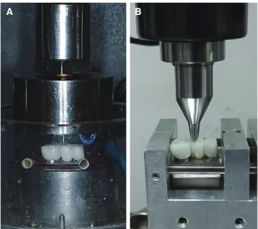
Cementation of the FDPs was done in the master model using G-CEM LinkAce resin cement (GC America, Alsip, IL, USA), and the samples were kept in distilled water at 37°C for more than 48 hours. To preload the FDPs, 50,000 times of mechanical loading were applied on the occlusal surface of the pontic under 50 N at 2 Hz, using dental chewing simulator (R&D Inc., Daejeon, Korea) (Fig. 4A).
The load was applied on the distal part of occlusal surface of the pontic by using a stainless steel sphere with a 4 mm diameter. Lastly, the FDPs were fixed onto the testing jig at 10° angle against the long axis, and load to fracture was applied using a universal testing machine (Instron 8871, Instron Co., Norwood, MA, USA) (Fig. 4B). The crosshead speed was set to 0.255 mm/min, and loading was executed to the previously preloaded site, using a stainless steel sphere with a 4 mm diameter, until the FDPs were frac- tured.16 The fracture resistance was recorded when the FDPs
were fractured; in this present study, fracture was defined as an abrupt force decrease, and the maximum force prior to the sudden decline was recorded as fracture resistance. The fracture pattern was classified into complete fracture, veneer chipping, and interfacial fracture, and the numbers of frac- ture were counted for each group. The specimens of LZ group were coated with platinum, and their microstructure of fractured surface was observed, using FE-SEM (JSM- 7401F, Jeol, Tokyo, Japan) (×180, ×250, and ×700).
To evaluate the statistical significance of the fracture resistance of the FDPs by material, SPSS ver. 23.0 (SPSS Inc., Chicago, IL, USA) was used. For the comparison of the three groups, the Kruskal-Wallis and Mann-Whitney U tests with Bonferroni’s correction (α = .05/3 = .017) were employed. All statistical analyses were done at the 5% sig- nificance level.
RESULTS
Preloading process did not induce fracture on all FDPs. The statistical analysis showed a significant difference between the LZ group (4943.87 ± 1243.70 N) and the ML group (2872.61 ± 658.78 N) as well as between the ML and MZ groups (4948.02 ± 974.51 N) (P < .05), but it did not exhib- it a significant difference between the LZ and MZ groups (P
> .05) (Fig. 5). In terms of fracture pattern, three cases of veneer chipping and two interfacial fractures in the LZ group and complete fracture in all the specimens of ML and MZ groups were observed (Fig. 6, Table 2). The frac- ture lines of the specimens in the LZ group were formed vertically along the load direction, and the fracture pattern
Fig. 5. Box plots of fracture resistance for each
experimental group. The same lowercase letter suggests no significant difference found among the groups (P >
.05). LZ: lithium disilicate glass ceramic pressed on zirconia, ML: monolithic lithium disilicate, MZ:
monolithic zirconia.
Fig. 4. Preparation of the test set-up. (A) Repetitive preloading in a chewing simulator, (B) Fracture load test in a universal testing machine.
A B
was clearly seen at the pontic where a direct force was given.
In the ML and MZ groups, most of the fracture patterns were also formed in the mesiodistal direction, centering on the pontic where a direct force was given. The specimen that experienced veneer chipping in the LZ group was observed with FE-SEM, displaying no pore, defect, or void between zirconia core and lithium disilicate glass ceramic veneer (Fig. 7).
Table 2. Fracture modes of the three experimental groups (n = 5)
Group Complete fracture Veneer chipping Interfacial fracuture
LZ - 3 2
ML 5 - -
MZ 5 - -
LZ: lithium disilicate glass ceramic pressed on zirconia, ML: monolithic lithium disilicate, MZ: monolithic zirconia.
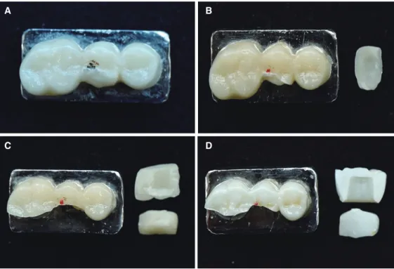
Fig. 6. Typical failure types of the experimental groups after the fracture load test. (A) Veneer chipping of the LZ group, (B) Interfacial fracture of the LZ group, (C) Complete fracture of the ML group, (D) Complete fracture of the MZ group.
LZ: lithium disilicate glass ceramic pressed on zirconia, ML: monolithic lithium disilicate, MZ: monolithic zirconia.
A B
C D
Fig. 7. SEM image of the specimen with veneer chipping in LZ group. (A) No pore, defect, or void observed between zirconia core and lithium dislicate glass ceramic veneer, (B) Magnified image of area inside rectangle in Fig. 7A, (C) Magnified image of area inside rectangle in Fig. 7B (original magnification: ×180, ×250, ×700, respectively). Zr:
zirconia coping, Li: lithium disilicate glass ceramic veneer.
A B C
DISCUSSION
Preclinical in vitro tests for dental materials are important in evaluating mechanical performance and compatibility of materials in mouth.18 The conventional laboratory tests apply static loading until failure using a universal testing machine. These tests provide information on material strength, potential risk of failure, and deformation of the material. However, they cannot sufficiently predict the long- term performance of dental restorations.19 The intraoral environments that need to be considered for dental restora- tions include humidity, pH, and cyclic loading. Therefore, studies must reproduce experimental conditions similar to the clinical situations in order to create the failure pattern in actual clinical practice.18,19
In the present study, the FDPs were designed to mimic the clinical situation anatomically as much as possible.
Herein, the loading stainless steel ball was able to fit in a cavity formed in the middle of the pontic, and a three-point contact between the occlusal surface and steel ball was achieved successfully.17 Clinically, the mechanical failure of dental prostheses occurs a long time after their application, indicating that fatigue failure accounts for a larger propor- tion of the failure cases than acute overload.20 Damage is accumulated by repetitive contact between maxillary and mandibular teeth, and the lifetime and survival rate of the prosthesis are reduced.21 Therefore, this study performed a fatigue testing in order to replicate the clinical settings as much as possible.18 The preload force was set at 50 N, based on the previous studies.6,22 50,000 cycles were applied during the preload to replicate the average number of mastication in 4 weeks.23 Moreover, the preloading was conducted in dry environment. Thermal cycling with water removes water from the surface of specimens and leads to further aging of material. In addition, wet environment may cause subcritical crack growth, change in ceramic structure, and superficial phase transitions.24 Therefore, less fracture resistance might be speculated if the experimental condition was set in wet environment. Furthermore, as most of the previous studies reported a chewing rate in humans under 2 Hz, the test fre- quency of this study was set at 2 Hz.25,26 Nevertheless, because it is impossible for in vitro studies to replicate clini- cal scenario completely, the result of the experiment may be different from that in the actual clinical setting. For instance, the present experimental model did not employ periodontal ligaments while the periodontal ligaments around roots are responsible for stress distribution in natural dentition.27 As shown in the previous study, the models without periodon- tal ligament demonstrated a greater fracture resistance than the models with periodontal ligament.22 In addition, natural tooth would have replicated the clinical settings more pre- cisely if chosen as an abutment of this study. Yet, the pres- ent study chose metal alloy abutment since natural teeth have different size, shape, and quality, and the preparation cannot be standardized.28-30 In addition, natural teeth with lower elastic modulus can be fractured near cervical area.31,32 Therefore, the present study utilized metal alloy abutment
with higher elastic modulus and fracture resistance for test- ing fracture resistance of the experimental groups. Resin cement with strong rigidity was also used to lower a chance of failure by luting agent.33 Moreover, the influence of elas- tic modulus of abutment on the fracture resistance of den- tal prosthesis was demonstrated in many in vitro studies,29,31,34 and some clinical studies reported a higher failure rate of crown in response to lower elastic modulus of abutment material.35 Therefore, based on the earlier studies, the exper- imental outcomes of the present study are thought to mani- fest higher fracture resistance than in the clinical situation.
However, the present study rather investigated relative com- parison among the fracture resistances of the experimental groups, regardless of the experimental condition. Therefore, these factors may not affect the relative comparison among the groups.
The ML group in this experiment was lithium disilicate glass ceramic, a kind of particle-filled glass that has been reported to have a higher fracture resistance than leucite- reinforced glass ceramic or feldspathic ceramic.36 The rea- son for this seems to be that lithium disilicate crystal is more efficient in promoting crack deflection and crack branching compared to glass ceramic including the leucite or fluorapatite crystal phase.37,38 The MZ group consists of the typical yttria-stabilized tetragonal zirconia polycrystalline (Y-TZP) and provides high toughness and fracture load through the mechanism called “transformation toughen- ing.”39 In other words, when the phase changes from the tetragonal phase to the monoclinic phase, 3 - 5% volume expansion occurs, which gives rise to internal stress.40 Stabilizing agents such as calcium, magnesia, yttrium, and ceria are also added in order to stabilize the tetragonal phase at room temperature and to control the volume expansion.41 In this study, the fracture resistance of the MZ group (4948.02 ± 974.51 N) was significantly larger than that of the ML group (2872.61 ± 658.78 N), which is consistent with the results of other previous studies.1,14,42 This is because polycrystalline materials are less vulnerable to fatigue degradation than glass ceramic.43 In a previous study, it was confirmed that the crack propagation of lithium disili- cate glass ceramic appeared only within the residual glass matrix and did not spread through the crystal.38 Additionally, such result can be partially explained by the flexural strengths of lithium disilicate glass ceramic and zirconia (440 and 1250 MPa, respectively). In this study, the fracture resistance of the monolithic zirconia FDPs (4948.02 ± 974.51 N) and the lithium disilicate glass ceramics pressed on zirconia-based FDPs (4943.87 ± 1243.70 N) did not show a statistically significant difference, which is similar to the result of another study that compared crowns.14 Also, a comparable level of fracture resistance in both lithium desil- icated glass ceramics pressed on zirconia-based FDPs and monolithic zirconia FDPs accounts that a higher fracture resistance of FDPs with ceramic core can be obtained from the core material.30
Fracture can be roughly divided into delamination (core exposure and adhesive facture), cohesive fracture within the
veneer porcelain (chipping), cracks extending to the frame- work (radial cracks), and complete fracture (catastrophic fracture, bulk fracture, and total fracture).11 In this study, the ML and MZ groups, which were monolithic FDPs, showed complete fracture while the LZ group showed veneer chip- ping and interfacial fracture. From the clinial perspective, chipping is mainly observed at the site where a high contact force is applied, and it appears more when air bubbles are present inside the veneer layer. In fact, during the fabrica- tion of the conventional feldspathic ceramic veneer, pores are inevitably generated, but the fabrication through press system prevents the formation of these microstructural defects.44 As evident in Fig. 7, the secure contact between lithium disilicate glass ceramic veneers and zirconia core may be responsible for the high fracture resistance. Yet, considering that, in some specimens, interfacial fracture was observed between the core and the veneer and that the stan- dard deviation of the fracture resistance of the LZ group was greater than those of the two other groups, further studies must be performed on the reasons for the higher fracture resistance.
Meanwhile, many studies on the occlusal force reported that gender, age, and measurement site including the anteri- or and posterior regions resulted in the considerably differ- ent values. Dental restoration must be able to support occlusal force greater than 1000 N because the occlusal force exceeds more than 1000 N in parafuction.30,39 Within the limitation of this study, as all the groups showed a frac- ture resistance of more than 1000 N, the fracture resistance can be clinically accepted.
As the limitation of this study, the oral environment dis- plays thermal stress due to the presence of water while the present experiment was performed in dry environment. In addition, mechanical property of the metal alloy abutment was different from that of natural teeth, the load was exe- cuted in a single direction, and the absence of periodontal ligament did not represent the clinical settings completely.
Moreover, there are limitations because of the small num- ber of specimens, and there is a need for additional experi- mental groups such as CAD-fabricated lithium disilicate veneer luted or fused on zirconia-framework. Therefore, further studies should be executed to investigate the addi- tional groups of FDPs mentioned above and long-term effects of cyclic loading under thermal stress along with clinical studies.
CONCLUSION
Within the limitations of this study, such as the small num- ber of the specimens, lithium disilicate glass ceramics pressed on zirconia-based FDPs showed comparable frac- ture resistance to monolithic zirconia FDPs and more out- standing fracture resistance than monolithic lithium disili- cate FDPs.
ORCID
Jae-Won Choi https://orcid.org/0000-0001-6786-9251 Jung-Bo Huh https://orcid.org/0000-0001-7578-1989 REFERENCES
1. Lüthy H, Filser F, Loeffel O, Schumacher M, Gauckler LJ, Hammerle CH. Strength and reliability of four-unit all-ceram- ic posterior bridges. Dent Mater 2005;21:930-7.
2. Datla SR, Alla RK, Alluri VR, Babu JP, Konakanchi A. Dental ceramics: Part II - Recent advances in dental ceramics. Am J Mater Eng Technol 2015;3:19-26.
3. Venclíkova Z, Benada O, Bártova J, Joska L, Mrklas L.
Metallic pigmentation of human teeth and gingiva: morpho- logical and immunological aspects. Dent Mater J 2007;26:96- 104.
4. Donovan TE. Factors essential for successful all-ceramic res- torations. J Am Dent Assoc 2008;139:14S-8S.
5. Sailer I, Fehér A, Filser F, Gauckler LJ, Lüthy H, Hämmerle CH. Five-year clinical results of zirconia frameworks for pos- terior fixed partial dentures. Int J Prosthodont 2007;20:383-8.
6. Schultheis S, Strub JR, Gerds TA, Guess PC. Monolithic and bi-layer CAD/CAM lithium-disilicate versus metal-ceramic fixed dental prostheses: comparison of fracture loads and failure modes after fatigue. Clin Oral Investig 2013;17:1407- 13.
7. Rekow ED, Silva NR, Coelho PG, Zhang Y, Guess P, Thompson VP. Performance of dental ceramics: challenges for improvements. J Dent Res 2011;90:937-52.
8. Kim HK, Kim SH. Effect of the number of coloring liquid applications on the optical properties of monolithic zirconia.
Dent Mater 2014;30:e229-37.
9. Takeichi T, Katsoulis J, Blatz MB. Clinical outcome of single porcelain-fused-to-zirconium dioxide crowns: a systematic re- view. J Prosthet Dent 2013;110:455-61.
10. Sailer I, Gottnerb J, Kanelb S, Hammerle CH. Randomized controlled clinical trial of zirconia-ceramic and metal-ceramic posterior fixed dental prostheses: a 3-year follow-up. Int J Prosthodont 2009;22:553-60.
11. Silva NR, Bonfante EA, Rafferty BT, Zavanelli RA, Rekow ED, Thompson VP, Coelho PG. Modified Y-TZP core design improves all-ceramic crown reliability. J Dent Res 2011;90:
104-8.
12. Baltzer A. All-ceramic single-tooth restorations: choosing the material to match the preparation-preparing the tooth to match the material. Int J Comput Dent 2008;11:241-56.
13. Schmitter M, Schweiger M, Mueller D, Rues S. Effect on in vitro fracture resistance of the technique used to attach lithi- um disilicate ceramic veneer to zirconia frameworks. Dent Mater 2014;30:122-30.
14. Kim SY, Choi JW, Ju SW, Ahn JS, Yoon MJ, Huh JB. Fracture strength after fatigue loading of lithium disilicate pressed zir- conia crowns. Int J Prosthodont 2016;29:369-71.
15. Guess PC, Zhang Y, Thompson VP. Effect of veneering techniques on damage and reliability of Y-TZP trilayers. Eur J Esthet Dent 2009;4:262-76.
16. Ambre MJ, Aschan F, Vult von Steyern P. Fracture strength of yttria-stabilized zirconium-dioxide (Y-TZP) fixed dental prostheses (FPDs) with different abutment core thicknesses and connector dimensions. J Prosthodont 2013;22:377-82.
17. Wimmer T, Erdelt KJ, Eichberger M, Roos M, Edelhoff D, Stawarczyk B. Influence of abutment model materials on the fracture loads of three-unit fixed dental prostheses. Dent Mater J 2014;33:717-24.
18. Nawafleh N, Hatamleh M, Elshiyab S, Mack F. Lithium disili- cate restorations fatigue testing parameters: A systematic re- view. J Prosthodont 2016;25:116-26.
19. Kelly JR. Clinically relevant approach to failure testing of all- ceramic restorations. J Prosthet Dent 1999;81:652-61.
20. Wiskott HW, Nicholls JI, Belser UC. Stress fatigue: basic prin- ciples and prosthodontic implications. Int J Prosthodont 1995;8:105-16.
21. Zhang L, Wang Z, Chen J, Zhou W, Zhang S. Probabilistic fa- tigue analysis of all-ceramic crowns based on the finite ele- ment method. J Biomech 2010;43:2321-6.
22. Beuer F, Steff B, Naumann M, Sorensen JA. Load-bearing ca- pacity of all-ceramic three-unit fixed partial dentures with dif- ferent computer-aided design (CAD)/computer-aided manu- facturing (CAM) fabricated framework materials. Eur J Oral Sci 2008;116:381-6.
23. McLaren EA, Giordano RA. Zirconia-based ceramics:
Material properties, esthetics, and layering techniques of a new veneering porcelain, VM9. Quintessence Dent Technol 2005;28:99-112.
24. Preis V, Weiser F, Handel G, Rosentritt M. Wear performance of monolithic dental ceramics with different surface treat- ments. Quintessence Int 2013;44:393-405.
25. DeLong R. Intra-oral restorative materials wear: rethinking the current approaches: how to measure wear. Dent Mater 2006;22:702-11.
26. Woda A, Mishellany A, Peyron MA. The regulation of masti- catory function and food bolus formation. J Oral Rehabil 2006;33:840-9.
27. Rees JS. An investigation into the importance of the peri- odontal ligament and alveolar bone as supporting structures in finite element studies. J Oral Rehabil 2001;28:425-32.
28. Cho L, Song H, Koak J, Heo S. Marginal accuracy and frac- ture strength of ceromer/fiber-reinforced composite crowns:
effect of variations in preparation design. J Prosthet Dent 2002;88:388-95.
29. Rosentritt M, Naumann M, Hahnel S, Handel G, Reill M.
Evaluation of tooth analogs and type of restoration on the fracture resistance of post and core restored incisors. J Biomed Mater Res B Appl Biomater 2009;91:272-6.
30. Tinschert J, Natt G, Mautsch W, Augthun M, Spiekermann H.
Fracture resistance of lithium disilicate-, alumina-, and zirco- nia-based three-unit fixed partial dentures: a laboratory study.
Int J Prosthodont 2001;14:231-8.
31. Scherrer SS, de Rijk WG. The fracture resistance of all-ce- ramic crowns on supporting structures with different elastic moduli. Int J Prosthodont 1993;6:462-7.
32. Potiket N, Chiche G, Finger IM. In vitro fracture strength of teeth restored with different all-ceramic crown systems. J
Prosthet Dent 2004;92:491-5.
33. Bindl A, Lüthy H, Mörmann WH. Strength and fracture pat- tern of monolithic CAD/CAM-generated posterior crowns.
Dent Mater 2006;22:29-36.
34. Rosentritt M, Behr M, Gebhard R, Handel G. Influence of stress simulation parameters on the fracture strength of all- ceramic fixed-partial dentures. Dent Mater 2006;22:176-82.
35. Sorensen JA, Choi C, Fanuscu MI, Mito WT. IPS Empress crown system: three-year clinical trial results. J Calif Dent Assoc 1998;26:130-6.
36. Clausen JO, Abou Tara M, Kern M. Dynamic fatigue and fracture resistance of non-retentive all-ceramic full-coverage molar restorations. Influence of ceramic material and prepa- ration design. Dent Mater 2010;26:533-8.
37. Della Bona A, Mecholsky JJ Jr, Anusavice KJ. Fracture behav- ior of lithia disilicate- and leucite-based ceramics. Dent Mater 2004;20:956-62.
38. Apel E, Deubener J, Bernard A, Höland M, Müller R, Kappert H, Rheinberger V, Höland W. Phenomena and mechanisms of crack propagation in glass-ceramics. J Mech Behav Biomed Mater 2008;1:313-25.
39. Rodríguez V, Castillo-Oyagüe R, López-Suárez C, Gonzalo E, Peláez J, Suárez-García MJ. Fracture load before and after ve- neering zirconia posterior fixed dental prostheses. J Prosthodont 2016;25:550-6.
40. Garvie RC, Hannink RH, Passcoe RT. Ceramic Steel? Nature 1975;258:703-4.
41. Luthardt RG, Sandkuhl O, Reitz B. Zirconia-TZP and alumi- na-advanced technologies for the manufacturing of single crowns. Eur J Prosthodont Restor Dent 1999;7:113-9.
42. Weyhrauch M, Igiel C, Scheller H, Weibrich G, Lehmann KM.
Fracture strength of monolithic all-ceramic crowns on titani- um implant abutments. Int J Oral Maxillofac Implants 2016;
31:304-9.
43. Belli R, Petschelt A, Hofner B, Hajtó J, Scherrer SS, Lohbauer U. Fracture rates and lifetime estimations of CAD/CAM all- ceramic restorations. J Dent Res 2016;95:67-73.
44. Guess PC, Zavanelli RA, Silva NR, Bonfante EA, Coelho PG, Thompson VP. Monolithic CAD/CAM lithium disilicate ver- sus veneered Y-TZP crowns: comparison of failure modes and reliability after fatigue. Int J Prosthodont 2010;23:434-42.