Microtensile bond strength and
micromorphologic analysis of surface-treated resin nanoceramics
Joon-Ho Park, Yu-Sung Choi*
Department of Prosthodontics, College of Dentistry, Dankook University, Cheonan, Republic of Korea
PURPOSE. The aim of this study was to evaluate the influence of different surface treatment methods on the microtensile bond strength of resin cement to resin nanoceramic (RNC). MATERIALS AND METHODS. RNC onlays (Lava Ultimate) (n=30) were treated using air abrasion with and without a universal adhesive, or HF etching followed by a universal adhesive with and without a silane coupling agent, or tribological silica coating with and without a universal adhesive, and divided into 6 groups. Onlays were luted with resin cement to dentin surfaces. A microtensile bond strength test was performed and evaluated by one-way ANOVA and Tukey HSD test (α=.05). A nanoscratch test, field emission scanning electron microscopy, and energy dispersive X-ray spectroscopy were used for micromorphologic analysis (α=.05). The roughness and elemental proportion were evaluated by Kruskal–Wallis test and Mann–Whitney U test. RESULTS. Tribological silica coating showed the highest roughness, followed by air abrasion and HF etching. After HF etching, the RNC surface presented a decrease in oxygen, silicon, and zirconium ratio with increasing carbon ratio. Air abrasion with universal adhesive showed the highest bond strength followed by tribological silica coating with universal adhesive. HF etching with universal adhesive showed the lowest bond strength. CONCLUSION. An improved understanding of the effect of surface treatment of RNC could enhance the durability of resin bonding when used for indirect restorations. When using RNC for restoration, effective and systemic surface roughening methods and an appropriate adhesive are required. [J Adv Prosthodont 2016;8:275-84]
KEYWORDS: Ceramics; Computer-Aided Design-Computer-Aided Manufacturing (CAD-CAM); Morphologic analysis; Surface treatment; Tensile bond strength
INTRODUCTION
Technological developments in materials and devices pro- vide dentists with new and advanced options for indirect prosthetic treatments. Computer-aided design-computer- aided manufacturing (CAD-CAM) technologies continue to
advance and offer more options to dentists for manufactur- ing dental prosthetics. Designing and milling protocols can be performed in a dental laboratory or clinic, reducing treatment time and removing the need for temporary chair- side restorations.1 Recently, the entire restoration making process has been implemented in a digital workflow envi- ronment, without the need for creating physical models.2
The CAD-CAM block materials for restoration have favorable mechanical and physical properties in comparison with laboratory-processed composites3,4: remarkable reduc- tion in voids, flaws, and cracks,5 fewer discolorations,6 and higher abrasion resistance.7 Various materials have been used with CAD-CAM machining and commercially provided to dentists: yttria stabilized zirconia (e.g., IPS e.max ZirCAD, Ivoclar Vivadent, Schaan, Lichtenstein),8,9 feldspathic porce- lain (e.g., VITABLOCS Mark II, VITA, Bad Säckingen, Germany),10 glass ceramic (e.g., IPS e.max CAD, Ivoclar Vivadent, Schaan, Lichtenstein),11 and resin composites (e.g., Paradigm MZ100, 3M ESPE, St. Paul, MN, USA).11
Corresponding author:
Yu-Sung Choi
Department of Prosthodontics, College of Dentistry, Dankook University, 119, Dandae-ro, Dongnam-gu, Cheonan 31116, Republic of Korea Tel. +82415501979: e-mail, [email protected]
Received May 23, 2016 / Last Revision August 1, 2016 / Accepted August 10, 2016
© 2016 The Korean Academy of Prosthodontics
This is an Open Access article distributed under the terms of the Creative Commons Attribution Non-Commercial License (http://creativecommons.
org/licenses/by-nc/3.0) which permits unrestricted non-commercial use, distribution, and reproduction in any medium, provided the original work is properly cited.
The authors would like to thank Prof. Jong-Soo Kim and Prof. Dong-Hun Baek for supporting this research with laboratory supplies and the present research was conducted by the research fund of Dankook University in 2014.
The new material Lava Ultimate (3M ESPE, St. Paul, MN, USA), called resin nanoceramic (RNC), is a resin- ceramic composite (primarily ceramic). This material is pro- duced using nanomer and nanocluster fillers with a whole nanoceramic material content of 80 wt%. The nanomers are silica and zirconia with diameters of 20 nm and 4 to 11 nm, respectively.
Nanoclusters have structural integrity that permits a high proportion of ceramic filler to be contained into the blocks, providing great strength, fracture resistance, and wear resistance. The material has excellent polish retention for lasting esthetics and eliminates the need for a firing step after milling.12 The Lava Ultimate material was reported to have better performance than ceramics when applying an ultrathin (e.g. 0.5 mm) restoration.13 This material also showed equivalent fracture resistance to glass ceramics and a fundamental balance similar to enamel structures with a flexural modulus in the identical range as dentin.14 However, at present, few data are accessible in the scientific literature for RNC, in particular regarding resin-bonding protocols.
Resin bonding is an important step for the procedure and longevity of indirect restorations.15,16 It is crucial that the adhesive bond is durable to provide high retention,17 prevention of microleakages, and improvement of marginal adaptation.18 A strong resin bond depends on the chemical adhesion between the cement and restoration, and on the micromechanical interlocking produced by surface rough- ening.19 Current roughening techniques are: (1) grinding,20 (2) abrasion with rotary instruments,21,22 (3) air abrasion,23-26 (4) acid etching,27 and (5) a combination of these tech- niques.
Air abrasion is required for achieving enough bond strength between resins and high-strength ceramics rein- forced with either zirconia or alumina.23 Surface modifica- tion of alumina has been mostly achieved using a particle size of 50 μm during air abrasion.24 The abrasive process eliminates loose contaminant layers, and the roughened sur- face supplies some degree of mechanical interlocking with the adhesive. The increased roughness increases the surface area for bonding.25
Silica coating is an air abrasion method also known as Cojet or Rocatec. Restorations are sprayed with alumina particles (around 110 μm) modified with silicic acid,24 resulting in the deposition of a molecular coating of alumi- na with silicic acid on the bonding surface. The surface is then coated with silane to make it more chemically reactive to the resin.26,28
Acid etching with solutions of ammonium bifluoride or hydrofluoric acid (HF) can achieve a suitable surface rough- ness and texture.23 It was reported that 2.5 - 5% HF solu- tions applied for 2 - 3 minutes were the most successful.29-31 However, the HF etching leaves an amorphous sediment of fluoride on tooth structures, which may negatively affect the bonding.32 Moreover, alumina increases the strength of the ceramic but it is highly chemically resistant and does not etch well.24
Various methods are used to measure bond strength
(BS), including shear (SBS), microshear (μSBS), tensile (TBS), and microtensile (μTBS) bond strength tests and pull-out tests; the most common methods are TBS and SBS tests.33-35 The advantages of the TBS test are that it uses a small quantity of material and quite even stress distribution can be obtained.35 The advantage of the SBS test is that it is easy to use,33,35 but the stresses developing at the bond site are more complicated.33,34 Therefore, the reliability of the test is questionable.33-35 In contrast, the μTBS test shows more consistent stress distribution during loading and, therefore, higher bond strength values with less cohesive failures can be obtained.35-37
Although resin-bonding protocols for silica-based23 and zirconia.19,38 ceramics are well known, there are few studies of laboratory-processed composites.39 Studies regarding bond strength and surface treatment of RNC material are rare.16 Therefore, the aim of this study was to evaluate the influence of different surface treatment methods on the microtensile bond strength of resin cement to RNC using a μTBS test.
MATERIALS AND METHODS
Thirty healthy human molars were collected for this study, obtained and used according to a protocol approved by the Institutional Review Board at Dankook University (IRB No. 1503/003/001). The teeth were stored in 0.1% thymol solution at 4°C and used within 1 month of extraction.
Coronal dentin surfaces were exposed by sectioning occlu- sal enamel and dentin with a water-cooled diamond saw (METSAW-HS, R&B Inc., Daejeon, Korea). The dentin surfaces were roughened using a wet 600-grit SiC paper (DBG Supplies, Doral, FL, USA) mounted on a disc polish- ing machine (J-POS2, JISICO, Seoul, Korea) for 30 seconds to produce uniform smear layers. Before the bonding pro- cedure, the dentin surfaces were washed with distilled water and dried with oil-free air.
Thirty onlays (10 × 10 × 5 mm) were cut from CAD- CAM RNC blocks (Lava Ultimate, 3M ESPE, St. Paul, MN, USA) with a diamond saw (METSAW-HS, R&B Inc., Daejeon, Korea). The surfaces to be bonded were polished with a precision lapping machine (SPL-15, Okamoto Machine Tool Works Ltd., Shin-Yokohama, Yokohama, Japan) with diamond pastes of 6 μm and finally 1 μm. The onlays were then ultrasonically cleaned in distilled water for 3 minutes. After air-drying, the RNC onlays were divided into six groups of five onlays each and received one of the surface treatments.
1) Group A
Surfaces were air-abraded with 50 μm aluminum oxide glass spheres using a sandblasting unit (Basic classic, Renfert GmbH, Hilzingen, Germany) for 15 seconds at 2 bars pressure, 10 mm away from the surface.25,38 2) Group AB
Air abrasion was performed as mentioned above fol- lowed by a bonding agent. A universal adhesive (SinglebondUniversal Adhesive, 3M ESPE, St. Paul,
MN, USA) was applied and light cured for 10 sec- onds with an LED curing lamp (Demi Plus, Kerr, Orange, CA, USA).
3) Group HB
Surfaces were acid-etched with 4% HF (4% Porcelain Etchant, Bisco Inc., Schaumburg, IL, USA) for 5 minutes. After water washing and air-drying, the Singlebond Universal Adhesive was applied. A group using 4% HF acid etching without universal adhesive was planned. However, a pilot test showed excessive- ly weak bond strengths and these samples were ruled out for μTBS testing.
4) Group HSB
A silane solution (RelyX Ceramic Primer, 3M ESPE, St. Paul, MN, USA) was applied for 60 seconds before applying the adhesive in the protocol of Group HB.
5) Group T
Surfaces were air-abraded with 110 μm silica-coated alumina oxide (Rocatec Plus, 3M ESPE, St. Paul, MN, USA) for 15 seconds at 2 bar pressure, 10 mm away from the surface to form a silica layer.
6) Group TB
After sandblasting with Rocatec Plus the universal adhesive was applied as described above.
To analyze the texture, roughness, and composition of RNC surfaces to be bonded for each group, various surface analyses were undertaken. The group with non-treated sur- faces (Group N) and the group with their surfaces treated with 4% HF etching (Group H) were included for a com- parison between conditions of before and after surface treatment.
A nano-indenter (TI 750 Ubi, HYSITRON, Minneapolis, MN, USA) was used for measuring the RNC surface rough- ness, with a 100 nm Berkovich tip with 142.3° angle at a peak force of 2000 μN. The lateral displacement was 20
μm, and the scratch time was 30 seconds. The tests were performed 10 times at different points for statistical analysis.
The surface textures of all samples were examined with FESEM. Observations were performed using an S-4300 instrument (Hitachi, Tokyo, Japan) at 15 - 20 kV, working distance of 16 - 18 mm, and magnifications from ×5000.
An integrated EDS system was used at 15 kV to measure the elemental composition of treated RNC surfaces. The test was performed 10 times at different points.
The RelyX resin cement was used for this study and prepared on a mixing pad. The thin layer of cement was applied to the surfaces to be bonded, and each RNC block was luted on the dentin surface while maintaining a contin- uous pressure of 1 kg over 6 minutes. After curing, the bonded specimens were soaked in distilled water and stored in a laboratory incubator (IB-11E, JEIO TECH, Daejeon, Korea) for 24 hours at 37°C until the μTBS test was per- formed.
After storage, the onlays were sectioned in both x and y directions across the bonded interface into beams (2 ± 0.3
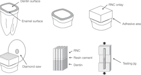
× 2 ± 0.3 mm) using a diamond saw (METSAW-HS, R&B Inc., Daejeon, Korea) under constant water cooling. The exact dimensions of the cross-sectional area of the inter- face were measured by a digital caliper (Absolute Digimatic, Mitutoyu, Tokyo, Japan) to calculate the formal bond strength (in MPa). Thirty samples were selected for each group. For the μTBS tests, each sample was attached with cyanoacrylate resin (Zapit, DVA, Corona, CA, USA) and tested until failure using a microtensile tester (Bisco Inc., Schaumburg, IL, USA) at a speed of 0.5 mm/min. Each specimen was evaluated with a stereomicroscope (SZ-PT, Olympus, Tokyo, Japan), and the failure modes were classi- fied as cohesive (in RNC, dentin, or cement), adhesive (between RNC/cement or dentin/cement), or mixed. The failed surfaces were evaluated with FESEM. The entire pro- tocol of this study is shown in Fig. 1.
Fig. 1. Schematic illustration of the protocol used in this study: (A) Occlusal enamel and dentin surfaces were sectioned and polished. (B) Roots were sectioned parallel to the occlusal dentin surface. (C) RNC onlays were cemented over the dentin surface. (D) Specimens were sectioned with a diamond saw. (E) Beam-shaped specimens were fabricated. (F) µTBS was evaluated.
Diamond saw Dentin surface
Enamel surface
RNC onlay
Adhesive area
RNC Resin cement
Dentin Testing jig
The results of μTBS tests on the samples with different surface treatments were statistically analyzed by one-way ANOVA. Subsequent comparisons were performed using the Tukey HSD test. The roughness and EDS analysis data were analyzed using the Kruskal-Wallis and Mann–Whitney U tests. All statistical tests were performed using the Statistical Package for the Social Science (SPSS v18.0, SPSS Inc., Chicago, IL, USA). P-values less than .05 were consid- ered statistically significant.
RESULTS
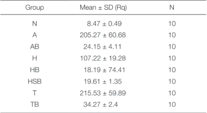
Here, we demonstrated that the nanoscratch test was useful for characterizing the roughness of the RNC samples. The roughness values of the surface of RNC onlays are pre- sented in Table 1. The Kruskal–Wallis test presented signif- icant differences among the experimental groups (Table 2).
Group T showed the highest roughness (215.53 ± 60.68 nm) followed by Group A (205.27 ± 60.68 nm). There was no significant difference between Group A and T. However, the surface with 4% HF etching had significantly lower rough- ness than air-abraded samples, Group A and T (P < .05).
Groups AB, HB, HSB, and TB had low roughness values.
Images of the scratched surfaces of RNC onlays are presented in Fig. 2. The surfaces of Group N samples had an uneven texture that was deeply furrowed. The surface of Group H had a crater-like texture. Group A and T showed
Table 1. Surface roughness data
Group Mean ± SD (Rq) N
N 8.47 ± 0.49 10
A 205.27 ± 60.68 10
AB 24.15 ± 4.11 10
H 107.22 ± 19.28 10
HB 18.19 ± 74.41 10
HSB 19.61 ± 1.35 10
T 215.53 ± 59.89 10
TB 34.27 ± 2.4 10
Table 2. Results of the Kruskal-Wallis test for surface- treated RNC groups
Roughness
Chi-square 73.130
df 7
Asymp. Sig. <.001*
* denotes significant difference at level of 0.05
Fig. 2. Surface roughness profiles from the nanoscratch tests. (A) Group N, (B) Group A, (C) Group AB, (D) Group H, (E) Group HB, (F) Group HSB, (G) Group T, and (H) Group TB.
A B
C D
E F
G H
a texture with sharp valleys. Group AB, HB, HSB, and TB had similar textures, a smooth wave pattern from the uni- versal adhesive (UA). Regardless of the roughening tech- nique, after applying the UA, the surfaces presented similar texture and roughness.
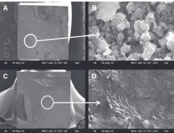
FESEM images of the surface topography of the RNC onlays are shown in Fig. 3. In the photomicrographs of RNC surfaces without any treatment (Group N), there were many fine irregular silica and zirconia fillers in the dense organic resin matrix. Pores in the resin matrix were observed on the surface of Group H. In Group A, rough resin and filler particles were observed. In Group T, the silica coat was visible on the surface. Smooth surfaces caused by the UA were observed in Group AB, HB, HSB, and TB.
After air abrasion, the surface was rougher and irregular particles were mixed in the matrix. After etching with HF, honeycomb-like pores were observed, similar to conven- tional ceramics. Borges et al.40 assessed the surface topogra- phy of different ceramics after treatment with either HF etching or airborne aluminum oxide particle abrasion. They reported highly modified surfaces on IPS Empress after HF etching, and dense pores on the surface were observed.
However, for zirconia, there was no change in the superfi- cial structure; many studies have reported that zirconia is not easily etched by HF.38 Swift et al.41 reported a significant decrease in the bond strength after HF etching of a glass- filled hybrid composite. They explained the decrease by the etching effect of HF absorbed in the resin matrix, causing softening and possibly a total dissolution of exposed glass particles. Here, fewer pores were formed after HF etching than for conventional glass ceramics. These samples had an insufficient roughness for resin bonding to RNC. Moreover, RNC includes resin zirconia nanomers in a resin matrix, which may decrease the etching effect and bond strength.
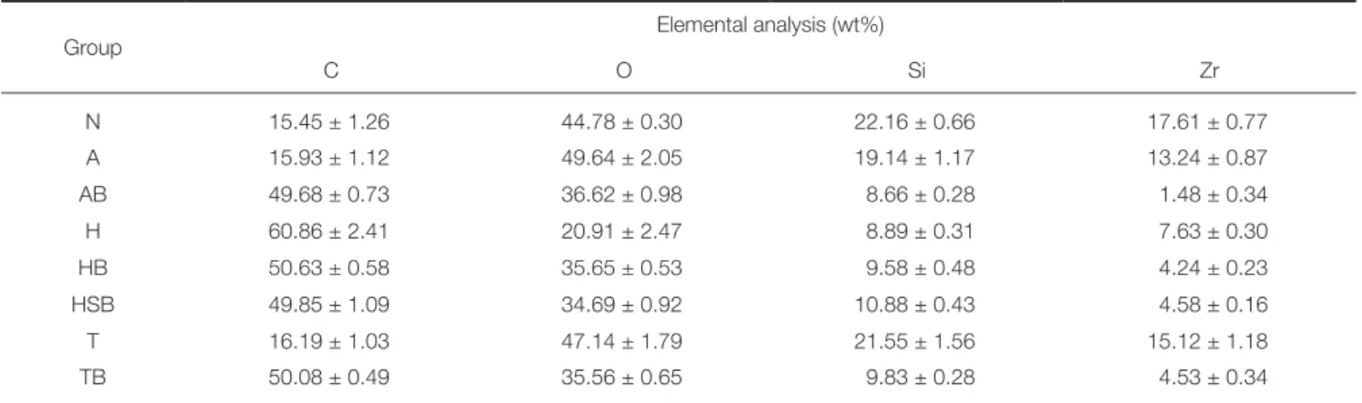
Elemental analysis using EDS showed concentrations of oxygen, carbon, silicon, and zirconium, as shown in Table 3.
For the groups without UA, there were significant chemical differences in the surfaces etched with 4% HF for all ele- ments. A decrease in oxygen, silicon, and zirconium ratio and an increase in carbon ratio were observed. However, for samples treated with UA, no significant differences were observed. Moreover, for Group A, a small quantity of alu- minum was observed (2.53 wt%), and some remaining fluo- ride (1.77 wt%) from the HF was detected for Group H samples.
EDS generally has a penetration depth of a few microm- eters depending on the material analyzed.42,43 Kern and Thompson42 claimed that this depth is appropriate for eval- uating chemical changes of a ceramic induced by sandblast- ing (surface roughening and powder particles mechanically embedded in the ceramic).
Fig. 3. Scanning electron microscopy images (×5000 magnification). (A) Group N, (B) Group A, (C) Group AB, (D) Group H, (E) Group HB, (F) Group HSB, (G) Group T, and (H) Group TB.
A B
C D
E F
G H
Table 3. Elemental concentrations from EDS analyses
Group Elemental analysis (wt%)
C O Si Zr
N 15.45 ± 1.26 44.78 ± 0.30 22.16 ± 0.66 17.61 ± 0.77
A 15.93 ± 1.12 49.64 ± 2.05 19.14 ± 1.17 13.24 ± 0.87
AB 49.68 ± 0.73 36.62 ± 0.98 8.66 ± 0.28 1.48 ± 0.34
H 60.86 ± 2.41 20.91 ± 2.47 8.89 ± 0.31 7.63 ± 0.30
HB 50.63 ± 0.58 35.65 ± 0.53 9.58 ± 0.48 4.24 ± 0.23
HSB 49.85 ± 1.09 34.69 ± 0.92 10.88 ± 0.43 4.58 ± 0.16
T 16.19 ± 1.03 47.14 ± 1.79 21.55 ± 1.56 15.12 ± 1.18
TB 50.08 ± 0.49 35.56 ± 0.65 9.83 ± 0.28 4.53 ± 0.34
The results of μTBS tests are presented in Table 4.
Group AB showed the highest bond strength (7.64 ± 3.37 MPa) followed by Group SB (7.41 ± 2.68 MPa). Group HB had the lowest bond strength (5.33 ± 2.40 MPa). The one- way ANOVA test showed that the differences in bond strength between groups had statistical significance (P <
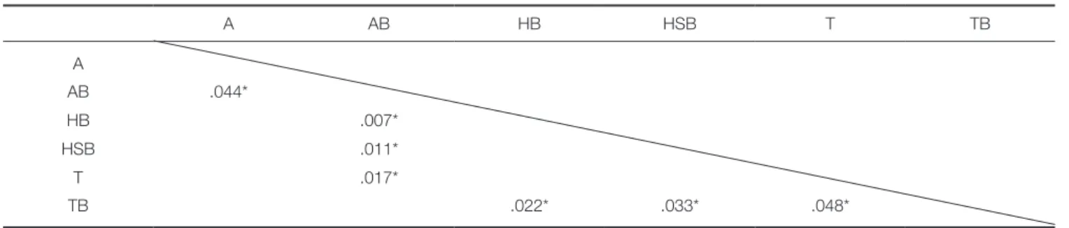
.05) (Table 5). Table 6 presents the results of the Tukey HSD test for the experimental groups.
There were no significant differences among Group A, HB, HSB, and TB or between Group AB and TB. Comparison of Groups A and AB and Groups T and TB showed that the UA increased the bond strength between the RNC and resin cement. However, the silane coupling agent had no effect (there was no significant difference between Groups H and HB). In Group AB, HB, and TB, air abrasion with alumina and the tribological silica coating showed similar bond strengths, but HF etching showed a lower bond strength.
The conventional test methods to evaluate the bond strength are the SBS or TBS test.35 However, as bonding
techniques and materials have improved, the bond strength has become sufficiently high to cause cohesive failures in dentin.37 This means that dentin breaks from dentin, leaving the resin–dentin interface intact. Pashley et al.44 reported that the frequency of cohesive failures of dentin can be as high as 80% when the bond strength reaches 25 MPa. Such failures interrupt the measurement of bond strength at the bonding interface and do not mean that the resin-dentin bonds are uniformly stronger than the intrinsic strength of dentin; however, the bond that is stressed is non-uniform and concentrated at the highly focused region where a crack in the dentin is present.37
The failure mode distribution is shown in Fig. 4. A high percentage of adhesive failure and non-cohesive failure
Table 4. Means and standard deviations for microtensile bond strength
Group Mean ± SD (MPa) N
A 5.72 ± 1.93 30
AB 7.64 ± 3.37 30
HB 5.33 ± 2.40 30
HSB 5.42 ± 2.75 30
T 5.51 ± 2.08 30
TB 7.41 ± 2.68 30
Table 5. Results of one-way ANOVA for experimental groups
Source Sum of Squares df Mean Square F Sig.
Between 167.364 5 33.473 5.214 <.001*
Within 1117.143 174 6.420
Total 1285.507 179
* denotes significant difference at level of 0.05
Table 6. Results of Tukey HSD test for experimental groups
A AB HB HSB T TB
A
AB .044*
HB .007*
HSB .011*
T .017*
TB .022* .033* .048*
* denotes pair of groups significantly different at level of 0.05
Fig. 4. Prevalence of failure modes after microtensile bond strength tests.
were observed for all groups. Figures 5 to 7 show SEM images of samples after fracture. Figures 5A, 5C, 6C, and 7C show cohesive failures and Figures 6A and 7A illustrate adhesive failures for the RNC surface. Similar to the SEM analysis of the RNC surface, irregular inorganic particles were observed in Group A and detached adhesive and pores were observed in Group HB. The silica coating was also observed in Group T. Group AB, HB, HSB, and TB showed tags and wave patterns caused by the UA.
The μTBS test offers versatility that cannot be achieved using conventional methods. Despite being labor-intensive it has great potential for providing insights into the strength of adhesion.37,44 Sano et al.36 mentioned that the μTBS test was useful because small surface areas can be tested, which showed adhesive failures at the bonded interface. Scherrer et al.34 argued that only mixed failures or adhesive failures with small resin segment should be considered for the bond strength computation. Our specimens mostly showed adhe- sive failure on the RNC surface, and no cohesive failure was observed. Therefore, these results may be useful for the analysis of resin bond strength of RNC.
Discussion
The aim of this study was to evaluate the influence of dif- ferent surface treatments on the resin bond strength of RNC. It was found that the bond strength between RNC and resin cement can be affected by the specific surface treatment, such as mechanical treatment and application of adhesive material. A strong resin bond depends on micro- mechanical interlocking and chemical bonding to the sur- face of the restoration, which requires cleaning and rough- ening for adequate surface activation.23 Modern surface conditioning methods require airborne particle abrasion of the surface before bonding.25 Here, air abrasion with UA resulted in the highest bond strength. There was no signifi- cant difference between air abrasion and a tribological silica coating. Comparing the air-abraded samples, HF etching, and tribological silica coating followed by UA, and HF etched surface with UA showed the lowest bond strengths.
Therefore, air abrasion and the tribological silica coating seemed to increase the bond strength more. Imamura et al.45 Fig. 5. Scanning electron microscopy images of fractured
RNC specimens: (A) Group A (×40 magnification), (B) Group A (×20,000 magnification), (C) Group AB (×40 magnification), and (D) Group AB (×20,000
magnification).
A B
C D
Fig. 6. Scanning electron microscopy images of fractured RNC specimens: (A) Group HB (×40 magnification), (B) Group HB (×20,000 magnification), (C) Group HSB (×40 magnification), and (D) Group HSB (×20,000
magnification).
A B
C D
Fig. 7. Scanning electron microscopy images of fractured RNC specimens: (A) Group T (×40 magnification), (B) Group T (×20,000 magnification), (C) Group TB (×40 magnification), and (D) Group TB (×20,000
magnification).
A B
C D
reported that air abrasion and the Rocatec system were very effective in increasing the bond strength of a laboratory- processed composite resin. However, these methods can induce a high loss of material,42 so excessive use should be avoided.
HF etching can achieve a suitable surface texture and roughness,23 creating a honeycomb-like topography on the ceramic surface that is ideal for micromechanical bonding.40 A chemical reaction between HF and silica in the feldspath- ic ceramics occurs, forming hexafluorosilicate, which is removed by water.46
6H2F2 + 6SiO2 → 2H2SiF6 + 4H2O Formula (1) Here, we observed that HF etching showed a lower roughness and bond strength than air abrasion and Rocatec treatment. In general, increasing surface roughness through mechanical surface treatment is more effective for increas- ing the bond strength than chemical bonding,16 which is corroborated by this study.
Here, we showed that there is a need to apply additional adhesive to increase the bond strength between RNC and resin cement. In this study, application of the UA increased the bond strength significantly. Stawarczyk et al.47 reported that when repairing RNC with a direct composite, universal adhesives (Scotchbond Universal and Futurabond U) with phosphoric acid monomers performed better than an adhe- sive based exclusively on methacrylic monomers, and this could be explained by the presence of 10-methacryloxydec- yl dihydrogen phosphate (MDP) monomers in Singlebond Universal. MDP was necessary to achieve a durable resin bond for zirconia.38 Lava Ultimate is composed of resin matrix, silica, and zirconia nanomers, so it can describe the higher bond strength when the UA was used.
There was no significant difference in the bond strength when the silane coupling agent was used in this study. One end of a silane molecule is organically functional, and can be polymerized with an organic matrix (e.g., a methacrylate).
The other end is generally composed of alkoxy groups, which can react with a hydroxylated surface, such as porce- lain.19 In most studies, silane treatment yielded further increased bond strength.16 In contrast, D’Alcangelo and Vanini48 stated that silane did not have a significant effect on resin bonds, especially HF-etched composite restora- tions. Here, the silane coupling agent was used with HF etching, and there was no significant effect on the bond strength. The silica coating remained when the tribological silica (Rocatecsystem) was used. Before forming the silica coat, a roughness increase is achieved by alumina particles modified with silicic acid.24 In this study, there was no sig- nificant difference between the bond strengths or rough- ness values of air-abraded or tribological silica coated sam- ples. Therefore, it seemed that the efficacy of silica coating for RNC was minor.
The manufacturer of Lava Ultimate states that it can be used for inlays, onlays, single/implant crowns, and veneers.
Behr et al.49 mentioned that the required tooth and resin bond strength is at least 10 MPa; all samples tested here showed a lower bond strength. Lebon et al.50 investigated
the roughness of dental materials, including RNC, after milling. Three commonly used milling tools with average diamond grit sizes of 105, 78, and 43.5 µm were used. They reported that there is a quasilinear correlation between dia- mond grit size and milled surface roughness. The rough- ness (Ra) of RNC after milling was approximately 2 to 10 µm. However, in this study, the RNC onlays were polished with diamonds of 1 µm grit and the roughness (Rq) was 8.47 ± 0.49 nm. Therefore, higher surface roughness and resin bond strength are expected in clinical situations.
Moreover, when using RNC in certain cases that need more retention or veneers, it will be necessary to use accurate, effective, and systemic surface roughening methods and appropriate adhesives. In addition, more studies are neces- sary to compare the effects of various surface treatment methods and primers or adhesives on the resin bond strength of RNC.
CONCLUSION
We conclude that HF etching is not an appropriate method for RNC surface conditioning. When using RNC for resto- ration, effective and systemic surface roughening methods and an appropriate adhesive are required.
ORCID
Yu-Sung Choi http://orcid.org/0000-0003-0053-7441 REFERENCES
1. Alt V, Hannig M, Wöstmann B, Balkenhol M. Fracture strength of temporary fixed partial dentures: CAD/CAM versus directly fabricated restorations. Dent Mater 2011;27:
339-47.
2. Schoenbaum TR. Dentistry in the digital age: an update.
Dent Today 2012;31:108, 110, 112-3.
3. Poticny DJ, Klim J. CAD/CAM in-office technology: innova- tions after 25 years for predictable, esthetic outcomes. J Am Dent Assoc 2010;141:5S-9S.
4. Stawarczyk B, Ender A, Trottmann A, Özcan M, Fischer J, Hämmerle CH. Load-bearing capacity of CAD/CAM milled polymeric three-unit fixed dental prostheses: effect of aging regimens. Clin Oral Investig 2012;16:1669-77.
5. Tinschert J, Zwez D, Marx R, Anusavice KJ. Structural reli- ability of alumina-, feldspar-, leucite-, mica- and zirconia- based ceramics. J Dent 2000;28:529-35.
6. Stawarczyk B, Sener B, Trottmann A, Roos M, Ozcan M, Hämmerle CH. Discoloration of manually fabricated resins and industrially fabricated CAD/CAM blocks versus glass- ceramic: effect of storage media, duration, and subsequent polishing. Dent Mater J 2012;31:377-83.
7. Stawarczyk B, Özcan M, Trottmann A, Schmutz F, Roos M, Hämmerle C. Two-body wear rate of CAD/CAM resin blocks and their enamel antagonists. J Prosthet Dent 2013;
109:325-32.
8. Luthardt RG, Holzhüter MS, Rudolph H, Herold V, Walter
MH. CAD/CAM-machining effects on Y-TZP zirconia.
Dent Mater 2004;20:655-62.
9. Lin WS, Ercoli C, Feng C, Morton D. The effect of core ma- terial, veneering porcelain, and fabrication technique on the biaxial flexural strength and weibull analysis of selected den- tal ceramics. J Prosthodont 2012;21:353-62.
10. Addison O, Cao X, Sunnar P, Fleming GJ. Machining vari- ability impacts on the strength of a ‹chair-side› CAD-CAM ceramic. Dent Mater 2012;28:880-7.
11. El Zohairy AA, De Gee AJ, Mohsen MM, Feilzer AJ.
Microtensile bond strength testing of luting cements to pre- fabricated CAD/CAM ceramic and composite blocks. Dent Mater 2003;19:575-83.
12. 3M ESPE. Lava ultimate technical product profile: 2011.
Available at: http://multimedia.3m.com/mws/media/
781394O/lava-ultimate-technical-product-profile golbal.pdf 13. Chen C, Trindade FZ, de Jager N, Kleverlaan CJ, Feilzer AJ.
The fracture resistance of a CAD/CAM Resin Nano Ceramic (RNC) and a CAD ceramic at different thicknesses.
Dent Mater 2014;30:954-62.
14. Joda T, Huber S, Bürki A, Zysset P, Brägger U. Influence of Abutment Design on Stiffness, Strength, and Failure of Implant-Supported Monolithic Resin Nano Ceramic (RNC) Crowns. Clin Implant Dent Relat Res 2015;17:1200-7.
15. Vargas MA, Bergeron C, Diaz-Arnold A. Cementing all-ce- ramic restorations: recommendations for success. J Am Dent Assoc 2011;142:20S-4S.
16. Spitznagel FA, Horvath SD, Guess PC, Blatz MB. Resin bond to indirect composite and new ceramic/polymer materials: a review of the literature. J Esthet Restor Dent 2014;26:382- 93.
17. el-Mowafy O. The use of resin cements in restorative dentist- ry to overcome retention problems. J Can Dent Assoc 2001;
67:97-102.
18. Sorensen JA, Kang SK, Avera SP. Porcelain-composite inter- face microleakage with various porcelain surface treatments.
Dent Mater 1991;7:118-23.
19. Thompson JY, Stoner BR, Piascik JR, Smith R. Adhesion/ce- mentation to zirconia and other non-silicate ceramics: where are we now? Dent Mater 2011;27:71-82.
20. Semmelman JO, Kulp PR. Silane bonding porcelain teeth to acrylic. J Am Dent Assoc 1968;76:69-73.
21. Jochen DG, Caputo AA. Composite resin repair of porcelain denture teeth. J Prosthet Dent 1977;38:673-9.
22. Ferrando JM, Graser GN, Tallents RH, Jarvis RH. Tensile strength and microleakage of porcelain repair materials. J Prosthet Dent 1983;50:44-50.
23. Blatz MB, Sadan A, Kern M. Resin-ceramic bonding: a re- view of the literature. J Prosthet Dent 2003;89:268-74.
24. Ozcan M. Evaluation of alternative intra-oral repair tech- niques for fractured ceramic-fused-to-metal restorations. J Oral Rehabil 2003;30:194-203.
25. Amaral R, Ozcan M, Bottino MA, Valandro LF. Microtensile bond strength of a resin cement to glass infiltrated zirconia- reinforced ceramic: the effect of surface conditioning. Dent Mater 2006;22:283-90.
26. Ozcan M. The use of chairside silica coating for different
dental applications: a clinical report. J Prosthet Dent 2002;87:
469-72.
27. Bailey LF, Bennett RJ. DICOR surface treatments for en- hanced bonding. J Dent Res 1988;67:925-31.
28. Egilmez F, Ergun G, Cekic-Nagas I, Vallittu PK, Ozcan M, Lassila LV. Effect of surface modification on the bond strength between zirconia and resin cement. J Prosthodont 2013;22:529-36.
29. Sorensen JA, Engelman MJ, Torres TJ, Avera SP. Shear bond strength of composite resin to porcelain. Int J Prosthodont 1991;4:17-23.
30. Chen JH, Matsumura H, Atsuta M. Effect of different etch- ing periods on the bond strength of a composite resin to a machinable porcelain. J Dent 1998;26:53-8.
31. Chen JH, Matsumura H, Atsuta M. Effect of etchant, etching period, and silane priming on bond strength to porcelain of composite resin. Oper Dent 1998;23:250-7.
32. Szep S, Gerhardt T, Gockel HW, Ruppel M, Metzeltin D, Heidemann D. In vitro dentinal surface reaction of 9.5%
buffered hydrofluoric acid in repair of ceramic restorations: a scanning electron microscopic investigation. J Prosthet Dent 2000;83:668-74.
33. Braga RR, Meira JB, Boaro LC, Xavier TA. Adhesion to tooth structure: a critical review of “macro” test methods.
Dent Mater 2010;26:e38-49.
34. Scherrer SS, Cesar PF, Swain MV. Direct comparison of the bond strength results of the different test methods: a critical literature review. Dent Mater 2010;26:e78-93.
35. Papia E, Larsson C, du Toit M, Vult von Steyern P. Bonding between oxide ceramics and adhesive cement systems: a sys- tematic review. J Biomed Mater Res B Appl Biomater 2014;102:395-413.
36. Sano H, Shono T, Sonoda H, Takatsu T, Ciucchi B, Carvalho R, Pashley DH. Relationship between surface area for adhe- sion and tensile bond strength-evaluation of a micro-tensile bond test. Dent Mater 1994;10:236-40.
37. Pashley DH, Carvalho RM, Sano H, Nakajima M, Yoshiyama M, Shono Y, Fernandes CA, Tay F. The microtensile bond test: a review. J Adhes Dent 1999;1:299-309.
38. Blatz MB, Phark JH, Ozer F, Mante FK, Saleh N, Bergler M, Sadan A. In vitro comparative bond strength of contempo- rary self-adhesive resin cements to zirconium oxide ceramic with and without air-particle abrasion. Clin Oral Investig 2010;14:187-92.
39. Soares CJ, Soares PV, Pereira JC, Fonseca RB. Surface treat- ment protocols in the cementation process of ceramic and laboratory-processed composite restorations: a literature re- view. J Esthet Restor Dent 2005;17:224-35.
40. Borges GA, Sophr AM, de Goes MF, Sobrinho LC, Chan DC. Effect of etching and airborne particle abrasion on the microstructure of different dental ceramics. J Prosthet Dent 2003;89:479-88.
41. Swift EJ Jr, Brodeur C, Cvitko E, Pires JA. Treatment of composite surfaces for indirect bonding. Dent Mater 1992;8:
193-6.
42. Kern M, Thompson VP. Sandblasting and silica coating of a glass-infiltrated alumina ceramic: volume loss, morphology,
and changes in the surface composition. J Prosthet Dent 1994;71:453-61.
43. Hantsche H. Comparison of basic principles of the surface- specific analytical methods: AES/SAM, ESCA (XPS), SIMS, and ISS with X-ray microanalysis, and some applications in research and industry. Scanning 1989;11:257-80.
44. Pashley DH, Sano H, Ciucchi B, Yoshiyama M, Carvalho RM. Adhesion testing of dentin bonding agents: a review.
Dent Mater 1995;11:117-25.
45. Imamura GM, Reinhardt JW, Boyer DB, Swift EJ Jr.
Enhancement of resin bonding to heat-cured composite res- in. Oper Dent 1996;21:249-56.
46. Janda R, Roulet JF, Wulf M, Tiller HJ. A new adhesive tech- nology for all-ceramics. Dent Mater 2003;19:567-73.
47. Stawarczyk B, Krawczuk A, Ilie N. Tensile bond strength of resin composite repair in vitro using different surface prepa- ration conditionings to an aged CAD/CAM resin nanoc- eramic. Clin Oral Investig 2015;19:299-308.
48. D’Arcangelo C, Vanini L. Effect of three surface treatments on the adhesive properties of indirect composite restora- tions. J Adhes Dent 2007;9:319-26.
49. Behr M, Proff P, Kolbeck C, Langrieger S, Kunze J, Handel G, Rosentritt M. The bond strength of the resin-to-zirconia interface using different bonding concepts. J Mech Behav Biomed Mater 2011;4:2-8.
50. Lebon N, Tapie L, Vennat E, Mawussi B. Influence of CAD/
CAM tool and material on tool wear and roughness of dental prostheses after milling. J Prosthet Dent 2015;114:236-47.