10.3803/jkes.2009.24.3.206
Introduction
1)Congenital abnormalities in pituitary development typically exhibit a phenotype of combined pituitary hormones deficiency (CPHD), which often includes short stature[1].
The occurrence of spontaneous linear growth despite the lack of growth hormone (GH), termed ‘growth without GH phenomenon', has been observed in some patients after the removal of tumor(s) in the hypothalamic-pituitary area[2~5]. This syndrome is also observed in a small number of patients with hypopituitarism[6~12]. Possible mechanisms that may induce linear growth have been observed in several patients; however, serum levels of
접수일자: 2009년 3월 14일 통과일자: 2009년 5월 18일
책임저자: 함종렬, 경상대학교 의과대학 내과학교실
factors underlying these mechanisms (i.e., hyperinsulinemia, elevated insulin-like growth factor (IGF)-I, and/or increased prolactin levels) have not been demonstrated in most cases[5~10].
Here, we describe a 22-year-old male diagnosed as CPHD with a hypoplastic pituitary gland. The patient presented with short stature at the age of 18 and subsequently presented with continuous linear growth, insulin resistance, and increased IGF-II levels.
Case
Patient: 22-year-old male Chief complaint: micropenis
Present illness: Four years ago, when the patient complained of short stature and the absence of puberty, he
성장 호르몬과 인슐린유사성장인자-I의 결핍에도 과속 선형 성장한 1예
경상대학교 의학전문대학원 내과학교실, 건강과학연구원, 울산대학교 의과대학 서울아산병원 소아과교실1
이경주․함종렬․정태식․정정화․김수경․백종하․이원현․유한욱
1․정순일
The Case of Accelerated Linear Growth Despite Growth Hormone and Insulin-like Growth Factor-I Deficiency
Kyeong Ju Lee, Jong Ryeal Hahm, Tae Sik Jung, Jung Hwa Jung, Soo Kyoung Kim, Jong Ha Baek, Won Hyun Lee, Han-wook Yoo1, Soon Il Chung
Department of Internal Medicine, School of Medicine, Gyeongsang Institute of Health Science, Gyeongsang National University;
and Department of Pediatrics1, Asan Medical Center, University of Ulsan College of Medicine
ABSTRACT
Here we describe a male patient who attained normal height despite combined hypopituitarism with an abnormal growth hormone-insulin-like growth factor (IGF)-I axis. When he was an 18-year-old, he presented with a short stature and underdeveloped external genitalia. The patient had not undergone normal pubertal development and he displayed a height below the fifth percentile. Hormonal and radiological studies revealed the findings of severe anterior pituitary hormone deficiency and an atrophic pituitary gland. There had been no recent follow-ups with the patient or medical treatment since that time. In the current presentation, the patient, now 22 years of age, had attained normal height, yet he remained prepubertal and showed manifestations of delayed bone age and combined hypopituitarism. In addition, the patient’s IGF-II levels were increased for his age. (J Korean Endocr Soc 24:206~211, 2009)
ꠏꠏꠏꠏꠏꠏꠏꠏꠏꠏꠏꠏꠏꠏꠏꠏꠏꠏꠏꠏꠏꠏꠏꠏꠏꠏꠏꠏꠏꠏꠏꠏꠏꠏꠏꠏꠏꠏꠏꠏꠏꠏꠏꠏꠏꠏꠏꠏꠏꠏꠏꠏꠏꠏꠏꠏꠏꠏꠏꠏꠏꠏꠏꠏꠏꠏꠏꠏꠏꠏꠏꠏꠏꠏꠏꠏꠏꠏꠏꠏꠏꠏꠏꠏꠏꠏꠏꠏꠏꠏꠏꠏꠏꠏꠏꠏꠏꠏꠏ Key Words: growth hormone, hypopituitarism, insulin-like growth factor-II
was diagnosed as CPHD with GH deficiency. At that time the patient's height was 162 cm [below the fifth percentile, body mass index (BMI): 21.6 kg/m2], and he displayed delayed puberty at Tanner stage 1. The patient's 55-year-old father was 177 cm tall and the 53-year-old mother was 163 cm tall. His target height as determined by his parent's heights was 176.5 cm using the Bayley-Pinneau method [(father's height + mother's height + 13) / 2]. The patient was born full-term in the hospital by spontaneous vaginal delivery with breech presentation.
The medical record and Apgar scores from the birth were not available. According to his mother, the patient's birth height and weight were within the normal range for his age. The postnatal course was uneventful; however, the patient was small for his age during infancy and was always the smallest boy (below the third percentile) in his classes during school (Fig. 2). The patient was within the normal range of intelligence quotient throughout his school days.
Past medical history: The patient was diagnosed as having hepatitis B antigenemia at 14 years of age.
Fig. 1. A photograph of the patient. Height was 176 cm and body weight was 72 kg (body mass index: 23.2 kg/m2).
Waist/hip and upper/lower body ratios were 0.92 and 0.60, respectively. The patient presented with a micropenis, small scrotum, and no pubic hairs.
Fig. 2. Patient growth curve. Until 16 years of age, the subject's height-for-age and weight-for-age remained below the 10th percentile. At age 18, the subject's height-for-age remained below the third percentile; however, the weight-for-age increased to the 25th percentile. At age 22, the patient gained mid-parental height and his weight-for-age increased to the 75th percentile.
Closed squares represent height and closed circles represent weights, which were measured at 14, 15, 16, 17, 18 and 22 years of age.
Family history: The patient also had one younger sister and his family history was non-specific.
Physical examination: During the most recent medical visit, the patient showed an continuous growth rate (176 cm), and weight gain (72 kg) with body mass index (23.2 kg/m2) (Fig. 1). The waist/hip and upper/lower body ratios were 0.92 and 0.60, respectively. Examination of the penis, scrotum, axillary hair, and pubic area revealed that the patient remained in Tanner stage 1. Left and right testicular volumes were 2 cc and 1 cc, respectively.
Laboratory findings: Laboratory blood findings were as follows: WBC, 4.1 × 10⁹/L; hemoglobin, 10.8 g/dL;
hematocrit, 29%; platelets, 1.5 × 10⁹/L; total protein, 74 (67~86 g/L); albumin, 42 (40~50 g/L); total bilirubin, 6.8 (5.1~22 μmol/L); aspartate aminotransferase, 0.63 (0.12~0.70 μkat/L); alanine aminotransferase, 0.58 (0.20~0.65 μkat/L);
blood urea nitrogen, 3.7 (2.5~7.1 mmol/L); creatinine, 88 (53~106 μmol/L); sodium, 139.9 mmol/L; potassium, 4.1 mmol/L; chloride, 105.1 mmol/L; and urine specific gravity, 1.010. The patient had positive HBs Ag, but negative HBe Ag. Alfa-fetoprotein levels were normal and thyroid hormone profiles were as follows: free T4, 4.0 (10.3~21.9 pmol/L); free T3, 2.2 (3.7~6.5 pmol/L); thyroid stimulating hormone (TSH), 10.46 (0.34~4.25 mIU/L); and autoantibodies, negative. Basal hormonal profiles were as follows; free testosterone, 0.52 (163~847 pmol/L); total testosterone, 0.12 (9.36~37.10 nmol/L); dehydroepiandrosterone -sulfate, 80 (100~6,190 μg/L); estrone, 6.6 (55~240 pmol/L);
IGF-I, 45 (normal, 182~780 μg/L); and IGF-II, 3,000 (447~1,039 ng/mL). The patient’s glucose homeostasis model assessment (HOMA) index was 3.02 (normal <
2.60) [calculated by fasting plasma insulin level (13.80 μU/mL) × fasting plasma glucose level (89 mg/dL) / 22.5]. Combined pituitary stimulation tests performed during the previous and current visit indicated that the patient had CPHD (Table 1). The patient’s karyotype was
46 XY. We performed pituitary transcription factor gene mutation analysis (Hesx1, SOX3, LHX3, PROP1 and PIT1); however, the results were negative.
Imaging findings: Sellar MRI performed during the current visit showed findings similar to the previous ones obtained four years before, i.e., a hypoplastic stalk and hypophyseal gland (Fig. 3). The patient had an intact olfactory bulb and no evidence of optic nerve hypoplasia or defects in the midline structures. A 99m-technetium scan revealed a normal-sized thyroid and normal uptake ratio. Dual energy X-ray absorptiometry (DXA) showed low bone mineral density of the femur neck (Z-score -1.7) and lumbar spine (Z-score -3.7) below the expected range for his age at 27.1% of total fat tissues. The patient's bone age was 14 to 15 years and the femoral epiphyseal plates were not closed.
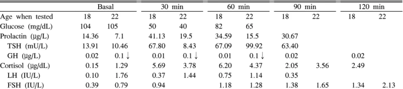
Clinical course: The patient has not complained of posterior pituitary hormone deficiency symptoms such as polyuria and polydipsia since diagnosis. The presence of Table 1. Combined pituitary stimulation tests performed during the first and second visits
Basal 30 min 60 min 90 min 120 min
Age when tested 18 22 18 22 18 22 18 22 18 22
Glucose (mg/dL) 104 105 50 40 82 65
Prolactin (μg/L) 14.36 7.1 41.13 19.5 34.59 15.5 30.67
TSH (mU/L) 13.91 10.46 67.80 8.43 67.09 99.92 63.40
GH (μg/L) 0.02 0.1↓ 0.01 0.1↓ 0.01 0.1↓ 0.02 0.02
Cortisol (μg/dL) 0.15 1.29 5.69 3.78 6.20 4.37 2.05 3.56 2.49 LH (IU/L) 0.10 1.76 0.37 1.44 0.75 1.14 0.35
FSH (IU/L) 0.39 0.79 0.94 1.18 1.28 1.38 1.65 1.34 2.13 FSH, follicle stimulating hormone; GH, growth hormone; LH, luteinizing hormone; TSH, thyroid stimulation hormone.
Fig. 3. Sellar MRI findings. Sagittal view shows an interrupted or thin hypoplastic stalk (thick arrow) and an aplastic or hypoplastic hypophyseal gland (thin arrow).
hepatitis B antigenemia and highly elevated levels of IGF-II prompted us to order liver computed tomography (CT), ultrasonography and colonoscopy. These studies did not detect any lesions except mild fatty liver and hepatosplenomegaly. At present, the patient is being followed as an outpatient and is taking oral prednisolone acetate 2.5 mg and testosterone undecanoate 160 mg daily.
He has no specific complaints.
Discussion
The patient was a typical case of idiopathic CPHD without germline mutations or the presence of polymorphisms in five pituitary transcription factors.
However, he attained normal height over the course of four years without hormonal or medical treatment. His appearance and intelligence were normal. On the first visit, the patient presented with short stature, small testes, and a lack of body hair. When the patient returned four years later, he showed typical eunuchoid (ratio of upper/lower segment: 0.60) and prepubertal body features.
All parameters, including bone age and serum levels of testosterone, luteinizing hormone (LH), follicle-stimulating hormone (FSH), and IGF-I, were below the normal ranges.
Growth without GH syndrome has been described in some children following pituitary or hypothalamic operation.
In some cases, puberty has occurred with transcriptional factor mutations and CPHD[2~12]. However, in most cases, the exact mechanisms, or possible factors involved, have not been identified[5~10]. Although our patient did not show central obesity, had normal body fat content as determined by DXA, and showed no family history of type 2 diabetes mellitus, the HOMA index indicated an insulin resistant state. Insulin resistance in this patient may be associated with GH deficiency. The hyperinsulinemia, combined with the permissive effects of low sex hormone concentrations on delayed fusion of the epiphyseal plates, might induce accelerated bony growth of the lower extremities[5,6,13~15]. Interestingly, on the second visit after the patient had attained normal height, serological assays revealed elevated IGF-II levels. This is the first reported case of growth with GH deficiency combined with elevated levels of IGF-II. However, IGF-II was not measured four years ago when the patient showed short stature.
IGF regulates cell growth and differentiation in humans
and many species. The anabolic functions of GH are largely mediated by IGF-I, thus designating IGF-I as a major determinant of somatic growth[16]. IGF-I levels are more GH-dependent than IGF-II levels and are more likely to reflect subtle differences in GH secretory patterns. The main pathogenic mechanisms of IGF-II over-expression are loss of IGF-II imprinting and gene duplication[16].
Sonic hedgehog, which plays a key role in regulating vertebrate organogenesis, can also transcriptionally activate the IGF-II gene[16]. IGF-II is widely expressed during murine embryonic development and is particularly important in placental growth[17]. IGF-II-null mice, which possess a normal GH-IGF-I system, are growth-impaired at birth but subsequent growth proceeds at normal rates[18]. IGF-II is virtually dispensable for post-natal development in mice. IGF-II can stimulate cellular proliferation and differentiation through insulin receptor and IGF-I receptor binding[16]. IGF-II also promotes the proliferation and differentiation of myocytes and bone cells in vitro[19,20]. IGF-II exerts its growth-promoting activity predominantly during fetal development. However, the elevated IGF-II levels suggest that it has postnatal activity.
Patients with severe liver disease, such as liver cirrhosis, may have lower serum IGF-I levels than healthy individuals[21]. It was not likely that our patient’s liver function caused a decreased serum IGF-1 level, because our patient was a hepatitis B carrier with normal liver function (Child-Pugh score 5). IGF-1 variant exhibits very weak binding to IGF-binding proteins and is substantially more potent than the natural growth factor[22]. We could not completely exclude the possibility that the IGF-1 variant induced continuous linear growth in the patient.
In conclusion, this report describes a 22-year-old male with accelerated growth despite CPHD. We suggest that elevated IGF-II may have contributed to normal growth, in part, following prolonged hypogonadism. The role of elevated IGF-II production and its possible effects on the patient’s growth remain to be evaluated.
요 약
성장호르몬의 결핍이 포함된 복합 뇌하수체기능저하증 환자에서 간혹 정상적인 선형 성장이 이루어졌다는 보고가 있다. 그 원인으로 고인슐린혈증, 성호르몬 결핍, 인슐린유 사성장인자-I의 증가 또는 고프로락틴혈증 등이 추정되고 있
으나 특별한 기전을 밝히지 못하는 경우가 대부분이다. 저자 들이 처음 환자를 진료했을 당시 18세 남자로 뇌하수체 형 성저하증, 복합 뇌하수체기능저하증과 Tanner I기의 성발달 을 보였다. 당시 환자의 키는 162 cm이었고 부모의 키를 이 용한 Bayley-Pinneau 방법에 의한 환자의 예상 키는 176.5 cm였다. 이후 환자는 병원을 다니지 않았고 특별한 치료를 받은 적도 없이 지내다가 22세에 다시 병원을 방문하게 되 었는데 환자의 키가 176 cm로 과속 선형 성장되어 있었다.
환자의 호르몬검사에서 성장호르몬과 인슐린유사성장인자-I 의 결핍을 비롯한 복합 뇌하수체기능저하증이 계속 있었으 나 인슐린유사성장인자-II가 3,000 ng/mL로 매우 높게 증가 되어 있었다. 인슐린유사성장인자-II가 인슐린 수용체나 인 슐린유사성장인자-I 수용체를 통하여 세포의 분열이나 증식 을 촉진할 수 있다고 알려져 있다. 환자가 18세 이후 4년에 걸쳐 14 cm의 지속적인 선형 성장은 고인슐린혈증, 성선기 능저하증과 함께 인슐린유사성장인자-II의 증가가 그 원인으 로 추정되어 보고하는 바이다.
참 고 문 헌
1. Low MJ: Neuroendocrinology. In: Kronenbeg HM, Melmed S, Polonsky KS, Larsen PR ed. Williams textbook of endocrinology. 11th ed. pp85-154, Philadelphia, WB Sounders Co., 2008
2. Kenny FM, Guyda HJ, Wright JC, Friesen HG:
Prolactin and somatomedin in hypopituitary patients with “catch up” growth following operations for craniopharyngioma. J Clin Endocrinol Metab 36:378 -380, 1973
3. Blethen SL, Weldon VV: Outcome in children with normal growth following removal of a craniophary- ngioma. Am J Med Sci 292:21-24, 1986
4. Tiulpakov AN, Mazerkina NA, Brook CG, Hindmarsh PC, Peterkova VA, Gorelyshev SK: Growth in children with craniopharyngioma following surgery. Clin Endocrinol (Oxf) 49:733-738, 1998
5. Bucher H, Zapf J, Torresani T, Prader A, Froesch ER, Illig R: Insulin-like growth factors I and II, prolactin, and insulin in 19 growth hormone-deficient children with excessive, normal, or decreased longitudinal growth after operation for craniopharyngioma. N Engl J Med 309:1142-1146, 1983
6. Bistritzer T, Chalew SA, Lovchik JC, Kowarski AA:
Growth without growth hormone: the “invisible” GH syndrome. Lancet 1:321-323, 1988
7. Wada S, Minagawa A, Imamaki K, Suda S, Yamanaka
K, Iitaka M, Katayama S: A patient of hypogonado- tropic hypogonadism accompanied by growth hormone deficiency and decreased bone mineral density who attained normal growth. Intern Med 39:641-645, 2000 8. Lazar L, Dan S, Phillip M: Growth without growth
hormone: growth pattern and final height of five patients with idiopathic combined pituitary hormone deficiency. Clin Endocrinol (Oxf) 59:82-88, 2003 9. Papastathopoulou L, Tzanela M, Vlassopoulou V,
Vassiliadi D, Thalassinos N: Untreated hypopituitarism due to absence of the pituitary stalk with normal adult height: report of two cases. Endocrine 29:175-179, 2006
10. Pavlou M, Tsatsoulis A, Efstathiadou Z, Bitsis S, Papadopoulou ZL: A study of the growth-promoting and metabolic effects of growth hormone (GH) in a patient with the “growth without GH” syndrome.
Growth Horm IGF Res 11:225-230, 2001
11. Geffner ME, Lippe BM, Bersch N, Van Herle A, Kaplan SA, Elders MJ, Golde DW: Growth without growth hormone: evidence for a potent circulating human growth factor. Lancet 1:343-347, 1986 12. Murashita M, Tajima T, Nakae J, Shinohara N,
Geffner ME, Fujieda K: Near-normal linear growth in the setting of markedly reduced growth hormone and IGF-1. A case report. Horm Res 51:184-188, 1999 13. Groop L, Segerlantz M, Bramnert M: Insulin sensitivity
in adults with growth hormone deficiency and effect of growth hormone treatment. Horm Res 64:45-50, 2005
14. Lee PA, Witchel SF: The influence of estrogen on growth. Curr Opin Pediatr 9:431-436, 1997
15. Hill DJ, Milner RD: Insulin as a growth factor. Pediatr Res 19:879-886, 1985
16. Chao W, D'Amore PA: IGF2: epigenetic regulation and role in development and disease. Cytokine Growth Factor Rev 19:111-120, 2008
17. Constância M, Hemberger M, Hughes J, Dean W, Ferguson-Smith A, Fundele R, Stewart F, Kelsey G, Fowden A, Sibley C, Reik W: Placental-specific IGF-II is a major modulator of placental and fetal growth. Nature 417:945-948, 2002
18. Baker J, Liu JP, Robertson EJ, Efstratiadis A: Role of insulin-like growth factors in embryonic and postnatal growth. Cell 75:73-82, 1993
19. Wilson EM, Hsieh MM, Rotwein P: Autocrine growth
factor signaling by insulin-like growth factor-II mediates MyoD-stimulated myocyte maturation. J Biol Chem 278:41109-41113, 2003
20. Minuto F, Palermo C, Arvigo M, Barreca AM: The IGF system and bone. J Endocrinol Invest 28:8-10, 2005
21. Assy N, Hochberg Z, Amit T, Shen-Orr Z, Enat R,
Baruch Y: Growth hormone-stimulated insulin-like growth factor (IGF) I and IGF-binding protein-3 in liver cirrhosis. J Hepatol 27:796-802, 1997
22. Tomas FM, Knowles SE, Chandler CS, Francis GL, Owens PC, Ballard FJ: Anabolic effects of insulin-like growth factor-I (IGF-I) and an IGF-I variant in normal female rats. J Endocrinol 137:413-421, 1993