173
Immune Network
Cell Response to Minor Histcompatibility Antigen, H60
Kyoung Min Jung and Eun Young Choi
Graduate Program of Immunology, Seoul National University College of Medicine, Seoul, Korea
ABSTRACT
Background: We studied the role for expression of CD40 and CD40L by CD4 and CD8 T cells in the generation of CD8 T cell response to minor histocompatibility anti- gen, H60. H60 is a cellular antigen to which CD8 responses require CD4 T cell help.
Methods: CD40- or CD40L-deficient mice were adoptively transferred with normal CD4 or CD8 T cells or with memory CD4 or CD8 T cells, and were immunized with male H60 congenic splenocytes to induce CD8 T cell response to H60. Peripheral blood CD8 T cell from the immunized mice were stained with the H60 tetramer. Results: CD8 T cell response to H60 was not induced in both CD40- and CD40L-deficient mice.
Adoptive transfer of CD40+/+ CD8 T cells into CD40-deficient mice did not compensate the defect in inducing CD8 T cell response to H60, while the H60-specific CD8 T cells were activated in the CD40-deficient mice that were adoptively transferred with CD40+/+
CD4 T cells. Adoptive transfer of CD40L+/+ CD4 T cells into CD40L-deficient mice induced primary CD8 T cell response for H60 and the presence of CD40L+/+ CD4 T cells was required even for memory CD8 T cells response to H60. Conclusion: Our results suggest that the CD40-CD40L interaction mediates the delivery of CD4 T cell help to naïve and memory H60-specific CD8 T cells. While the expression of CD40L by CD4 T cells is essential, signaling through CD40 on CD8 T cells is not required for the induction of CD8 T cell response to H60. (Immune Network 2007;7(4):
173-178)
Key Words: CD8 T cell response, CD4 help, CD40, CD40L, minor histocompatibility antigen
Correspondence to: Eun Young Choi, Center for Animal Resource Development, Seoul National University College of Medicine, 103, Daehangno, Jongno-gu, Seoul 110-799, Korea (Tel) 82-2-740-8530, (Fax) 82-2-740-8538, (E-mail) [email protected]
This study was supported by a grant from Korea Science and Engine- ering Foundation (R01-2006-000-10565-0).
Introduction
Minor histocompatibility (H) antigens are perceived as foreign during allogeneic transplantation due to naturally occurring polymorphism (1). The T cells re- sponses directed to minor H antigens contribute to graft rejection after MHC-matched transplantation or graft-versus-host disease, a major complication after bone marrow transplantation between MHC-matched individuals, which is a standard procedure for the treat- ment of hematological malignancy (2). In these T cell responses, CD8 cytotoxic T cells are major effectors
killing the target peptide/MHC complexes, usually mi- nor H antigen-originated peptides, expressed on the grafts or on the recipient tissues (2,3). In this respect, understanding the nature of CD8 T cell responses to minor H antigens is essential for controlling immune rejection and treatment of GVHD.
Priming CD8 T cells specific for cellular antigens requires help from CD4 T cells (4-6). The CD4 T cell helper function during CD8 T cell response is known to involve CD40-CD40L interactions which are cen- tered on antigen presenting cells (APCs) that allow cross-talk between CD4 and CD8 T cells and APCs (7-9). CD40L is expressed on the surface of activated CD4 T cells and is involved in the development into effectors (8,10). Ligation of CD40 on the surface of APCs such as dendritic cells (DC), macrophages and B cells increases their antigen-presentation and co-sti- mulatory capacity (11,12). However, some controversy
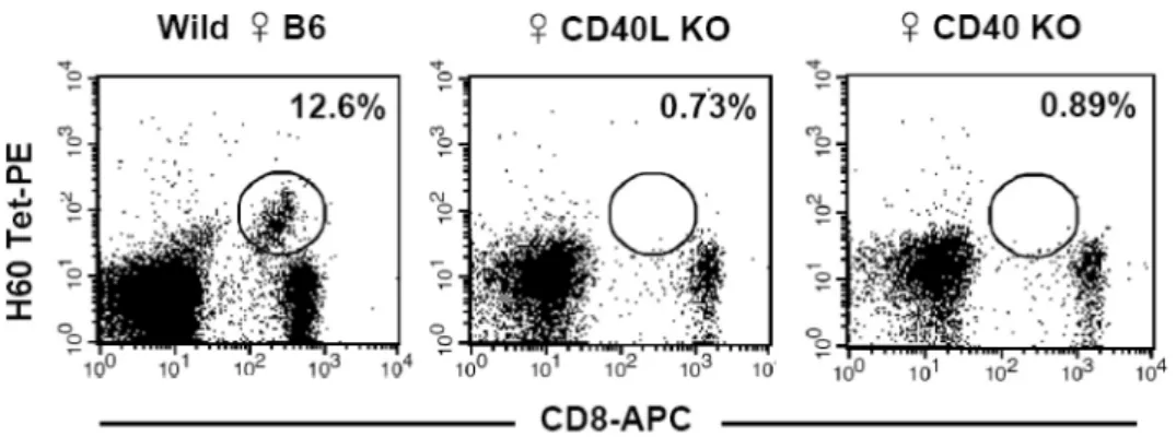
Figure 1. Primary CD8 T cell response in CD40-deficient and CD40L-deficient mice. Normal female B6 (positive control) and CD40- and CD40L-deficient mice were injected intraperitoneally with 2×10⁷ male H60 congenic splenocytes. PBLs from the immunized mice were stained with H60-tetramer-PE and APC-conjugated anti-CD8 mAb on day 10 post-immunization. The percentages of tetramer-binding cells in CD8 T cell population were marked. The representative FACs data from three independent experiments are shown.
remains regarding the role for CD40 expression on APCs versus their expression on CD8 T cells. In some models, namely the influenza-specific CD8 T cell me- mory model, CD40 expression by APCs influenced the outcome of CD8 T cell responses (13,14). However, this was not the case fo other models, which also shows the importance of CD40 expression by CD8 T cells (15,16).
Minor H antigen, H60, is a representative CD4 help-dependent cellular antigen. In this study, we in- vestigated the role for expression of CD40 and CD40L molecules by CD4 and CD8 T cells in the induction of primary and memory CD8 T cell response to H60.
Our results show that CD40 expression by CD8 T cells is not essential in receiving CD4 help, while CD40L expression by CD4 T cells and, thereby, CD40L-CD40 interaction are required for both pri- mary and secondary response generation.
Materials and Methods
Mice. C57BL/6 (B6), B6.PL congenic, B6. CD40/, and B6.CD40L/ mice were purchased from The Jack- son Laboratory (Bar Harbor, ME, USA). H60 congenic mouse strain, B6.C-H60C/DCR, was kindly provided by Dr. Derry Roopenian (The Jackson Laboratory).
The mice were maintained under specific pathogen- free conditions in the Center for Animal Resource Development of Seoul National University College of Medicine, and were used for experiments at the ages between 8 and 12 weeks.
Immunization. A suspension of splenocytes was pre- pared from male H60 congenic mice and 2×10⁷ cells
were intraperitoneally injected into each responder mouse. Immunized mice were eye-bled for flow cy- tometry on day 10 post-immunization and on day 7 post-immunization to detect CD8 T cells binding to H60-tetramer during primary and memory response, respectively.
Adoptive transfer. Single cell suspension of spleens from normal female B6 mice was prepared and CD4+ T cells or CD8+ T cells were purified by magnetic cell sorting (MACS). 1×10⁶ purified CD4 or CD8 T cells in 0.2 ml 1×PBS were injected into the tail vein of CD40- or CD40L-deficient mice. Two days after the transfer, the mice were immunized with male H60 congenic splenocytes. For the adoptive transfer of me- mory CD4 and CD8 T cells, female B6.PL congenic mice were immunized with H60 congenic splenocytes 70 days prior to the purification of CD4 and CD8 T cells. The purified memory B6.PL CD4 and CD8 T cells or naïve B6.PL CD4 T cells were injected into the tail vein of CD40L-deficient mice two days before the immunization with male H60 congenic splenocytes.
Flow cytometry. Fresh peripheral blood leukocytes (PBLs) were incubated at 4oC for 30 min in staining buffer (1×PBS with 0.1% bovine serum albumin and 0.1%
sodium azide) containing PE-labeled H60/H-2Kb tet- ramer and saturating amounts APC-conjugated mouse CD8 mAb (BD Pharmingen, San Diego, CA, USA) and mouse CD11a-FITC (BD Pharmingen). Synthetic peptides LTFNYRNL were used for production of H60-tetramer. The stained cells were analyzed using a FACSCalibur equipped with CellQuest software (BD Pharmingen).
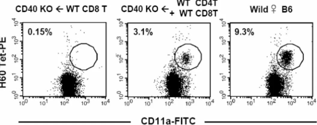
Figure 2. Induction of primary CD8 T cell response in the CD40-deficient mice that were adoptively transferred with normal CD4 or CD8 T cells. CD4 or CD8 T cells were purified from wild type female B6 mice and CD8 T cells either by themselves or in combination with CD4 T cells were injected (1×10⁶) into tail vein of female CD40-deficient mice. Two days after the adoptive transfer, the mice were immunized with male H60 congenic splenocytes and PBLs from the immunized mice were stained with H60-tetramer-PE, anti-CD8-APC, and anti-CD11a-FITC. The percent values of activated (CD11a+) H60-tetramer-binding cells in CD8 T cell population were noted in the FACs data. Two mice were included in each group and the experiments were repeated twice. Representative FACs data gated on CD8 are shown.
Results
H60-specific CD8 T cell response requires CD40 and CD40L expression. Since CD8 T cell response to H60 is a re- sponse against a cellular antigen, we questioned as to whether CD40 and CD40L molecules would play roles in inducing H60-specific CD8 T cell response. To test this, we immunized CD40L-deficient or CD40-defi- cient mice with splenocytes from male H60 congenic mice. As a control, normal female B6 mice were im- munized with the male H60 splenocytes. Activation of CD4 T cells that provide help to H60-specific CD8 T cells is mediated by recognition of HY-epitope that originates from Y chromosome (17). Therefore, the in- jection of normal female B6 mice with male H60 sple- nocytes results in activation and proliferation of H60- sepcific CD8 T cells that receive help from HY-re- active CD4 T cells, and H60-specific CD8 T cells are detectable with a peak frequency (8~15% of periph- eral blood CD8 T cells) on day 10 post-immunization under normal circumstances. However, when the male H60 splenocytes were injected into female CD40-defi- cient or female CD40L-deficient mice, H60-tetramer binding CD8 T cells were not detectable (Fig. 1), demonstrating that CD40 and CD40L molecules were involved in H60-specific CD8 T cell response.
Expression of CD40 by CD8 T cells does not play significant role in H60-specific CD8 T cell response. To examine whe- ther signaling through CD40 directly on CD8 T cells would be involved in receiving CD4 T cell help, H60-
specific CD8 T cell response was induced in CD40-de- ficient mice that were adoptively transferred with nor- mal (CD40+/+) B6 CD8 T cells by injecting male H60 splenocytes. In the PBLs from the CD40-deficient mice that were adoptively transferred with CD40+/+ CD8 T cells, we could not detect H60-tetramer binding CD8 T cells (Fig. 2). However, unexpectedly, in the blood from the CD40-deficient mice that were adoptively transferred with CD40+/+ CD4 and CD8 T cells, H60- specific CD8 T cells were detected, even though it was at a lower level (~3% of peripheral CD8 T cells) than that usually detected in normal hosts immunized with H60 splenocytes. These results demonstrate that CD40 expression on CD8 T cells does not play a pivotal role in receiving CD4 T cell help for immune response induction.
CD40L expressed by CD4 T cells is essential for induction of CD8 T cell response for H60. Next, we investigated the role for CD40L expression by CD4 T cells or by CD8 T cells in the induction of primary CD8 T cell response for H60. Since it has been reported that CD40L molecule was expressed in activated CD8 T cells as well as in activated CD4 T cells (18,19), we wondered whether CD40L expression by CD8 T cells would have any role in inducing CD8 T cell response.
CD40L-deficient mice were adoptively transferred with CD4 T cells or CD8 T cells from normal female B6 mice (CD40L+/+) and immunized with male H60 con- genic splenocytes.
On day 10 post-immunization, we were able to de-
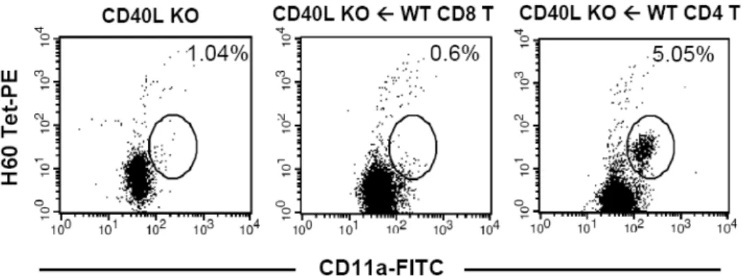
Figure 3. Primary CD8 T cell response in CD40L-deficient mice that were adoptively transferred with normal CD4 or CD8 T cells.
1×10⁶ CD4 or CD8 T cells purified from wild type female B6 mice were adoptively transferred to CD40L-deficient mice. Two days after the adoptive transfer, the mice were immunized with male H60 congenic splenocytes and PBL from the immunized mice were stained with H60-tetramer-PE, anti-CD8-APC, and anti-CD11a-FITC. The percent values of activated (CD11a+) H60-tetramer- binding cells in CD8 T cell population are noted in the FACs data. Two mice were included in each group and the experiments were repeated twice. Representative FACs data gated on CD8 are shown.
tect H60-tetramer binding cells in the blood from the CD40L-deficient mice that were adoptively transferred with CD40L+/+ CD4 T cells (5% of peripheral blood CD8 T cells), but not in the CD40L-deficient mice adoptively transferred with CD40L+/+ CD8 T cells, which did not show CD8 T cell response specific for H60 (Fig. 3). This demonstrates that CD40L expressed by CD8 T cells is also dispensable for receiving CD4 help, and suggests that CD40L expression on CD4 T cells is a limiting factor in providing help to H60- specfic CD8 T cells. This most likely happens by inter- action with CD40 expressed on APCs.
CD40-CD40L interaction is necessary for memory CD8 T cell response to H60. Based on the results above, we con- cluded that CD40-CD40L interaction was required for the induction of CD8 T cell response to H60, much like the case for other CD8 T cell responses to cellular antigen. Next, we wondered whether CD40L-CD40 interaction would still participate in generating memo- ry CD8 T cell response for H60 and, if it did, whether CD4 memory T cells would deliver more efficient help to H60 memory response than naïve CD4 T cells.
To test this, we immunized female B6.PL congenic mice with male H60 congenic splenocytes and waited until the primary CD8 T cell response subsided (up to 70 days). 71 days after immunization, CD4 T cells and CD8 T cells were purified from the immunized female B6.PL mice and were adoptively transferred in- to CD40L-deficient hosts. To compare the efficiency in delivering help to memory CD8 T cells between memory and naïve CD4 T cells, naïve CD4 T cells
purified from female B6.PL congenic mice were also included.
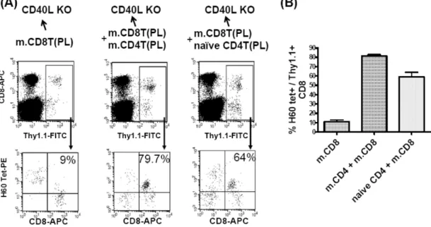
On day 7 post-immunization with male H60 con- genic splenocytes, peripheral CD8 T cells from the im- munized mice were stained with H60-tetramer to de- tect H60-specific CD8 memory T cells. By gating on Thy1.1 (of PL. origin), it was possible to distinguish memory CD8 T cells of PL origin from CD8 T cells of host origin. In the CD40L-deficient hosts that were adoptively transferred with CD40L+/+ memory Thy1.1+ CD8 T cells only, the percentages of H60-tetramer binding Thy1.1+ CD8 T cells were significantly low (~10% of Thy1.1+ CD8 T cells), compared to those in the CD40L-deficient mice co-transferred with mem- ory (~80% of Thy1.1+ CD8 T cells) or naïve (~60%
of Thy1.1+ CD8 T cells) CD40L+/+ CD4 T cells (Fig.
4). This result indicated that CD40L expression on CD4 T cells and, therefore, CD40L-CD40 interaction are required even for CD8 memory response. When the efficiency in help delivery was compared between me- mory and naïve CD4 T cells in the CD40L-deficient hosts, the H60-reactive CD8 T cells were activated more efficiently in the presence of memory CD40L+/+
CD4 T cells than in the presence of naïve CD40L+/+
CD4 T cells. However, naïve Thy1.1+ CD40L+/+ CD4 T cells were still potent enough to generate H60-spe- cific CD8 T memory response in the CD40L-deficient hosts.
Discussion
In this study, we showed that CD40-CD40L inter-
Figure 4. Memory CD8 T cell response in CD40L-deficient mice that were adoptively transferred with memory CD8 T cells by themselves or in combination with memory or naïve CD4 T cells. Th1.1+ CD4 or CD8 T cells were purified from female B6.PL mice that were pre-immunized with male H60 congenic splenocytes 70 days earlier. (A) Memory Thy1.1+ CD8 T cells (1×10⁶) were injected into tail vein of female CD40-deficient mice alone or with memory (1×10⁶) or naïve (1×10⁶) Thy1.1+ CD4 T cells.
Two days after the adoptive transfer, the mice were immunized with male H60 congenic splenocytes and PBL from the immunized mice were stained with H60-tetramer-PE, anti-CD8-APC, and anti-Thy1.1-FITC. The percent values of H60-tetramer-binding cells in Thy1.1+ CD8 T cell population are shown in the FACs data. Two mice were included in each group and the experiments were repeated twice. Representative data are shown. (B) The mean percentages of H60-tetramer-positive cells in Thy1.1+ CD8 T cells from each condition are plotted. The graph shows one result out of two independent experiments.
action was required for the delivery of CD4 T cell help necessary for induction of H60-specific CD8 T cell memory response as well as primary response.
CD40-CD40L-mediated direct interaction between CD4 and CD8 T cells has been raised as a new me- chanism for CD4 help delivery to CD8 T cells (15).
However, the role for CD40 expression by CD8 T cells in receiving help from CD4 T cells was found to be insignificant in our study. Rather, our results demonstrated that CD40 expression by CD4 T cell could compensate the loss of immunity due to the lack of CD40 expression in the hosts, albeit at a minimal level. Since CD40 expression was detected in activated CD4 T cells by RT-PCR (15), it is possible that CD40 molecules expressed by CD4 T cells may play some role in CD8 T cell response, which has not yet been identified in normal condition but can be exaggerated when the cellular milieu is devoid of CD40 expression.
This notion is supported by a report that function of CD40-deficient CD8 T cells and normal CD8 T cells were equivalent when they are primed in a CD40-suf- ficient environment (14). Combined with our results, it is suggested that CD8 T cell responses are facili-
tated by CD40 signaling but CD40 expression is not required on CD8 T cells.
Although the expression of CD40L molecules in ac- tivated CD8 T cells has been reported (18, 19), the presence of CD40L+/+ CD8 T cells in the CD40L-de- ficient hosts did not induce H60-specific CD8 T cell response. Only when CD40L+/+ CD4 T cells were present in the CD40L-deficient hosts, were primary and secondary CD8 T cell responses generated at sig- nificant levels. These results suggest CD8 T cells play only a passive role and that the two players, CD4 T cells and APCs are major contributors to the induction of CD8 T cell responses in the CD4-CD8-APC cluster.
Regarding the CD4 help requirement for memory CD8 T cell response, the widely held view is that once CD8 T cell are helped during the primary response, the CD4 help is not necessary for memory CD8 T cell responses (20-22). Our result from this study is against this view, demonstrating that memory Thy1.1+ CD8 T cells themselves, even though they were helped once during primary response, did not show significant memory response to H60 in the ab- sence of CD40L+/+ CD4 T cells in the CD40L-defi-
cient hosts.
In summary, we showed that CD40-CD40L inter- action mediates CD4 T cell help for the generation of CD8 T cell response to H60 and expression of CD40L on CD4 T cells, not the expressions of CD40 or CD40L by CD8 T cells, is essential for both pri- mary and memory H60-specific CD8 T cell responses.
These results will provide basic information on the na- ture of CD8 T cell response to minor H antigen and may be of help in designing ways to control immune rejection or GVHD.
References
1. Wallny HJ, Rammensee HG: Identification of classical minor histocompatibility antigen as cell-derived peptide. Nature 343;
275-278, 1990
2. Hambach L, Goulmy E: Immunotherapy of cancer through targeting of minor histocompatibility antigens. Curr Opin Imunol 17;202-210, 2005
3. Blair AN, Goulden J, Libri NA, Oakhill A, Pamphilon DH:
Immunotherapeutic strategies in acute lymphoblastic leukae- mia relapsing after stem cell transplantation. Blood 19;289- 300, 2005
4. Antoniou A, McCormick D, Scott D, Yeoman H, Chandler P, Mellor A, Dyson J: T cell tolerance and activation to a transgene-encoded tumor antigen. Eur J Immunol 26;1094- 1102, 1996
5. VanderVegt FP, Johnson LL: Induction of long-term H-Y-spe- cific tolerance in female mice given male lymphoid cells while transiently depleted of CD4+ or CD8+ T cells. J Exp Med 177;1587-1592, 1993
6. Janssen EM, Lemmens EE, Wolfe T, Christen U, von Herrath MG, Schoenberger SP: CD4+ T cells are required for secon- dary expansion and memory in CD8+ T lymphocytes. Nature 421;852-856, 2003
7. Noelle RJ: CD40 and its ligand in host defense. Immunity 4;415-419, 1996
8. Grewal IS, Flavell RA: CD40 and CD154 in cell-mediated immunity. Ann Rev Immunol 16;111-135, 1998
9. Lanzavecchia A: Immunology. Licence to kill. Nature 393;
413-414, 1998
10. van Essen D, Kikutani H, Gray D: CD40 ligand-transduced co-stimulation of T cells in the development of helper function.
Nature 378;620-623, 1995
11. Koch F, Stanzl U, Jennewein P, Janke K, Heufler C, Kampgen E, Romani N, Schuler G: High level IL-12 production by mu- rine dendritic cells: upregulation via MHC class II and CD40 molecules and downregulation by IL-4 and IL-10. J Exp Med 184;741-746, 1996
12. Stout RD, Suttles J, Xu J, Grewal IS, Flavell RA: Impaired T cell-mediated macrophage activation in CD40 ligand-defi- cient mice. J Immunol 156;8-11, 1996
13. Lefrancois L, Altman JD, Williams K, Olson S: Soluble anti- gen and CD40 triggering are sufficient to induce primary and memory cytotoxic T cells. J Immunol 164;725-732, 2000 14. Lee BO, Hartson L, Randall TD: CD40-deficient, influenza-
specific CD8 memory T cells develop and function normally in a CD40-sufficient environment. J Exp Med 198;1759-1764, 2003
15. Bourgeois C, Rocha B, Tanchot C: A role for CD40 expression on CD8+ T cells in the generation of CD8+ T cell memory.
Science 297;2060-2063, 2002
16. Ozaki ME, Coren BA, Huynh TN, Redondo DJ, Kikutani H, Webb SR: CD4+ T cell responses to CD40-deficient APCs: defects in proliferation and negative selection apply only with B cells as APCs. J Immunol 163;5250-5256, 1999 17. Scott D, Addey C, Ellis P, James E, Mitchell MJ, Saut N, Jurcevi S, Simpson E: Dendritic cells permit identification of genes encoding MHC class II-restricted epitopes of trans- plantation antigens. Immunity 12;711-720, 2000
18. Whitmire JK, Flavell RA, Grewal IS, Larsen CP, Pearson TC, Ahmed R: CD40-CD40 ligand costimulation is required for generating antiviral CD4 T cell responses but is dispensable for CD8 T cell responses. J Immunol 163;3194-3201, 1999 19. Hermann P, Van-Kooten C, Gaillard C, Banchereau J, Blan- chard D: CD40 ligand-positive CD8+ T cell clones allow B cell growth and differentiation. Eur J Immunol 25;2972-2977, 1995
20. Matloubian M, Concepcion RJ, Ahmed R: CD4+ T cells are required to sustain CD8+ cytotoxic T-cell responses during chronic viral infection. J Virol 68;8056-8063, 1994 21. Belz GT, Wodarz D, Diaz G, Nowak MA, Doherty PC: Com-
promised influenza virus-specific CD8(+)-T-cell memory in CD4(+)-T-cell-deficient mice. J Virol 76;12388-12393, 2002 22. Sun JC, Bevan MJ: Defective CD8 T cell memory following
acute infection without CD4 T cell help. Science 300;339-342, 2003