https://doi.org/10.5468/ogs.2017.60.3.303 pISSN 2287-8572 · eISSN 2287-8580
Introduction
Delayed postpartum hemorrhage occurs between 24 hours and 12 weeks postpartum. Delayed postpartum hemorrhage may be caused by various factors, such as retained products of conception, infection, abnormal involution of the placen- tal site or uterine artery pseudoaneurysm (UAP) [1]. When a punctured or lacerated artery is not completely sealed, blood may escape and diffuse into adjacent tissues and col- lect in perivascular areas. If the blood collection maintains a link with the parent vessel, it may cause a pseudoaneurysm.
A pseudoaneurysm boundary forms with the peripheral thrombus and part of the three arterial layers [2]. UAP is rare, although a recent study reported that it occurs at an actual ratio of 2 to 3 instances per 1,000 deliveries [3]. UAP may occur as a consequence of intra-cesarean section vascular trauma, traumatic abortion/delivery or uncomplicated spon- taneous vaginal delivery [3]. UAP often leads to a delayed postpartum hemorrhage that ruptures within the uterine cav- ity. However, there was few report of case in which muscle layer rupture was the cause of postpartum hemoperitoneum.
Therefore, we report a case of delayed postpartum hemo- peritoneum caused by intramyometrial pseudoaneurysm rup- ture after vaginal delivery.
Case report
A 30-year-old woman (gravida 2, para 1) visited the emer- gency department on postpartum day 8 with lower abdomi- nal cramping as her major complaint. She had a normal spon- taneous vaginal delivery at 39 weeks of pregnancy. When she arrived at the emergency department, her status was stable.
Her blood pressure was 120/70 mmHg; pulse rate was 78 beats/min; and respiration rate was 20 breaths/min. Blood tests revealed normal outcomes, including hemoglobin (10.3 g/dL) and platelet (308,000/μL) measurements. She had no remarkable medical or surgical history, except one spontane-
Delayed postpartum hemoperitoneum due to uterine artery pseudoaneurysm rupture
Kyu-Sang Kyeong
1, Ji-Yeon Moon
1, Song-Hwa Chae
1, Seung Hwa Hong
1, Minho Kang
2, Eun-Hwan Jeong
1Departments of 1Obstetrics and Gynecology, 2Radiology, Chungbuk National University Hospital, Chungbuk National University College of Medicine, Cheongju, Korea
A 30-year-old woman experienced severe abdominal pain 8 days after vaginal delivery. The patient was diagnosed with hemoperitoneum due to rupture of the left uterine artery pseudoaneurysm, which was confirmed via ultrasound with color Doppler and computed tomography scans. This patient was treated with bilateral uterine artery embolization to maintain fertility. A uterine artery pseudoaneurysm that causes delayed postpartum hemorrhage can occur after cesarean section or vaginal delivery. A uterine artery pseudoaneurysm can be fatal, so its detection and diagnosis are critical. Herein, we report a case of delayed postpartum hemoperitoneum due to uterine artery pseudoaneurysm rupture.
Keywords: Postpartum period; Hemoperitoneum; Aneurysm, false; Embolization
Articles published in Obstet Gynecol Sci are open-access, distributed under the terms of the Creative Commons Attribution Non-Commercial License (http://creativecommons.
org/licenses/by-nc/3.0/) which permits unrestricted non-commercial use, distribution, and reproduction in any medium, provided the original work is properly cited.
Copyright © 2017 Korean Society of Obstetrics and Gynecology Received: 2016.9.19. Revised: 2016.10.31. Accepted: 2016.10.31.
Corresponding author: Kyu-Sang Kyeong
Department of Obstetrics and Gynecology, Chungbuk National University Hospital, Chungbuk National University College of Medicine, 776 1(il)sunhwan-ro, Heungdeok-gu, Cheongju 28644, Korea
Tel: +82-43-269-6057 Fax: +82-43-275-7359 E-mail: [email protected]
http://orcid.org/0000-0001-8208-3361
ous abortion without a coagulation disorder.
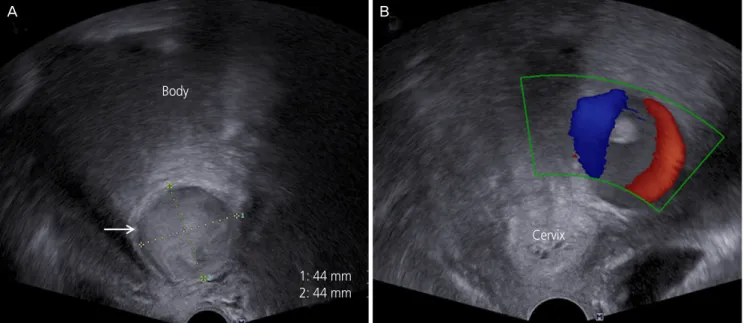
Via an ultrasound examination, a 53×44×44-mm-size pul- sating hypoechoic cyst was identified in the myometrium of the left lower segment (Fig. 1A). Based on a color Doppler ex- amination, turbulent artery-like flow with a ‘to-and-fro’ pat- tern was observed (Fig. 1B). In the pelvis, a small hematoma (mildly echogenic fluid) without para gutter fluid collection was present. Computed tomography (CT) scanning was per- formed, and the patient was diagnosed with hemoperitone-
um. The CT scan (intra-abdominal and pelvic cavity) revealed high density fluid collection and leakage of contrast media into the left side of the uterus (Fig. 2A). While undergoing the CT scan, she complained of pain aggravation, and her blood pressure decreased to 100/60 mmHg. Her pulse rate had in- creased to 98 beats/min. In a blood test conducted 2 hours later, her hemoglobin level had dropped to 6.5 g/dL. Bleed- ing of the left uterine artery was confirmed via arteriography, which was conducted based on the suspicion of pseudoaneu-
Fig. 1. (A) Transvaginal gray scale sonograms showing a hypoechoic lesion (arrow) in the myometrium of the left lower segment of the uterus. (B) Color Doppler signals showing the typical arterial ‘to-and-fro’ pattern.Body
1: 44 mm 2: 44 mm
Cervix
A B
Fig. 2. Computed tomography revealed (A) leakage of contrast media (arrow) from the left uterine artery (before embolization), (B) no evi- dence of contrast media leakage (after embolization).
A B