408
The Cardioprotective Effects of Resveratrol via Anti-Apoptosis in Hypoxic Injury of Myocardial Cells
Tae Yeol Kim, MD
1, Hae Min Chung, MD
1, Kye Hyang Lee, MD
1, Gyeong Hoon Lee, MD
1, Eun Jin Choi, MD
1, Jin Kyung Kim, MD
1, Hai Lee Chung, MD
1, Dong Suk Lee, MD
2, Eok Su Seo, MD
3, Woo Taek Kim, MD
1and Hye Jin Park, MD
11
Department of Pediatrics, School of Medicine, The Catholic University of Daegu, Daegu,
2
Department of Pediatrics and
3Opthalmology, Dongguk University College of Medicine, Gyeongju, Korea
ABSTRACT
Background and Objectives:Resveratrol (trans-3, 4’, 5-trihydroxy-stilbene), a naturally occurring polyphenolic phy- toalexin found abundantly in grape skins and red wines, has been reported to protect heart cells from ischemia/
reperfusion (I/R) injury through its significant antioxidant properties. Apoptosis of cardiac myocytes is also involved in several cardiovascular diseases, but it remains unknown whether the protective effects of resveratrol in hypoxic myocardial cell injury are mediated via suppression of apoptosis. In this study, we investigated whether resveratrol confers cardioprotection against hypoxia via anti-apoptosis in a hypoxic model of cultured H9c2 cardiomyoblasts.
Materials and Methods:H9c2 cardiomyoblasts were obtained from the Korean Cell Line Bank. The cultured cells were divided into four groups: a normal control group, a hypoxia group, a group treated with resveratrol (10 μg/mL) before hypoxic insult, and a group treated with resveratrol (10 μg/mL) after hypoxic insult. The control group was placed in 5% CO
2incubators, and the hypoxia and resveratrol-treated groups were placed in 1% O
2incubators.
Apoptosis was assayed by cytological analysis with Western blotting and real-time PCR for Bcl-2, Bax, and caspase-3.
Results:The expression of Bcl-2 was significantly decreased in the hypoxia group compared with the control group, and resveratrol treatment inhibited the hypoxia-induced decline of Bcl-2 in hypoxic myocardial cells. Conversely, the expressions of Bax and caspase-3 were significantly increased in the hypoxia group, while resveratrol inhibited the hypoxia-induced increase of Bax and caspase. In addition, hypoxia significantly increased the ratio of Bax/Bcl-2 expression, but it was significantly decreased in the resveratrol-treated group. Conclusion:The present study de- monstrates that the cardioprotective effects of resveratrol in hypoxic injury are mediated via the mechanisms of anti- apoptosis. (Korean Circulation J 2007;37:408-413)
KEY WORDS:Resveratrol;Apoptosis;Bax protein;Bcl-2;Caspase-3.
Introduction
Cardiovascular disease remains a leading cause of morbidity and mortality, even in developed countries.
In France, however, where red wine is commonly taken with meals, the mortality rate from coronary heart disease is approximately half that of other Western countries, despite the presence of similar cardiovas- cular risk factors.
1)This cardioprotective role of red wine is well-known worldwide as the “French paradox”.
It has been suggested that resveratrol(trans-3, 4’, 5-tri- hydroxystilbene), a polyphenolic phytoalexin that is fo- und in abundance in grape skins and red wines, may be the beneficial agent responsible for this protection.
2)Resveratrol is present in the cis and trans isoforms, the latter is the biologically active form.
Some in vivo and in vitro studies have suggested that resveratrol is able to protect against coronary heart dis- ease via significant antioxidant properties.
3)Resveratrol may also exert cardioprotective action through carious other mechanisms, including inhibition of platelet aggre- gation,
4)inhibition of endothelin-1 synthesis,
5)vasorel- axation,
6)and anti-inflammatory function.
7)Recently, resveratrol has been found to protect kidney, brain, and heart cells from ischemia/reperfusion(I/R) injury.
8-11)I/R injury is a major complication of anginal syn- dromes, myocardial infarction, cardiopulmonary bypass
Received:February 14, 2007 Revision Received:June 18, 2007 Accepted:July 2, 2007
Correspondence:Hye Jin Park, MD, Department of Pediatrics, School of Medicine, The Catholic University of Daegu, 3056-56 Daemyeong 4- dong, Nam-gu, Daegu 705-718, Korea
Tel: 82-53-650-4231, Fax: 82-53-622-4240
E-mail: [email protected]
surgery, and heart transplantation.
12)Previously, myo- cardial cell death after I/R injury was considered to be mediated mainly by necrosis.
13)14)However, it has re- cently been proposed that apoptosis can also contribute significantly to myocardial cell death after I/R injury.
15)As cardiomyocytes have very limited ability to regenerate, death of these cells due to apoptosis, as well as necrosis, may play an important role in myocardial ischemia.
12)16)A recent report suggested that resveratrol inhibits the mitochondrial steps of the apoptotic process in the rat brain after hypoxia-reoxygenation.
17)However, it re- mains unknown whether the protective effects of res- veratrol in hypoxic myocardial cells are mediated through suppression of apoptosis. In this study, we investigated whether resveratrol confers cardioprotection against hy- poxia via anti-apoptosis in an in vitro hypoxic model of cultured H9c2 cardiomyoblasts.
Materials and Methods Culturing of H9c2 cardiomyoblasts and drug administration
H9c2 cardiomyoblasts were obtained from the Korean Cell Line Bank(KCLB), and were maintained in Dul- becco’s modified Eagle’s medium(DMEM, GibcoBRL, NY, USA) supplemented with 10% fetal bovine serum (FBS, GibcoBRL, NY, USA) and antibiotic-antimycotic solution(GibcoBRL, NY, USA) in a 5% CO
2incubator at 37℃. Subcultured cells were grown for about 24 hours, to approximately 80% confluence prior to treatment.
The cultured cells were divided into four groups: a normal control group, a hypoxia group, a group treated with resveratrol(10 μg/mL) before hypoxic insult, and a group treated with resveratrol(10 μg/mL) after hy- poxic insult. We increased the resveratrol concentration in the hypoxic medium to 100 μg/mL for the evalua- tion of an adequate resveratrol dosage, but it did not affect our data(data not shown). The control cells were placed in 5% CO
2incubators, and the hypoxia and re- sveratrol-treated groups were placed in 1% O
2incuba- tors(94% N
2, 5% CO
2) for 24 hours, at which time the cells were collected and homogenized. The cell homo- genates were stored at -70℃ before further processing.
Protein extraction and Western blotting
H9c2 cells were lysed, and total protein was extracted using protein lysis buffer(50 mM, pH 8.0 Tris, 150 mM NaCl, 5 mM ethylenediaminetetraacetic acid(EDTA), 0.5% Nonidet P-40, 100 mM phenylmethylsulfonly fluo- ride(PMSF), 1 mg/mL leupeptin, 1 mg/mL aprotinin, 1 M 1, 4-dithio-DL-threitol(DTT)). Equal aliquots of proteins were boiled in loading buffer(100 mM Tris- HCl [pH 6.8], 200 mM DTT, 20% glycerol, 4% sodium dodecyl sulfate(SDS), and 0.2% bromopnenol blue) for 5 minutes. Samples containing equal amounts of pro-
tein(10 μg) were subjected to 12% SDS-polyacrylamide gel electrophoresis(SDS-PAGE), and proteins were then electro-transferred to polyvinylidine difluoride(PVDF) membranes. The membranes were blocked in TBS-T buffer(20 mM Tris-HCl [pH 7.5], 150 mM NaCl, 0.1%
Tween-20) containing 5% nonfat milk for 1 hour at room temperature. Proteins were visualized by specific primary antibodies against Bcl-2(Santa Cruz Biotech- nology, Santa Cruz, CA, USA), Bax(Cell Signaling Technology, Beverly, MA, USA), and caspase-3(Cell Signaling Technology, Beverly, MA, USA) at 4℃ over- night. After four washes in TBS-T buffer, the membranes were incubated with horseradish peroxidase-conjugated secondary antibodies(Santa Cruz Biotechnology, Santa Cruz, CA, USA) for 1 hour at room temperature. Im- munoreactivity was detected using the Enhanced Che- miluminescence(ECL) Plus Western Blotting Detection System(Amersham Biosciences, Piscataway, NJ, USA).
The intensities of the Western blot bands were mea- sured using a densitometer(Multi Gauge Software, Fuji Photofilm), and relative protein concentrations were expressed as the ratio of the signal intensity in the hy- poxic group to that of the control group.
RNA extraction and real-time PCR
Total RNA was extracted with TRIzol reagent(Invi- trogen Corporation, Carlsbad, CA, USA). In brief, total cultured cells were homogenized in 1 mL of TRIzol reagent, and total RNA was separated from DNA and proteins by extraction with chloroform and precipita- tion with isopropanol. The precipitate was washed twice in 75% ethanol, air-dried, and re-diluted in diethyl- pyrocarbonate(DEPC)-treated distilled water. The amount and purity of extracted RNA was quantified with a spec- trophotometer(Beckman Coulter, Fullerton, CA, USA).
The RNA was then stored at -70℃ pending further processing.
For reverse transcription, 1 μg total RNA was reverse transcribed for 1 hour at 37℃ in a reaction mixture containing 20 U RNase inhibitor(Promega, Madison, WI, USA), 1mM dNTP,(TaKaRa, Shiga, Japan), 0.5 ng Oligo(dT) 15 primer(Promega, Madison, WI, USA), 1
×RT buffer, and 200 U MMLV reverse transcriptase (Promega, Madison, WI, USA). The reaction mixture was then heated at 95℃ for 5 minutes to stop the rea- ction. The newly transcribed cDNA was then stored at -20℃ pending further processing.
Real-time PCR was performed in 48-well PCR pla- tes(Mini OpticonTM Real-Time PCR System, Bio-Rad, Hercules, CA, USA) using the Finnzymes DyNAmo SYBR green qPCR kit(Finnzymes, Beverly, MA, USA).
Amplification conditions are shown in Table 1. The
thermal profile was 95℃ for 15 minutes, followed by
40 cycles of denaturation, annealing, and extension as
indicated in Table 1. Real-time PCR data were analyzed
with LightCycler software(Bio-Rad, Hercules, CA, USA).
Each sample was run in triplicate.
Statistical analysis
Data were analyzed using the SPSS version 12 statis- tical analysis pack with Student’s t-test. For each test, the data are expressed as the mean SD, and p<0.05 was accepted as statistically significant.
Results
Imaging of H9c2 cells with treatment of resveratrol The H9c2 cells were observed by microscopy under high magnification(×400). The myocardial cells showed morphologically characterized changes of both apoptotic and necrotic cell death by hypoxia and resveratrol(10 μ g/mL), which inhibited hypoxia-induced myocardial cell death.
The cells in the control group (A) were well preserved.
Nuclear membranes were distinct, and the shape of the cytoplasm was normal. The cells in the hypoxia group (B) showed damage. Damaged cells showed cel- lular swelling, cell membrane disruption, and necrotic cellular debris. The nuclear shape was indistinct, and the cytoplasm showed numerous irregular vacuoles.
Cells treated with resveratrol before a hypoxic insult (C) were preserved almost identically to those in the control group. The cellular shape was generally regular.
The degree of cellular swelling and number of damaged cells were decreased compared to the hypoxia group, whereas cells treated with resveratrol after a hypoxic insult(D) were less well preserved than those treated with resveratrol prior to a hypoxic insult. The findings of cellular swelling and damaged cells are similar to the findings for the hypoxia group(Fig. 1).
Expressions of Bcl-2, Bax, and caspase-3 assayed by Western blotting with treatment of resveratrol in hypoxic injury of cultured H9c2 cells
The expression of Bcl-2, an anti-apoptotic marker, was significantly decreased in the hypoxia group(94.79) compared with the control group(100). Resveratrol treat- ment before hypoxic insult inhibited the hypoxia-in- duced decline in Bcl-2 immunoreactivity(96.74), but there was no significant inhibition of the hypoxia-in- duced decline in Bcl-2 in the resveratrol-treated group after hypoxic insult(91.64)(Fig. 2A). Conversely, the immunoreactivity of the pro-apoptotic marker, Bax, in
Table 1.
Primer pairs and annealing temperatures for real-time PCR Name Primer sequence (5’-3’) Annealing
F: TTGACGCTCTCCACACACATG Bcl-2
R: GGTGGAGGAACTCTTCAGGGA
57℃
F: TGCTGATGGCAACTTCAACT Bax
R: ATGATGGTTCTGATCAGCTCG
55℃
F: AATTCAAGGGACGGGTCATG Caspase-3
R: GCTTGTGCGCGTACAGTTTC
56℃
PCR: polymerase chain reaction
Fig. 1.
High magnification (×400) photomicrographs of cultured H9c2 cells. A: control group; nuclear membranes are distinct and the shape of the cytoplasm is regular. B: hypoxia group; the picture shows cellular swelling. Damaged cells show cell membrane disruption and necrotic cellular debris. Nuclear shape is indistinct, the cytoplasm shows numerous irregular vacuoles. C: resveratrol group treated before hypoxic exposure; the cellular shape is generally regular. The degree of cellular swelling and number of damaged cells are decreased compared to those of (B, D). Res- veratrol group treated after hypoxic exposure: findings of cellular swelling and damaged cells are similar to those of (B).
A B
C D
the hypoxia group(107.93) was significantly increased compared to that of the control group(100). The hypoxia- induced incline in Bax was significantly inhibited in both resveratrol groups(97.68, 93.59)(Fig. 2B). The im- munoreactivity of the other pro-apoptotic marker, cas- pase-3, was greater in the hypoxia group(105.73) than in the control group(100). The hypoxia-induced incline in caspase-3 was significantly inhibited in both resveratrol groups(93.82, 92.89)(Fig. 2C). The ratio of Bax/Bcl-2 expression was greater in the hypoxia group(1.14) than in the control group(1), and was significantly decreased in both resveratrol-treated groups(1.00, 1.02) compared with the hypoxia group(Fig. 2D).
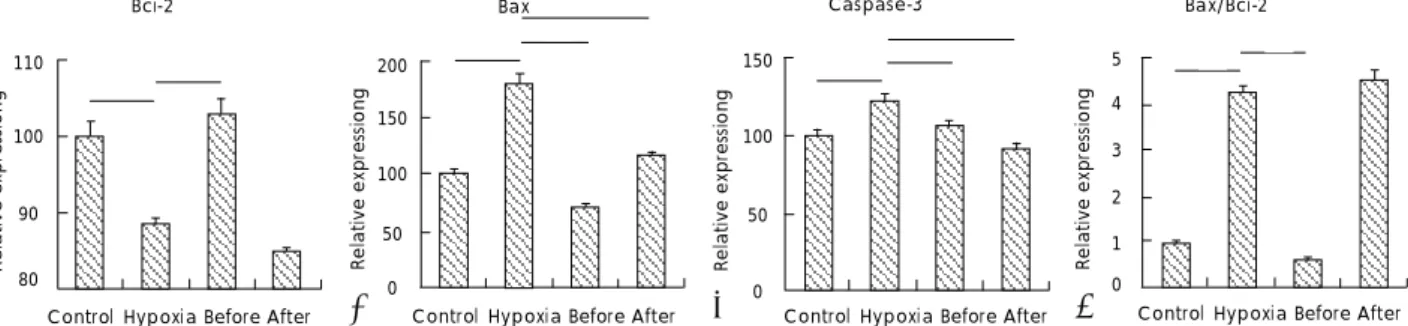
Expressions of Bcl-2, Bax, and caspase-3 measured by real-time PCR with treatment of resveratrol in hypoxic injury of cultured H9c2 cells
The mRNA expression of the apoptotic marker, Bcl-2, in the hypoxia group(42.34) was significantly decreased as compared with the control group(100). Treatment with resveratrol prior to hypoxic insult significantly inhi- bited the hypoxia-induced decline in Bcl-2 mRNA ex- pression(114.08), but there no significant change was observed in the resveratrol-treated group after hypoxic insult(25.17)(Fig. 3A). In contrast, the mRNA expression
of the pro-apoptotic marker, Bax, in the hypoxia group (179.98) was significantly increased compared to that in the control group(100). The hypoxia-induced incline in Bax was significantly inhibited in both resveratrol groups, and the inhibition was greater in the resver- atrol group treated before hypoxic insult(70.43) than in the group treated after hypoxic insult(114.65)(Fig.
3B). The mRNA expression of the other pro-apoptotic marker, caspase-3, was greater in the hypoxia group (121.42) than in the control group(100). The hypoxia- induced incline in caspase-3 was significantly inhibited in both of the resveratrol groups(105.70, 91.38)(Fig. 3C).
The ratio of Bax/Bcl-2 expression was greater in the hypoxia group(4.25) than in the control group (1), and was significantly decreased in the resveratrol group treated before hypoxic insult(0.62). However, there was no significant change in the group treated after hypoxic insult(4.55) compared with the hypoxia group(Fig. 3D).
Discussion
At the cellular/molecular level, resveratrol acts by modulating numerous intracellular target proteins,
18)but little is known about the exact cellular and molecular mechanisms by which resveratrol protects against coro-
Control Hypoxia Before After
Fig. 2.
Western blotting of Bcl-2 (A), Bax (B), and caspase-3 (C) from cultured H9c2 cells. The ratio of Bax/Bcl-2 (D) expression is also shown.
Resveratrol was administered at 10 μg/mL. Immunoblots from three independent experiments are shown. Densitometric analysis is also shown.
Data are presented as the ratios of band intensities compared with the control group. Before: resveratrol group treated before hypoxic exposure, After: resveratrol group treated after hypoxic exposure. *: p<0.05 compared with control.
1.2
1.1
1
Relative expressiong0.9
*
Bax/Bci-2110
100
90
Relative expressiong 80
* *
Bcl-2
110
100
90
Relative expressiong 80
Caspase-3
*
110*
100
90
Relative expressiong 80
* *
Bax
*
A
Control Hypoxia Before After Control Hypoxia Before After Control Hypoxia Before After
* *
B C D
*
*
200
150
100
50
0
Relative expressiong
* *
Bax
*
Control Hypoxia Before After 150
100
50
0
Relative expressiong
* *
Caspase-3
*
Control Hypoxia Before After 5 4
3
2
1 0
Relative expressiong
Control Hypoxia Before After
* *
Bax/Bci-2
Fig. 3.
Real-time PCR was performed for Bcl-2 (A), Bax (B), and caspase-3 (C) on DNA from cultured H9c2 cells. The ratio of Bax/Bcl-2 (D) expression is also shown. Resveratrol was administered at 10 μg/mL. Data are presented as the ratios of mRNA expression compared with those of the control group. Before: resveratrol group treated before hypoxic exposure, After: resveratrol group treated after hypoxic exposure. *: p<0.05 compared with control. PCR: polymerase chain reaction, DNA: deoxyribonucleic acid, mRNA: messenger ribonucleic acid.
110
100
90
Relative expressiong 80
* *
Bci-2
Control Hypoxia Before After