ABSTRACT
BACKGROUND/OBJECTIVES: The study was conducted to investigate the efficacy of the combination treatment of phytochemical resveratrol and the anticancer drug docetaxel (DTX) on prostate carcinoma LNCaP cells, including factors related to detailed cell death mechanisms.
MATERIALS/METHODS: Using 2-dimensional monolayer and 3-dimensional spheroid culture systems, we examined the effects of resveratrol and DTX on cell viability, reactive oxygen species (ROS) levels, mitochondrial membrane potential, apoptosis, and necroptosis by MTT, flow cytometry, and Western blotting.
RESULTS: At concentrations not toxic to normal human prostate epithelial cells, resveratrol effectively decreased the viability of LNCaP cells depending on concentration and time. The combination treatment of resveratrol and DTX exhibited synergistic inhibitory effects on cell growth, demonstrated by an increase in the sub-G
0/G
1peak, Annexin V-phycoerythrin positive cell fraction, ROS, mitochondrial dysfunction, and DNA damage response as well as concurrent activation of apoptosis and necroptosis. Apoptosis and necroptosis were rescued by pretreatment with ROS scavenger N-acetylcysteine.
CONCLUSIONS: We report resveratrol as an adjuvant drug candidate for improving the outcome of treatment in DTX therapy. Although the underlying mechanisms of necroptosis should be investigated comprehensively, targeting apoptosis and necroptosis simultaneously in the treatment of cancer can be a useful strategy for the development of promising drug candidates.
Keywords: Prostate cancer; resveratrol; docetaxel; apoptosis; necroptosis; oxidative stress
INTRODUCTION
Many natural bioactive substances derived from plants or microorganisms have shown promising potential for the prevention and treatment of various types of cancer. In particular, their use is advantageous in that they are safe to use and can reduce the high costs associated with the development of therapeutic drugs. Among them, phenylpropanoids are secondary metabolites produced by plants in response to various types of stress, including reactive oxygen species (ROS), pathogens or herbivore attack [1]. Stilbenes belongs to the family of phenylpropanoids containing a 1,2-diphenylethylene backbone. Resveratrol (trans-3,4′,5-
Original Research
Received: May 24, 2020 Revised: Jun 26, 2020 Accepted: Jul 28, 2020
§
Corresponding Author:
Yoon-Jin Lee
Department of Biochemistry, College of Medicine, Soonchunhyang University, 31 Soonchunhyang 6-gil, Dongnam-gu, Cheonan 31151, Korea.
Tel. +82-41-570-2440 Fax. 82-41-579-6080 E-mail. [email protected]
©2021 The Korean Nutrition Society and the Korean Society of Community Nutrition This is an Open Access article distributed under the terms of the Creative Commons Attribution Non-Commercial License (https://
creativecommons.org/licenses/by-nc/4.0/) which permits unrestricted non-commercial use, distribution, and reproduction in any medium, provided the original work is properly cited.
ORCID iDs Sang-Han Lee
https://orcid.org/0000-0001-6407-9959 Yoon-Jin Lee
https://orcid.org/0000-0001-6364-1284 Funding
This research was supported by the Basic Science Research Program through the National Research Foundation (NRF) of Korea, funded by the Ministry of Education (No. NRF- 2017R1D1A3B04031891).
Conflict of Interest
The authors declare no potential conflicts of interests.
Sang-Han Lee and Yoon-Jin Lee §
Department of Biochemistry, College of Medicine, Soonchunhyang University, Cheonan 31151, Korea
Synergistic anticancer activity of resveratrol in combination with
docetaxel in prostate carcinoma cells
Author Contributions
Conceptualization: Lee YJ, Lee SH; Data curation: Lee YJ, Lee SH; Formal analysis:
Lee YJ, Lee SH; Investigation: Lee YJ; Project administration: Lee YJ; Writing - original draft: Lee SH; Writing - review & editing: Lee YJ, Lee SH.
trihydroxystilbene, resveratrol [RSV]), a stilbene, consists of 2 phenolic rings linked by an ethylene bridge [2]. RSV shows various chemopreventive and chemotherapeutic properties resulting from anti-oxidative, anti-inflammatory, anti-carcinogenic and anti-proliferative activities [2,3]. For instance, it inhibits cancer cell growth and regulates apoptosis by regulating the action of various pro- and anti-apoptotic factors that affect apoptosis, reduce inflammatory reactions, and neutralize ROS [3,4]. In this process, several signaling pathways involved in cell survival and death are targets of its mechanisms of action. In addition to chemopreventive effects, many researchers have noted the role of RSV in enhancing the efficacy of traditional chemotherapy. In particular, RSV showed synergistic effects with anticancer agents in cells of several types of cancer including of the lung, cervical and prostate cancers [5-7]. Another study on RSV revealed that when combined with docetaxel (DTX), the synergistic effect of RSV on prostate cancer cells is induced by G
2/M cell cycle arrest and apoptosis via the p53/p21
WAF1/CIP1and p27
KIP1signaling [8].
Induction of apoptosis is one of the main criteria in evaluating the efficacy of candidates for the development of anticancer drugs. Evasion of apoptosis is known to be a prominent feature of tumorigenesis and progression in human cancer [9], and resistance to
conventional therapies is inevitably encountered during chemotherapy. Thus, a compound that can activate alternative cell death pathways to apoptosis would be extremely valuable.
Among forms of non-apoptotic cell death, necrosis has been considered to be an accidental, unregulated type of cell death that is triggered by various factors external to the cells or tissues, such as infection, toxins, insufficient blood perfusion, and ROS [10]. In some situations, necrosis can be executed and regulated in a manner similar to programmed cell death. Receptor-interacting protein kinase-3 (RIP3) and its substrate, the mixed lineage kinase domain-like protein (MLKL) were activated by phosphorylation to form a necrosome that subsequently triggered cell death [11,12]. This regulated form of necrosis is referred to as necroptosis and is specifically inhibited by necrostatin-1, a specific inhibitor of RIP1 activity [12]. Given the various forms of death that can occur in cancer cells under stressful conditions, it is important to identify the underlying cell death mechanisms and related regulators in developing new anticancer strategies.
DTX is the first-line treatment for metastatic and hormone-refractory prostate cancer; it is a taxane derivative of Taxus baccata, also known as the European yew tree, and it displays potent antitumor activity [13]. DTX inhibits microtubule depolymerization by binding to and stabilizing tubulin, resulting in growth arrest and apoptosis [14]. Combination therapy is believed to enhance cancer-killing actions through simultaneous targeting of multiple oncogenic factors, or more effective inhibition of multiple pathways, incuding antioxidant response, cell survival, hypoxia, apoptosis, angiogenesis, and epigenetic modulation pathways [15]. Various combinatorial trials have also been conducted to improve the efficacy of DTX in prostate cancer, and these include doxorubicin, prednisone, estramustine, and anthracycline [16-19], and research on the efficacy of neoadjuvants is ongoing.
Androgen deprivation therapy is one of the main treatment options for patients with
early-stage androgen-dependent prostate cancer, but almost all patients proceed to
develop androgen-independent prostate cancer in a few years. Progress to an incurable and
irreversible androgen-independent stage remains a major obstacle to effective treatment,
and a strong need for alternative treatment strategies is being emphasized. The LNCaP cells
used in this study are androgen-dependent prostate cancer cells, and have been used most
frequently in in vitro models for basic and preclinical studies of prostate cancer.
Using prostate carcinoma LNCaP cells, we present here a novel mechanism of cell death that is induced by the combination treatment of DTX and RSV targeting apoptosis as well as necroptosis, which commonly triggers ROS-induced DNA damage. Our data also provided the basis for a potential therapeutic strategy that uses RSV as an adjuvant in treating prostate carcinoma.
MATERIALS AND METHODS
Reagents and cell culture
Dimethylsulfoxide (DMSO), RSV, DTX, 3-(4,5-dimethylthiazol-2-yl)-2,5-diphenyltetrazolium bromide (MTT reagent), lactic acid, fluorescein diacetate (FDA), propidium iodide (PI), phosphate buffered saline (PBS), N-acetylcysteine (NAC), necrostatin-1, Q-VD-Oph-1, casein blocking buffer, 2′,7′-dichlorodihydrofluorescein diacetate (DCF-DA), rhodamine 123, and Tween-20 were purchased from Sigma-Aldrich (Merck KGaA, Darmstadt, Germany).
Antibodies to MLKL (catalog No. 14993), p-MLKL (Catalog No. 91689), RIP3 (catalog No.
13526), p-RIP3 (catalog No. 93654), p-ATM
Ser1981(catalog No. 5883), p-ATR
Ser428(catalog No. 2853), p-Histone H2A.X
Ser139(catalog No. 9718), Bax (catalog No. 5023), Bcl-2 (catalog No. 2820), poly ADP-ribose polymerase (PARP; catalog No. 9542), cleaved PARP (catalog No. 9541), and cleaved caspase-3 (catalog No. 9664) were purchased from Cell Signaling Technology, Inc. (Danvers, CO, USA). Goat anti-rabbit immunoglobulin G-horseradish peroxidase (IgG-HRP; catalog No. sc-2004), mouse anti-goat IgG-HRP (catalog No. sc- 2354), and goat anti-rabbit IgG-HRP (catalog No. sc-2005) were purchased from Santa-Cruz Biotechnology (Dallas, TX, USA). LNCaP (human prostate cancer) and human prostate epithelial cell (HPrEC) lines were acquired from the American Type Culture Collection (ATCC; Manassas, USA). HPrECs grew in prostate epithelial cell basal medium supplemented with prostate epithelial cell growth kit (ATCC). LNCaP cells grew in DMEM (Welgene Inc., Gyeongsan, Korea) supplemented with 5% fetal bovine serum.
Cell viability assay
Cells were seeded into 96-well plates at 5 × 10
4cells/mL in 100 μL complete medium overnight, after which, cells were treated with vehicle (DMSO), RSV, and DTX for the times indicated in the figure legends. MTT reagent was added to each well and incubated for 4 h at 37°C. Absorbance values were read at 540 nm using a GloMax-Multi microplate multimode reader (Promega Corporation, Durham, NC, USA). The percentage viability of cells was determined by comparison with the results obtained using vehicle-treated cells (100%).
The combination effect of the 2 drugs was evaluated using the combination index (CI) as described previously [20]. CI values < 1, equal to 1, and > 1 were defined as synergistic, additive, and antagonistic effects, respectively.
Cell cycle analysis
Cell cycle was analyzed by quantification of DNA content in cells stained with PI. Briefly,
cells were harvested, fixed with 70% ethanol, and left at −20°C overnight. After washing
and resuspension in 1× PBS, the Muse
TMcell cycle reagent (Merck Millipore, Burington,
MA, USA) was added to the cells. The DNA distribution from 10,000 cells was analyzed by
a MACSQuant analyzer and MACSQuantify
TMsoftware version 2.5 (Miltenyi Biotec GmbH,
Bergisch, Germany).
Annexin V-phycoerythrin (V-PE) binding assay
Apoptotic and necrotic cell distributions were determined using the Muse
TMAnnexin V &
Dead Cell Assay kit (Merck KGaA). Briefly, cells were harvested by centrifugation and labeled with Annexin V-PE and 7-amino-actinomycin D for 20 min at room temperature in the dark.
Cells (5 × 10
3) were then analyzed by Muse
TMcell analyzer (Merck KGaA).
Western blot analysis
Cells were washed and lysed with 1× RIPA buffer and protein quantitation was performed using a BCA protein assay kit (Thermo Fisher, Waltham, MA, USA). Cell lysates containing 40 μg of protein were loaded on 4–12% NuPAGE gel (Thermo Fisher) for electrophoresis and transferred to polyvinylidene difluoride membrane (GE Healthcare Life Science, Munich, Germany). After blocking with 1× casein blocking buffer (Sigma-Aldrich) for 2 h, the membrane was incubated with various primary antibodies overnight at 4°C. After washing with 1× PBS with Tween-20, the membranes were incubated for 2 h at room temperature with secondary antibodies coupled to HRP for protein detection. Reactive proteins were visualized on an X-ray film using enhanced chemiluminescence detection kit (Cyanagen Srl, Bologna, Italy). The membrane was re-probed with antibodies to anti-ß-actin (Sigma-Aldrich) and served as a loading control.
Measurement of ROS and mitochondrial membrane potential
Cells were seeded in 6-well plates at 10
5cells/mL in 2 mL complete medium/well overnight, after which cells were treated with the vehicle, RSV, DTX or RSV/DTX in complete DMEM for the indicate times in figure legends. Cells were harvested by centrifugation and then stained with 10 µM DCF-DA and 30 nM rhodamine 123 to measure the levels of ROS and mitochondrial membrane potential (ΔΨm), respectively, in the dark at 37˚C for 30 min. After washing cells with 1× PBS, the average fluorescence intensity of 10,000 cells was measured using the MACSQuant analyzer and MACSQuantify
TMsoftware version 2.5 (Miltenyi Biotec GmbH).
Spheroid culture and viability assay
Cells (10
4cells/well) were seeded in ultra-low attachment 96-well plates. The plates were
centrifuged at 1,000 rpm for 10 min to facilitate clustering of the cells into the wells, as described by Chambers et al. [21], and maintained in the complete DMEM. Spheroids were treated with curcumin for 48 h. Phase-contrast images were taken using a Leica inverted microscope.
Spheroid viability was determined using an enhanced cell viability assay kit (CellVia, Seoul, Korea). Briefly, 10 µL of Cellvia solution was added to each well, kept at room temperature for 1 h and then mixed by shaking for 1 min. Formazan formed in living cells was measured at 450 nm using a GloMax-Multi microplate multimode reader (Promega Corporation).
Spheroid staining
Cells were incubated with FDA (5 µg/mL) and PI (10 µg/mL) in the dark to stain viable and dead cells respectively. The FDA, a cell-permeable esterase substrate, indicates viable cells by assessing enzymatic activity and cell-membrane integrity, while PI passes through damaged areas of dead or dying cell membranes into the nucleus and binds to DNA. Cells were imaged using a Leica EL6000 fluorescence microscope and LAS version 4.3 software (Leica Microsystems Inc., Buffalo Grove, IL, USA).
Statistical analysis
We conducted statistical comparisons using analysis of variance and Tukey's post hoc
correction in SPSS 17.0 (SPSS, Inc., Chicago, IL, USA). Data were shown as mean ± SD for 3
independent experiments. P < 0.05 means statistical significance.
RESULTS
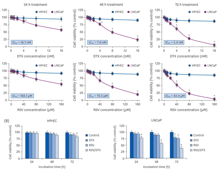
The combination of resveratrol and DTX causes synergistic cytotoxicity We first investigated the effects of RSV and anticancer drug DTX, alone or in combination, on the survival of prostate carcinoma LNCaP cells. Fig. 1A shows that RSV or DTX alone effectively decreased the viability of LNCaP cells depending on concentration and time;
IC
50values at 24 h, 48 h, and 72 h of RSV exposure were 183.1 μM, 72.5 μM, and 43.4 μM, respectively; and 19.7 nM, 7.9 nM, and 5.4 nM for DTX exposure, respectively. However, we observed little toxicity in normal HPrECs in response to RSV and DTX at the tested concentrations, indicating the selectivity of RSV and DTX to cancer cells. We then
investigated the cytotoxic effects of 20 μM RSV in combination with 2 nM DTX (RSV/DTX) on both cell types after 24, 48, and 72 h of exposure. Fig. 1B indicates that RSV augmented the cytotoxic effect of DTX on LNCaP cells at all time points tested, as compared to RSV or DTX alone. Next, we evaluated the CI to determine whether the combined effect of RSV and DTX
(A)
HPrEC LNCaP HPrEC LNCaP HPrEC LNCaP
HPrEC LNCaP HPrEC LNCaP HPrEC LNCaP
Cell viability (% control) Cell viability (% control)
125 100 75 50 25 0
125 100 75 50 25 0
0 4 8 12 16
DTX concentration (nM)
0 40 80 120 160
RSV concentration (µM)
Cell viability (% control) Cell viability (% control)
125 100 75 50 25 0
125 100 75 50 25 0
0 4 8 12 16
DTX concentration (nM)
0 40 80 120 160
RSV concentration (µM)
Cell viability (% control) Cell viability (% control)
125 100 75 50 25 0
125 100 75 50 25 0
0 4 8 12 16
DTX concentration (nM)
0 40 80 120 160
RSV concentration (µM) 24 h treatment
* *
*
*
*
*
*
*
*
* *
* *
*
*
*
*
*
48 h treatment 72 h treatment
IC
50= 19.7 nM IC
50= 7.9 nM IC
50= 5.4 nM
IC
50= 183.1 µM IC
50= 72.5 µM IC
50= 43.4 µM
(B)
125 100 75 50 25
0 24 48 72 24 48 72
Incubation time (h) HPrEC
Cell viability (% control)
Incubation time (h)
#
125 100 75 50 25 Cell viability (% control) 0
LNCaP
#
#
#
# #
Control
DTX RSV RSV/DTX Control
DTX RSV RSV/DTX
Fig. 1. Synergistic growth-inhibiting effects of RSV and DTX in LNCaP and HPrECs. (A) Cells were treated with increasing concentrations of RSV (0, 5, 10, 20, 40, 80, and 160 μM) and DTX (0, 0.5, 1, 2, 4, 8, and 16 nM) for the indicated times. (B) Cells were treated with RSV (20 μM) and DTX (2 nM) alone or in combination for 24, 48, and 72 h. The percentage of viable cells was then determined by MTT assay. Error bars represent the mean ± SD for 3 independent experiments.
RSV, resveratrol; DTX, docetaxel; RSV/DTX, the combination of resveratrol and docetaxel; HPrEC, human prostate epithelial cell.
* P < 0.05 compared with respective HPrEC cells;
#P < 0.05 compared with respective untreated cells.
was synergistic. The CI values at 24 h, 48 h, and 72 h of RSV/DTX were 0.73, 0.68, and 0.72, respectively, indicating that RSV cooperated synergistically with DTX to inhibit cell viability of LNCaP cells.
Resveratrol alone or in combination with DTX induces cell cycle transition delay at G2/M phase and DNA damage response (DDR)
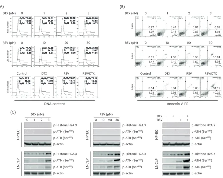
We conducted cell cycle analysis to elucidate how RSV/DTX decreased cell viability. The results of cell cycle analysis (Fig. 2A) showed that RSV/DTX caused G
2/M-phase accumulation with an increase in subG
0/G
1content, indicating apoptotic cell death. In Annexin V-PE staining assay, RSV/DTX remarkably increased the percentage of apoptotic cell populations (Annexin V-PE positive), as compared to RSV or DTX alone (Fig. 2B). Next, to investigate the ability of RSV/DTX to activate the DNA damage sensor kinases such as ataxia telangiectasia mutated (ATM) and ATM and RAD3-related (ATR), LNCaP cells were treated with RSV and DTX alone or in combination, after which the phosphorylated forms of ATM
Ser1981, ATR
Ser428,
Cell number
DNA content Annexin V-PE
Cell number
G0/G176.81 S 15.61 G2/M 7.59
G0/G177.31 S 14.81 G2/M 7.89
G0/G177.62 S 13.68 G2/M 8.70
G0/G175.66 S 13.63 G2/M 10.71
G0/G177.70 S 15.46 G2/M 6.84
G0/G177.88 S 14.08 G2/M 8.04
G0/G176.23 S 13.81 G2/M 9.96
G0/G175.02 S 13.67 G2/M 11.31
G0/G177.21 S 15.73 G2/M 7.06
G0/G177.54 S 14.00 G2/M 8.46
G0/G176.07 S 13.71 G2/M 10.22
G0/G172.11 S 13.49 G2/M 14.40
Cell number
0.27 1.07
3.47 2.15
6.01 2.87
8.09 4.94
7-AAD
0.12 1.47
4.33 2.19
6.53 3.52
10.81
7-AAD5.08
0.14 1.31
5.34 2.93
5.63 3.66
31.12 10.32
7-AAD
(A) (B)
DTX (nM)
RSV (µM) 0
0
1
10
2
20
3
Control DTX RSV RSV/DTX
30
DTX (nM)
RSV (µM) 0
0
1
10
2
20
3
Control DTX RSV RSV/DTX
30
(C)
0 1 2 3 0 10 20 30
DTX (nM)
p-Histone H2A.X
p-ATR (Ser
428) β-actin p-ATM (Ser
1981)
p-Histone H2A.X
p-ATR (Ser
428) β-actin p-ATM (Ser
1981) p-Histone H2A.X
p-ATR (Ser
428) β-actin p-ATM (Ser
1981)
p-Histone H2A.X
p-ATR (Ser
428) β-actin p-ATM (Ser
1981)
p-Histone H2A.X
p-ATR (Ser
428) β-actin p-ATM (Ser
1981) p-Histone H2A.X
p-ATR (Ser
428) β-actin p-ATM (Ser
1981)
HPrEC
DTX −
− +
− −
+ +
RSV + RSV (µM)
HPrEC HPrEC
LNCaP LNCaP LNCaP
Fig. 2. Cytotoxic effects induced by RSV and DTX in LNCaP cells. Cells were treated with indicated concentrations of RSV and DTX, otherwise RSV (20 μM) and DTX (2 nM) alone or in combination for 48 h. (A) Cell cycle distribution was determined by flow cytometry following PI (20 μg/mL) staining. (B) Apoptotic cell fraction was analyzed using Annexin V-PE binding assay. (C) The levels of DNA damage response proteins were assessed by Western blotting.
RSV, resveratrol; DTX, docetaxel; PI, propidium iodide; ATM, ataxia telangiectasia mutated; ATR, ataxia telangiectasia mutated and RAD3-related.
and histone H2A variant H2A.X
Ser139as a marker of DNA double-strand breaks were measured by immunoblotting. As shown in Fig. 2C, RSV/DTX potently increased phosphorylation of ATM
Ser1981, ATR
Ser428, and H2A.X
Ser139, suggesting that RSV/DTX triggers activation of the ATM/
ATR signaling pathway with DNA double-strand breaks.
Synergistic cytotoxicity of resveratrol and DTX concurrently induces apoptosis and necroptosis
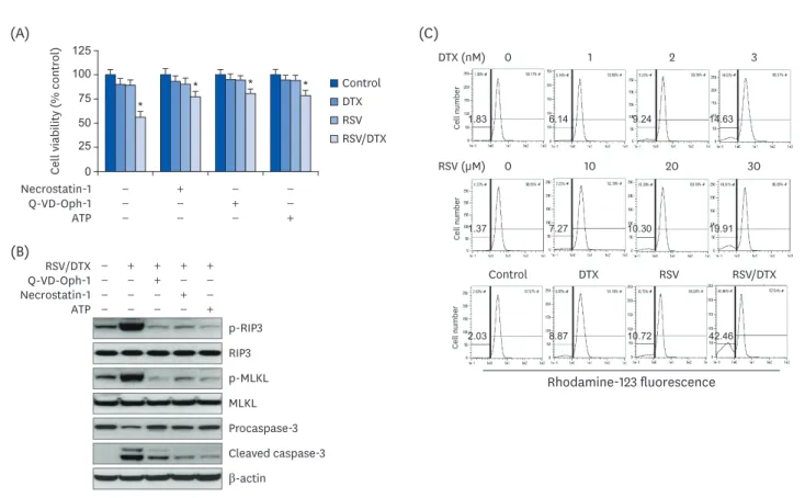
To elucidate the mechanisms underlying synergistic cytotoxicity of RSV/DTX, we measured levels of proteins involved in apoptosis and necroptosis by Western blotting. As shown in Fig. 3, RSV/DTX further increased the cleaved forms of caspase-3 and its substrate PARP as well as Bcl-2-associated X protein/B-cell lymphoma 2 (Bax/Bcl-2) ratio, compared with those induced by RSV and DTX alone. Furthermore, RSV/DTX increased the levels of p-MLKL and p-RIP3 as necroptosis mediators. Inhibition of either necroptosis or of apoptosis by pretreatment with necrostatin-1 or Q-VD-Oph-1, respectively, enhanced cell viability (Fig. 4A) and reversed the levels of apoptosis- or necroptosis-inducing molecules in LNCaP cells treated with RSV/DTX (Fig. 4B). This strongly suggests that RSV/DTX-induced cell death is mediated by
(A) (B)
0 1 2 3 0 1 2 3
DTX (nM)
Bcl-2
Procaspase-3 Cleaved caspase-3 BAX
β-actin PARP Cleaved PARP
HPrEC
DTX −
− +
− −
+ +
RSV + RSV (µM)
HPrEC LNCaP
LNCaP
p-RIP3
p-MLKL MLKL RIP3
Bcl-2
Procaspase-3 Cleaved caspase-3 BAX
β-actin PARP Cleaved PARP
p-RIP3
p-MLKL MLKL RIP3
Bcl-2
Procaspase-3 Cleaved caspase-3 BAX
β-actin PARP Cleaved PARP
p-RIP3
p-MLKL MLKL RIP3
Bcl-2
Procaspase-3 Cleaved caspase-3 BAX
β-actin PARP Cleaved PARP
p-RIP3
p-MLKL MLKL RIP3
Bcl-2
Procaspase-3 Cleaved caspase-3 BAX
β-actin PARP Cleaved PARP
p-RIP3
p-MLKL MLKL RIP3
Bcl-2
Procaspase-3 Cleaved caspase-3 BAX
β-actin PARP Cleaved PARP
p-RIP3
p-MLKL MLKL RIP3
HPrEC LNCaP
Fig. 3. Expression of apoptosis- and necroptosis-related proteins by RSV and DTX in LNCaP and HPrECs. (A) Cells were treated with increasing concentrations of RSV (0, 10, 20, and 30 μM) and DTX (0, 1, 2, and 3 nM) for 48 h. (B) Cells were treated with RSV (20 μM) and DTX (2 nM) alone or in combination for 48 h. The levels of apoptosis- and necroptosis-related proteins were assessed by Western blotting.
RSV, resveratrol; DTX, docetaxel; Bcl-2, B-cell lymphoma 2; BAX, Bcl-2-associated X protein; PARP, poly ADP-ribose polymerase; RIP3, receptor-interacting
protein kinase-3; MLKL, mixed lineage kinase domain-like protein; HPrEC, human prostate epithelial cell.
concurrent induction of both apoptosis and necroptosis. Previous evidence has demonstrated that cellular adenosine triphosphate (ATP) levels controlled both cell death processes [22].
To determine the role of ATP in the synergistic cytotoxicity of RSV/DTX, we examined the effects of ATP supplementation in RSV/DTX-treated LNCaP cells. Adding ATP improved cell viability (Fig. 4A) and decreased the levels of apoptosis- or necroptosis-inducing molecules (Fig. 4B). Next, we measured changes in ΔΨm by RSV/DTX to evaluate mitochondrial function.
The combination treatment caused a marked loss of ΔΨm in 42.46% of the cell population compared to RSV (10.72%) or DTX (8.87%) alone (Fig. 4C). Taken together, these findings indicated that RSV/DTX-induced cell death was caused, at least in part, by ATP depletion.
Excessive ROS is an upstream molecule that causes synergistic cytotoxicity of resveratrol and DTX
To investigate whether intracellular ROS was involved in the cytotoxicity induced by RSV/
DTX in LNCaP cells, we performed DCF fluorescence assay using flow cytometry. With the increasing RSV or DTX concentration, cellular ROS levels tended to gradually increase but showed a marked increase at 48 h of combination treatment (Fig. 5A). In addition, pretreatment of LNCaP cells with ROS scavenger NAC effectively improved a series of effects induced by RSV/DTX, including cellular ROS levels (Fig. 5B), percentage of cells showing ΔΨm loss ( Fig. 5C), Annexin V-PE positivity (Fig. 5D), and transition delay in the G
2/M phase of the cell cycle (Fig. 5E).
Cell viability (% control)
(C)
DTX (nM)
RSV (µM) 0
0
1
10
2
20
3
Control DTX RSV RSV/DTX
30
Cell numberCell number
Rhodamine-123 fluorescence
Cell number
1.37
6.14 1.83
7.27
9.24 14.63
10.30 19.91
8.87
2.03 10.72 42.46
125 100 75 50 25 0
(A)
− + − −
Necrostatin-1
Q-VD-Oph-1 − − + −
ATP − − − +
(B)
− − + − −
Necrostatin-1 Q-VD-Oph-1
− − − + −
ATP − − − − +
RSV/DTX − + + + +
p-RIP3 RIP3 p-MLKL MLKL
Cleaved caspase-3 β-actin
Procaspase-3
*
* * * Control
DTX RSV RSV/DTX
Fig. 4. Apoptosis, necroptosis, and oxidative mitochondrial dysfunction in LNCaP cell treated with RSV and DTX. Cells were pretreated with necrostatin-1 (25 μM), Q-VD-Oph-1(10 μM), and ATP (1 mM) 2 h prior to treatment with RSV (20 μM) and DTX (2 nM) in combination. (A) The percentage of viable cells was then determined by MTT assay. (B) The levels of apoptosis- and necroptosis-related proteins were assessed by Western blotting. Cells were treated with indicated concentrations of RSV and DTX, otherwise RSV (20 μM) and DTX (2 nM) alone or in combination for 48 h. (C) ΔΨm was measured after cells were stained with 30 nM rhodamine 123.
RSV, resveratrol; DTX, docetaxel; RSV/DTX, the combination of resveratrol and docetaxel; RIP3, receptor-interacting protein kinase-3; MLKL, mixed lineage kinase domain-like protein; ATP, adenosine triphosphate.
* P < 0.05 vs. respective untreated cells.
To determine whether the results of 2-dimensional (2D) monolayer culture matched in 3-dimensional (3D) culture, spheroids derived from LNCaP cells were pretreated with NAC for 2 h before being processed for another 48 h with RSV/DTX. Viable and dead cells were visualized under fluorescence microscope after double staining with FDA (green) and PI (red), respectively. RSV/DTX increased red fluorescence in the necrotic core with green fluorescence on the spheroid surface, which correlated with decreased spheroid viability;
however, this phenomenon was blocked by NAC pretreatment. Next, we investigated whether the expression of proteins related to apoptosis- and necroptosis are affected by RSV/DTX. As shown in Fig. 6C, RSV/DTX increased p-RIP3, p-MLKL, Bax, and cleaved form of caspase-3 and PARP proteins, and decreased Bcl-2, but NAC pretreatment significantly recovered changes in these proteins. Changes in cell viability and cell death-related protein levels observed in the 3D spheroid culture for RSV/DTX showed similar trends as those found in 2D monolayer culture.
DISCUSSION
Our studies showed that RSV synergistically augmented the anticancer effects of DTX by increasing both apoptosis and necroptosis in 2D monolayer and 3D spheroid culture models of prostate carcinoma LNCaP cells. Sensitizing effects of RSV to DTX were elicited at least in part through ROS-induced mitochondrial dysfunction and DNA damage, suggesting RSV as an adjuvant candidate for DTX therapy in prostate carcinoma.
NA C (+) NA C (−) NA C (+) NA C (−)
NA C (+) NA C (−) NA C (+) NA C (−)
Control RSV/DTX
Control RSV/DTX
Control RSV/DTX
Control RSV/DTX
DCF-DA fluorescence
Cell numberCell number
1.67 6.74 11.74 17.60
2.10 6.60 9.03 13.13
Cell number
1.26 9.07 10.26 42.81
0.26 1.62
30.59 8.84
7-AAD
0.20 0.93
19.03
7-AAD2.77
Annexin V-PE DNA content
Cell numberCell number
G0/G178.55 S 15.26 G2/M 6.18
G0/G175.59 S 13.61 G2/M 10.79
DCF-DA fluorescence
Cell number
1.46 22.17
1.71 42.18
Cell number
Rhodamine-123 fluorescence
Cell number
1.81
1.62 41.24
23.06
Cell number
G0/G177.05 S 16.01 G2/M 6.94
G0/G172.32 S 13.03 G2/M 14.65