307 http://dx.doi.org/10.4196/kjpp.2011.15.5.307
ABBREVIATIONS: 5-HT, 5-hydroxytryptamine; EFS, electrical field stimulation; CCK, cholecystokinin.
Received September 21, 2011, Revised October 14, 2011, Accepted October 17, 2011
Corresponding to: Yun-Lyul Lee, Department of Physiology, College of Medicine, Hallym University, 39, Hallymdaehak-gil, Chuncheon 200-702, Korea. (Tel) 82-33-248-2583, (Fax) 82-33-248-1640, (E-mail) [email protected]
Roles of Non-cholinergic Intrapancreatic Nerves, Serotonergic Nerves, on Pancreatic Exocrine Secretion in the Isolated Perfused Rat Pancreas
Zheng er Jiang1, Bich-Na Shin2, In-Hye Kim2, Hyun Joo Lee2, Jun-Hwan Yong3, Min-Jae Lee4, Moo-Ho Won5, and Yun-Lyul Lee2
1Department of Physiology, College of Nursing, Yanbian University, Yanji 133000, China, 2Department of Physiology, College of Medicine, Hallym University, Chuncheon 200-702, 3Department of Occupational Therapy, Dongnam Health College, Suwon 440-714, 4Department of Veterinary Lab. Animal Medicine & Science, College of Animal Resource Science, 5Department of Neurobiology, School of Medicine, Kangwon National University, Chuncheon 200-701, Korea
It has been rereported that axons which display 5-hydroxytryptamine (5-HT) immunoreactivity are abundant in the pancreas and the majority of serotonergic axons terminate within intrapancreatic ganglia, islet and acini. This histological result strongly suggests that intrapancreatic serotonergic nerves could affect to the pancreatic endocrine and exocrine secretion. Thus, this study was aimed to investigate whether intrapancreatic serotonergic nerves could affect pancreatic exocrine secretion and an action mechanism of the intrapancreatic serotonergic nerves. The rats were anesthetized with a single injection of urethane. The median line and the abdominal aorta was carefully dissected and cannulated with PE-50 tubing just above the celiac artery, and then tightly ligated just below the superior mesenteric artery. The pancreatic duct was also cannulated with Tygon microbore tubing.
With the addition of serotonin, pancreatic volume flow and amylase output were significantly inhibited electrical field stimulation (EFS). On the other hand, pancreatic volume flow and amylase output were significantly elevated in EFS with the addition of spiperone. EFS application, however, pancreatic volume flow and amylase output had no significant change in cholecystokinin (CCK) alone when serotonin was applied under a 5.6 mM glucose background. Pancreatic volume flow and amylase output under 18 mM glucose background were significantly elevated in CCK plus serotonin than in CCK alone.
These data suggest that intrapancreatic serotonergic nerves play an inhibitory role in pancreatic exocrine secretion and an important role in the insulin action or release.
Key Words: Intrapancreatic nerve, Electrical stimulation, 5-hydroxytryptamine, Cholecystokinin, Insulin
INTRODUCTION
Serotonin (5-hydroxytryptamine; 5-HT) is synthesized and released in platelets of enterochromaffin cells of the gastrointestinal mucosa. A small quantity of serotonin ex- ists in the central nervous system [1] and most of serotonin exists in the myenteric plexus of the gastrointestinal sys- tem [2]. Although most serotonin exists in the gastro- intestinal tract, the roles of serotonin in the gut are still unclear. It is mainly reported that roles of serotonin re- leased from enteric nervous system or enterochromaffin cells in the intestinal mucosa [3]. Thus this study was fo- cused to fined whether intrapancreatic serotonergic nerves could affect pancreatic exocrine secretion in the ex vivo model.
The pancreas is a unique organ which has both endocrine and exocrine functions in the body. Over 20 kinds of diges-
tive enzymes, including bicarbonate and water, are secreted from the exocrine part for digestion of foods. Moreover, in- sulin, glucagon, somatostatin and pancreatic polypeptide are also secreted from the endocrine part. In spite of these dual actions of the pancreas, the effect of serotonin on the pancreas has been controversial. It has been reported that axons which display 5-HT immunoreactivity are abundant in the pancreas and the majority of serotonergic axons ter- minate within intrapancreatic ganglia, islet, and acini [4].
These histological results strongly suggest that intra- pancreatic serotonergic nerves could affect pancreatic endo- crine and exocrine secretion.
Pancreatic exocrine secretion is regulated by the auto- nomic nerve system and gastrointestinal hormones. Recen- tly, it has been reported that pancreatic exocrine secretion is regulated by the interaction of hormones and the neural system [5]. In addition, pancreatic exocrine secretion is af- fected by pancreatic islet hormones such as insulin and, so- matostatin [6]. As seen in the above results and reports, pancreatic exocrine secretion is controled by a complex set of factors. Pancreatic exocrine secretion was enhanced by
electrical stimulation (EFS) of the vagus nerve, but this en- hanced effect was partly blocked by atropine, a muscarinic receptor antagonist [7]. EFS on rat pancreas enhanced pan- creatic exocrine secretion by activation of the intra- pancreatic nervous system [8,9]. Pre-treatment of atropine partly blocked this effect, but pre-treatment of tetrodotoxin, a nerve blocker, perfectly blocked it [10]. These results sug- gested that the non-cholinergic nerve system exists in the intrapancreatic nerve system, and it could affect the pan- creatic exocrine secretion. There are both peptidergic nerves and cholinergic nerves in the intrapancreatic nerve system [11]. The serotonergic nerve is involved in this pep- tidergic nerve system [12] and may participate in pancre- atic exocrine secretion. Because the axons of serotonergic nerve are distributed close to the non-serotonergic intra- pancreatic nerves (mainly cholinergic nerve), the serotoner- gic nerves can affect pancreatic exocrine secretion indirectly via the affect of the cholinergic nerve. In addition, the sero- tonergic nerve can affect pancreatic exocrine secretion di- rectly via the affect of the acini and islet cell because the axons of the serotonergic nerves are distributed near acini and islet cells [12].
Thus, this study was aimed to investigate whether intra- pancreatic serotonergic nerves could affect pancreatic exo- crine secretion and an action mechanism of the intra- pancreatic serotonergic nerves.
METHODS Experimental animals
Sprague-Dawley rats were used in the experiment which succeeded in experimental animal center of Hallym Univer- sity. The environment of breeding room was maintained at condition that temperature was 23±2oC and relative humid- ity was 55±10%. Artificial lighting maintained 12 hours per day. The rats were anesthetized with a single intraperi- toneal injection of 20% urethane (Sigma, USA) at a dose of 0.7 ml/100 g of body weight. The rats were sacrificed by an intravenous overdose injection of urethane after iso- lation of the pancreas. Food was forbidden from rats for 24 h before the experiment, but they were allowed to drink water freely.
Preparation of the isolated perfused pancreas The isolated pancreas was prepared according to a meth- od described previously [10]. The rats were anesthetized with a single injection of urethane. The median line and the abdominal aorta was carefully dissected and cannulated with PE-50 tubing (I.D. 0.58 mm, O.D. 0.97 mm; Clay Adams, USA) just above the celiac artery, and then tightly ligated just below the superior mesenteric artery. The pan- creatic duct was also cannulated with Tygon microbore tub- ing (I.D. 1.27 mm, O.D. 2.28 mm; Fisher Scientific Co, USA) to drain the perfusate. To prevent a duodenal secretion in- flow each beginning and ending of duodenum was cannu- lated with plastic tubing.
The pancreas was perfused with modified Krebs- Henseleit solution (pH 7.4, 305 mosmol/kg water) through the celiac and superior mesenteric arteries at a flow rate of 1.2 ml/min using a minipuls pump (Gilson, France). The perfusate contained 5.6 mM glucose (Sigma, USA), 0.1% bo- vine serum albumin (Sigma, USA) and 3% Dextran T-70
(Sigma, USA), and was continuously oxygenated with 95%
O2 and 5% CO2. Krebs-Henseleit solution basically consists of NaCl 191.5 mM, NaHCO3 25 mM, KCl 5.6 mM, MgCl2
1 mM, NaH2PO4 1.15 mM and CaCl2 2.5 mM.
The pancreas was isolated with the duodenum but sepa- rated from neighboring organs and tissues. The pancreas was placed in temperature-controlled experimental cham- ber at 37oC, which was also continuously supplied with Krebs-Henseleit solution at a flow rate of 0.35 ml/min and oxygenated. After equilibration period of 30 min, pancreatic juice was collected through the experiment every 15-min.
Effects of EFS on pancreatic exocrine secretion To investigate effects of intrapancreatic neuronal activa- tion, electrical field stimulation (EFS) was carried out with applying biphasic square waves (parameters: 12 V, 2 ms and 8 Hz for 45 min) to the isolated pancreas through a pair of coiled platinum electrodes immersed in the ex- perimental chamber 5 cm apart [10]. Pancreatic juice was collected in every 15-min period.
Effects of CCK on pancreatic exocrine secretion To confirm pancreatic exocrine secretion stimulated by CCK-8 in the isolated perfused rat pancreas, CCK-8 (Squibb, USA) at a concentration of 5 pmol added to the perfusate contained 5.6 mM glucose for 60 minutes. In or- der to find effects of endogenous insulin on pancreatic exo- crine secretion, a concentration of glucose in the perfusate was elevated from 5.6 mM to 18 mM before 45 min admin- istration of CCK-8.
Amylase assay
To investigate pancreatic digestive enzyme secretion, ac- tivity of representative enzyme α-amylase was measured.
α-amylase activity in the pancreatic juice was determined by method of Rick and Stegbauer [13]. In brief, 1 ml starch solution (1%, W/V) is put in 1 ml phosphate buffer (10 mM KH2PO4, 10 mM Na2HPO4, 10 mM NaCl, pH 6.9) and mixed with 1,000∼3,000 times diluted pancreatic juice (20μl).
Then it reacted in 37oC incubator for 10 minutes. One % starch solution was heated in 100oC for 20 minutes to melt and centrifuged in 4oC at a speed of 3,000 rpm for 20 mi- nutes then the supernatant was used. After reaction 2 ml dinitrosalicylic acid solution (1% 3,5 -dinitrosalycylic acid, 30% potassium sodium tartrate, 400 mM NaOH) was put in it and heated in 100oC for 5 minutes to appear color of maltose, the reactive product. The reactive tube was cooling in room temperature for 30 minutes then measured a de- gree of extinction at 546 nm by spectrophotometer (Kon- tron, Swiss). Amylase activity 1 unit decided amount of amylase which produce 1μm maltose for 1 minute with above condition and converted each 15-min (amylase out- put; U/15 min). Pancreatic juice was collected in poly- ethylene tube which has volume of 2.4μl/cm then measured length to determine volume flow. All reagents were bought from Sigma (USA).
Statistical analysis
All data are expressed as mean±SE. Student's t test or paired t test was used for statistical analysis of the data.
The difference was considered significant when the p value
Fig. 1. Effect of serotonin (5-HT, 2μM) on electrical field stimu- lation (EFS, 12 V, 2 ms, 8 Hz)-induced pancreatic volume flow and amylase output. Each points (○, EFS alone; ●, EFS+5-HT) re- present the mean±S.E. of 7 experiments. *The value is significantly (p<0.05) different from that of EFS alone.
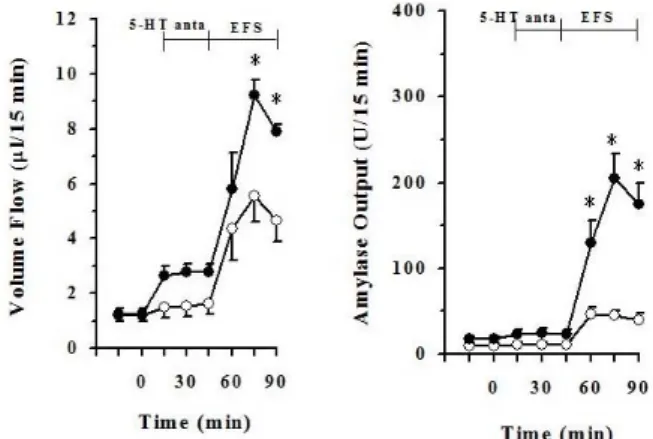
Fig. 2. Effect of spiperone hydrochloride (5-HT antagonist, 5μM) on electrical field stimulation (EFS, 12 V, 2 ms, 8 Hz)-induced pancreatic volume flow and amylase output. Each points (○, EFS alone; ●, EFS+5-HT antagonist) represent the mean±S.E. of 6∼7 experiments. *The value is significantly (p<0.05) different from that of EFS alone.
Fig. 3. Effect of spiperone hydrochloride (5-HT antagonist, 5μM) with atropine (ATR, 2μM) on electrical field stimulation (EFS, 12 V, 2 ms, 8 Hz)-induced pancreatic volume flow and amylase output.
Each points (○, EFS+ATR; ●, EFS+ATR+5-HT antagonist) represent the mean±S.E. of 6∼7 experiments. *The value is significantly (p<0.05) different from that of EFS plus atropine.
was <0.05.
RESULTS
Effects of serotonin on EFS-induced pancreatic exo- crine secretion
To investigate effects of serotonin (5-HT) on EFS-induced pancreatic exocrine secretion, serotonin (Sigma, USA) at a concentration of 2μM was perfused with the perfusate con- taining 5.6 mM glucose from 45 min earlier EFS applied to the end of the experiment, during total 90 min.
When EFS was applied to the pancreas as shown Fig.
1, pancreatic flow rate and amylase output were increased.
Pancreatic flow rate was elevated from 1.29±0.20μl/15 min in basal to 6.69±1.23μl/15 min in peak level. Amylase out- put was also elevated from 11.47±2.91μl/15 min to 49.71±8.24μl/15 min in peak level. When serotonin was added to the perfusate, however, the flow rate and amylase output were markedly decreased. Pancreatic flow rate was decreased from 6.69±1.23μl/15 min to 4.02±0.31μl/15 min in peak level. Amylase output was also decreased from 49.71±8.24μl/15 min to 21.58±2.92μl/15 min in peak level.
Effects of serotonin antagonist on EFS-induced pan- creatic exocrine secretion
To investigate effects of serotonin antagonist on EFS-in- duced pancreatic exocrine secretion, spiperone hydro- chloride (Tocris, USA), 5-HT2A serotonin antagonist, at a concentration of 5μM was perfused with the perfusate con- taining 5.6 mM glucose from 45 min earlier EFS applied to the end of the experiment.
When EFS applied to the pancreas as shown Fig. 2, pan- creatic flow rate and amylase output were increased. Pan- creatic flow rate was elevated from 1.18±0.09μl/15 min to 5.55±0.92μl/15 min in peak level. Amylase output was also elevated from 9.86±2.88μl/15 min to 45.74±9.42μl/15 min in peak level. Pancreatic flow rate of EFS plus spiperone, serotonin antagonist, was elevated from 1.24±0.20μl/15 min to 9.22±0.57μl/15 min in peak level. Amylase output of EFS plus spiperone was elevated from 17.56±2.81μl/15 min to 205.42±28.60μl/15 min in peak level. Pancreatic vol-
ume flow was significantly elevated in EFS plus spiperone from after 30 min EFS applied to the end of this experiment than in EFS alone (p<0.05). Pancreatic amylase output was significantly elevated in EFS plus spiperone than EFS alone during EFS applied 45 min (p<0.05).
Effects of serotonin antagonist with atropine on EFS- induced pancreatic exocrine secretion
To investigate effects of serotonin on EFS-induced pan- creatic exocrine secretion with atropine, atropine at a con- centration of 2μM was perfused with the perfusate con- taining 5.6 mM glucose with or without spiperone hydro- chloride.
Pancreatic flow rate of EFS plus atropine was from 0.76±0.09μl/15 min elevated to 2.99±0.51μl/15 min in peak level. Amylase output of EFS plus atropine was also elevated from 8.08±1.78μl/15 min to 23.66±8.37μl/15 min
Fig. 4. Effect of serotonin (5-HT, 2μM) on cholecystokinin (CCK, 5 pmol)-induced pancreatic volume flow and amylase output under 5.6 mM glucose background. Each points (○, CCK alone; ●, CCK+
5-HT) represent the mean±S.E. of 6 experiments. *The value is significantly (p<0.05) different from that of CCK alone.
Fig. 5. Effect of serotonin (5-HT, 2μM) on cholecystokinin (CCK, 5 pmol)-induced pancreatic volume flow and amylase output under 18 mM glucose background. Each points (○, CCK alone; ●, CCK+
5-HT) represent the mean±S.E. of 6 experiments. *The value is significantly (p<0.05) different from that of CCK alone.
in peak level. Pancreatic flow rate of EFS plus spiperone and atropine was elevated from 1.11±0.25μl/15 min to 6.67±0.65μl/15 min in peak level. Amylase output of EFS plus spiperone and atropine was also elevated from 10.74±2.35μl/15 min to 117.36±36.35μl/15 min in peak level (Fig. 3). Pancreatic volume flow and amylase output were significantly higher in EFS plus spiperone and atro- pine than in EFS plus atropine during EFS applied 45 min (p<0.05).
Effects of serotonin on CCK-induced pancreatic exo- crine secretion under 5.6 mM glucose background To investigate effects of serotonin on CCK-induced pan- creatic exocrine secretion, serotonin at a concentration of 2μM was perfused with the perfusate containing 5.6 mM glucose during 105 min from 45 min before administration of CCK-8 to the end of this experiment.
Administration of CCK to the perfusate stimulated the pancreatic flow rate and amylase output. Pancreatic flow rate was elevated from 1.08±0.24μl/15 min to 4.80±0.62μl/15 min in peak level. Amylase output was also elevated from 18.25±4.58μl/15 min to 48.49±9.51μl/15 min in peak level.
Pancreatic volume flow of CCK plus serotonin was elevated from 1.34±0.26μl/15 min to 5.92±0.54μl/15 in peak level.
Amylase output of CCK plus serotonin was also elevated from 14.09±4.53μl/15 min to 57.78±6.06μl/15 min in peak level. Pancreatic volume flow and amylase output had no significance between in CCK alone and in CCK plus seroto- nin under the 5.6 mM glucose background (Fig. 4).
Effects of serotonin on CCK-induced pancreatic exo- crine secretion under 18 mM glucose background To investigate effects of serotonin on CCK-induced pan- creatic exocrine secretion under 18 mM glucose back- ground, serotonin at a concentration of 2μM was perfused with the perfusate containing 18 mM glucose during 105 min from 45 min before administration of CCK-8 to the end of this experiment.
When CCK administrated to the perfusate under 18 mM glucose background as shown Fig. 5, pancreatic flow rate
and amylase output were increased. Pancreatic flow rate was elevated from 1.25±0.22μl/15 min to 8.10±0.98μl/15 in peak level. Amylase output was also elevated from 20.39±6.22μl/15 min to 140.07±21.91μl/15 min in peak level. These values represent that endogenous insulin po- tentiated the action of CCK in pancreatic flow rate and amylase output (Fig. 4, 5).
Pancreatic volume flow of CCK plus serotonin under 18 mM glucose background was elevated from 1.42±0.25μl/15 min to 5.04±0.73μl/15 in peak level. Amylase output of CCK plus serotonin under 18 mM glucose background was also elevated from 11.72±3.48μl/15 min to 35.73±8.57μl/15 min in peak level. As shown in Fig. 5, these values of pancreatic volume flow and amylase output under 18 mM glucose background were significantly inhibited by administration of serotonin (p<0.05).
DISCUSSION
In the present study, EFS-induced pancreatic exocrine se- cretion in the isolated perfused rat pancreas was inhibited by serotonin, including volume flow and amylase output.
Spiperone hydrochloride, a serotonin antagonist, signifi- cantly enhanced pancreatic secretion of both fluid and amylase. The previous report states that 5-HT3 receptor antagonists significantly increased pancreatic fluid and protein outputs [14]. These results indicate that serotonin is involved in regulation of pancreatic secretion. EFS-in- duced pancreatic exocrine secretion was completely blocked by tetrodotoxin and partially inhibited by atropine [10].
Similarly, spiperone hydrochloride-induced pancreatic exo- crine secretion was partially inhibited by atropine in this study. These results agree with previous reports that 5-HT1 receptor agonist produced a dose-related inhibition of pan- creatic exocrine secretion through a modulation of the vagal cholinergic pathway [15]. On the other hand, it is reported that 5-HT2 and 5-HT3 receptor antagonists inhibited pan- creatic exocrine secretion [16]. These results are in opposi- tion to our present study. The reason is that they put em- phasis on pancreatic exocrine secretion of fluid thus inves- tigated acid-induced pancreatic secretion. However, our
study put emphasis on pancreatic exocrine secretion of fluid and amylase output, therefore investigating EFS-induced pancreatic exocrine secretion.
This present study shows that pancreatic exocrine secre- tion in the isolated perfused rat pancreas had no significant difference between CCK alone administration and CCK plus serotonin administration when perfused with glucose at a dose of 5.6 mM. Interestingly, pancreatic exocrine se- cretion significantly inhibited by administration of seroto- nin when perfused with a high concentration of glucose, at a dose of 18 mM, prompted endogenous insulin secretion.
These data suggest that action of serotonin on pancreatic exocrine secretion is dependent on insulin. This is in agree- ment with the finding that the neural 5-HT1 receptor was involved in the control of endocrine pancreatic function [17].
The pancreas has a portal system called the 'insulo-acinar axis' between the endocrine part and the exocrine part. In a rat, about 6% of the total pancreatic blood flow passes through this insulo-acinar axis [18]. For this insulio-acinar axis, pancreatic islet hormones can affect pancreatic exo- crine secretion. Insulin enhanced the action of CCK [19,20]
and the interaction of CCK and secretin enhanced pancre- atic enzyme secretion mediated by insulin [21].
An action of serotonin on the insulin release or insulin action is still inconsistent. Both stimulatory [22,23] and in- hibitory effects have been published. The inhibitory reports used an in vitro perfused islet cell model, so this different method made different results. As an ex vivo model, the isolated perfused rat pancreas model was used in this study. Isolated perfused rat pancreas model could eliminate effects of all external nerves and other gastrointestinal hor- mones perfectly. It is quite effective to know the role of a single nerve fiber or hormone. The pancreas can maintain its exocrine secretion with perfect isolation of external nerves as intrapancreatic nerves continue basal activity [24]. Thus an isolated perfused rat model is the proper mod- el to study intrapancreatic nerves.
The controversial results might be based on the fact that serotonin plays a important role in insulin release. The present study also shows serotonin has a relationship with insulin. By using the isolated perfused rat pancreas model, all other effects can be eliminated. Therefore this study sug- gests serotonin has an inhibitory role in insulin release. On the contrary, 5-HT elicited marked increases in insulin se- cretion from normal pancreas but had an inhibitory effect on insulin secretion from diabetic pancreatic tissues [25].
This report shows another possibility that serotonin can have both an inhibitory and stimulatory effect on insulin release.
These data suggest that intrapancreatic serotonergic nerves play an inhibitory role in pancreatic exocrine secre- tion and an important role in the insulin action or release.
To elucidate these problem more clearly, a dose-dependent experiment of serotonin will be carried out. In addition, oth- er types of serotonergic antagonist will be administered to the pancreas to clarify detailed serotonin action on pancre- atic exocrine secretion. Moreover, this present study did not measure insulin volume in the perfusate. To measure accu- rate insulin volume will help to elucidate the insulin- sero- tonin relationship. Further study is necessary to study a insulin release for serotonin action mechanism.
ACKNOWLEDGEMENTS
This research was supported by Basic Science Research Program through the National Research Foundation of Korea (NRF) funded by the Ministry of Education, Science and Technology (KRF-2008-313-E00041). This research was partially supported by Hallym University Research Fund.
2010 (HRF-2010-034).
REFERENCES
1. Michaloudi EC, Papadopoulos GC. Atlas of the serotonin- containing cell bodies and fibers in the central nervous system of the hedgehog. J Hirnforsch. 1995;36:77-100.
2. Tack JF, Janssens J, Vantrappen G, Wood JD. Actions of 5-hydroxytryptamine on myenteric neurons in guinea pig gastric antrum. Am J Physiol. 1992;263:G838-846.
3. Spiller RC. Effects of serotonin on intestinal secretion and motility. Curr Opin Gastroenterol. 2001;17:99-103.
4. Kirchgessner AL, Liu MT, Raymond JR, Gershon MD.
Identification of cells that express 5-hydroxytryptamine1A receptors in the nervous systems of the bowel and pancreas.
J Comp Neurol. 1996;364:439-455.
5. Niebergall-Roth E, Singer MV. Central and peripheral neural control of pancreatic exocrine secretion. J Physiol Pharmacol.
2001;52:523-538.
6. Park HJ, Lee YL, Kwon HY. Effects of pancreatic polypeptide on insulin action in exocrine secretion of isolated rat pancreas.
J Physiol. 1993;463:421-429.
7. Nelson MT, Debas HT, Mulvihill SJ. Vagal stimulation of rat exocrine pancreatic secretion occurs via multiple mediators.
Gastroenterology. 1993;105:221-228.
8. Park HS, Park IS, Lee YL, Kwon HY, Park HJ. Effects of intrapancreatic neuronal activation on cholecystokinin-induced exocrine secretion of isolated perfused rat pancreas. Pflugers Arch. 1999;437:511-516.
9. Park HS, Kwon HY, Lee YL, Chey WY, Park HJ. Role of GRP- ergic neurons in secretin-evoked exocrine secretion in isolated rat pancreas. Am J Physiol Gastrointest Liver Physiol. 2000;
278:G557-562.
10. Park HS, Lee YL, Kwon HY, Chey WY, Park HJ. Significant cholinergic role in secretin-stimulated exocrine secretion in isolated rat pancreas. Am J Physiol. 1998;274:G413-418.
11. Hüchtebrock HJ, Niebel W, Singer MV, Forssmann WG.
Intrinsic pancreatic nerves after mechanical denervation of the extrinsic pancreatic nerves in dogs. Pancreas. 1991;6:1-8.
12. Kirchgessner AL, Gershon MD. Innervation of the pancreas by neurons in the gut. J Neurosci. 1990;10:1626-1642.
13. Rick W, Stegbauer H. a-Amylase; Measurement of reducing groups. Methods of Enzymatic Analysis. 2nd ed. Weinheim:
Verlag Chemie; 1974. 885-915 p.
14. Masuda M, Miyasaka K, Funakoshi A. Involvement of 5-hyd- roxytryptamine (5-HT)3 receptor mechanisms in regulation of basal pancreatic secretion in conscious rats. J Auton Nerv Syst.
1997;62:58-62.
15. Nagain-Domaine C, Presset O, Chariot J, Rozé C. Effect of su- matriptan on external pancreatic secretion and its interaction with endogenous norepinephrine in the rat. Pancreas. 1999;19:
56-61.
16. Li JP, Chang TM, Chey WY. Roles of 5-HT receptors in the release and action of secretin on pancreatic secretion in rats.
Am J Physiol Gastrointest Liver Physiol. 2001;280:G595-602.
17. Coulie B, Tack J, Bouillon R, Peeters T, Janssens J. 5-Hydroxy- tryptamine-1 receptor activation inhibits endocrine pancreatic secretion in humans. Am J Physiol. 1998;274:E317-320.
18. Lifson N, Lassa CV, Dixit PK. Relation between blood flow and morphology in islet organ of rat pancreas. Am J Physiol. 1985;
249:E43-48.
19. Park HJ, Lee YL. Effect of reserpine on pancreatic exocrine
secretion induced by mesencephalic reticular stimulation in rats. Korean J Physiol. 1988;22:101-109.
20. Lee KY, Lee YL, Kim CD, Chang TM, Chey WY. Mechanism of action of insulin on pancreatic exocrine secretion in perfused rat pancreas. Am J Physiol. 1994;267:G207-212.
21. Lee YL, Kwon HY, Park HS, Lee TH, Park HJ. The role of insulin in the interaction of secretin and cholecystokinin in exocrine secretion of the isolated perfused rat pancreas.
Pancreas. 1996;12:58-63.
22. Peschke E, Peschke D, Hammer T, Csernus V. Influence of melatonin and serotonin on glucose-stimulated insulin release from perifused rat pancreatic islets in vitro. J Pineal Res.
1997;23:156-163.
23. Choi KJ, Cho DS, Kim JY, Kim BJ, Lee KM, Kim SH, Kim DK, Kim SH, Park HS. Ca2+-induced Ca2+ release from internal stores in INS-1 rat insulinoma cells. Korean J Physiol Pharmacol. 2011;15:53-59.
24. Holst JJ. Neural regulation of pancreatic exocrine func-tion. In:
Go VLW ed, The Pancreas: Biology, Pathobiology, and Dis-ease.
2nd ed. New York: Raven Press; 1993. 381-402 p.
25. Adeghate E, Ponery AS, Pallot D, Parvez SH, Singh J.
Distribution of serotonin and its effect on insulin and glucagon secretion in normal and diabetic pancreatic tissues in rat.
Neuro Endocrinol Lett. 1999;20:315-322.