INTRODUCTION
Stenosis in the carotid or cerebral artery is a major cause of he-
modynamic disturbance and subsequent stroke in the brain.1-3 In recent years, magnetic resonance angiography (MRA) has been increasingly used as a non-invasive imaging modality for evaluating carotid arteries and intracranial cerebral arteries.4 On MRA of ischemic stroke patients, abnormality is frequently observed in the internal carotid artery (ICA) and middle cere- bral artery (MCA).5 Severity of intracranial stenosis evaluated on MRA is also applicable to guide revascularization or phar- macologic treatment.6,7 Additionally, long-term follow-up using MRA was reported to be effective in ischemic stroke patients with intracranial atherosclerosis.8 Although the application of MRA to cerebrovascular disease is still relatively limited due to various factors, including cost and socioeconomic factors,9 MRA
Hemodynamic Significance of Internal Carotid or Middle Cerebral Artery Stenosis Detected on Magnetic Resonance Angiography
Hyo Jung Seo1,2,3, Jefferson R. Pagsisihan4, Jin Chul Paeng1, Seung Hong Choi5, Gi Jeong Cheon1, June-Key Chung1, Dong Soo Lee1,2, and Keon Wook Kang1
Departments of 1Nuclear Medicine and 5Radiology, Seoul National University Hospital, Seoul;
2Department of Molecular Medicine and Biopharmaceutical Sciences, WCU Graduate School of Convergence Science and Technology, Seoul National University College of Medicine, Seoul;
3Department of Nuclear Medicine, Korea University College of Medicine, Seoul, Korea;
4Department of Nuclear Medicine, The Medical City, Pasig City, Philippines.
Purpose: We evaluated hemodynamic significance of stenosis on magnetic resonance angiography (MRA) using acetazolamide perfusion single photon emission computed tomography (SPECT).
Materials and Methods: Of 171 patients, stenosis in internal carotid artery (ICA) and middle cerebral artery (MCA) (ICA-MCA) on MRA and cerebrovascular reserve (CVR) of MCA territory on SPECT was measured using quantification and a 3-grade system.
Stenosis and CVR grades were compared with each other, and their prognostic value for subsequent stroke was evaluated.
Results: Of 342 ICA-MCA, 151 (44%) presented stenosis on MRA; grade 1 in 69 (20%) and grade 2 in 82 (24%) cases. Decreased CVR was observed in 9% of grade 0 stenosis, 25% of grade 1, and 35% of grade 2. The average CVR of grade 0 was significantly dif- ferent from grade 1 (p<0.001) and grade 2 stenosis (p=0.007). In quantitative analysis, average CVR index was -0.56±7.91 in grade 0, -1.81±6.66 in grade 1 and -1.18±5.88 in grade 2 stenosis. Agreement between stenosis and CVR grades was fair in patients with lateralizing and non-lateralizing symptoms (κ=0.230 and 0.346). Of the factors tested, both MRA and CVR were not significant prognostic factors (p=0.104 and 0.988, respectively), whereas hypertension and renal disease were significant factors (p<0.05, re- spectively).
Conclusion: A considerable proportion of ICA-MCA stenosis detected on MRA does not cause CVR impairment despite a fair correlation between them. Thus, hemodynamic state needs to be assessed for evaluating significance of stenosis, particularly in asymptomatic patients.
Key Words: Magnetic resonance angiography, stenosis, acetazolamide-challenged brain perfusion SPECT, cerebrovascular reserve, stroke
Yonsei Med J 2015 Nov;56(6):1686-1693
http://dx.doi.org/10.3349/ymj.2015.56.6.1686 pISSN: 0513-5796 · eISSN: 1976-2437
Received: November 20, 2014 Revised: January 12, 2015 Accepted: February 3, 2015
Corresponding author: Dr. Jin Chul Paeng, Department of Nuclear Medicine, Seoul National University Hospital, 101 Daehak-ro, Jongno-gu, Seoul 03080, Korea.
Tel: 82-2-2072-3793, Fax: 82-2-745-7690, E-mail: [email protected]
•The authors have no financial conflicts of interest.
© Copyright: Yonsei University College of Medicine 2015
This is an Open Access article distributed under the terms of the Creative Com- mons Attribution Non-Commercial License (http://creativecommons.org/ licenses/
by-nc/3.0) which permits unrestricted non-commercial use, distribution, and repro- duction in any medium, provided the original work is properly cited.
is commonly used as the first diagnostic imaging method in pa- tients suspected with cerebrovascular disease or even in pa- tients with vague neurologic symptoms.
However, stenosis in the ICA or MCA that is not related to any neurologic symptom is often observed on MRA. In such cases, as- sessment of cerebral hemodynamic state can give additional and complementary information. Single photon emission computed tomography (SPECT) for brain perfusion has been widely used for assessing cerebrovascular reserve (CVR), which can be mea- sured by rest and acetazolamide SPECT.10 Brain perfusion and CVR are more direct indices for hemodynamic state than vascu- lar stenosis. CVR can be used for identifying patients who are at higher risk of ischemic stroke without surgical revascularization,11 and also for predicting prognosis in acute stage of stroke.12 As a prognostic factor inducing subsequent stroke, however, image findings of MRA and SPECT have not been fully understood.
The purpose of this study was to investigate the hemodynamic significance of the carotid or cerebral artery stenosis detected on MRA. For this purpose, vascular stenosis on MRA was com- pared with CVR on brain perfusion SPECT. Additionally, clinical significance of the image findings were evaluated in terms of prognosis for subsequent stroke, in comparison with other es- tablished clinical risk factors.
MATERIALS AND METHODS
Patients
From the image database of our institution, patients who un- derwent both MRA and brain perfusion SPECT between 2009 and 2011 were retrospectively enrolled in this study. Among them, the following criteria were used for selection: 1) time in- terval between MRA and SPECT less than 60 days, 2) absence of previous cerebral infarction in the MCA territory, and 3) clinical follow-up after imaging study for more than 3 months.
Clinical information was obtained from thorough review of medical records, in terms of demographic information, neuro- logic symptoms, and events of stroke. When a patient had later- alizing neurologic symptoms such as unilateral weakness, pa- ralysis, and paresthesia, symptoms were attributed to ischemia in one of the bilateral MCA territory. Non-lateralizing symp- toms such as sudden explosive headache, syncope and col- lapse, dizziness and giddiness, or pathologic amnesia were at- tributed to both sides of hemispheres. A patient was regarded asymptomatic when there was no significant neurologic symp- tom at the time of MRA and SPECT, and during at least 1-month follow-up. The occurrence of stroke was determined based on follow-up medical records and brain imaging studies, and the onset date of new ischemic symptoms was regarded as the date of stroke occurrence.
The design of this study and exemption of the informed con- sent were approved by the Institutional Review Board of our in- stitution.
MRA
Brain magnetic resonance image (MRI), including time-of- flight (TOF)-MRA, was performed using a 3-T machine (Sigma, GE Medical Systems, Milwaukee, WI, USA) and an 8-channel head coil. The target MCA for high-resolution MRI was deter- mined based on review of TOF-MRA images. The black-blood technique with preregional fat saturation pulses of 80-mm thickness was used to saturate incoming arterial flow. The high- resolution MRI sequences included T1-weighted images (repe- tition time, 600 ms; echo time, 12 ms), T2-weighted images (repetition time, 2910 ms; echo time, 70 ms), proton density im- ages (repetition time, 2500 ms; echo time, 30 ms), and T1-weight- ed images with gadolinium enhancement. All images were ob- tained with the following parameters: field of view, 120×120 mm; slice thickness, 2 mm; matrix size, 384×269; and number of excitations, 4.13
MRA images were reviewed by an experienced neuroradiolo- gist, in terms of the ICA and MCA. The severity of a stenotic le- sion of the ICA was measured according to the formula of the North American Symptomatic Carotid Endarterectomy Trial (NASCET) method, in which % stenosis was calculated as [1- (minimum residual lumen diameter/distal internal carotid lu- men diameter)]×100.14-16 The % stenosis of MCA was defined as [1-(narrowest diameter/adjacent normal diameter)]×100, where the adjacent normal diameter was measured at a point distal to the stenosis, or at a point proximal to the stenosis in case of total occlusion. Because the MCA territory was the main target of analysis, stenosis in the ipsilateral ICA and/or MCA (ICA-MCA) was assessed using a 3-grade system: 0, no stenosis; 1, a single lesion of mild to moderate stenosis (<75% stenosis); 2, a single lesion of severe stenosis (≥75% stenosis) or multiple lesions.
The cutoff of 75% was adopted from the NASCET, in which a stenosis over 70% was related to an increased risk of subse- quent stroke. Aneurysms in the cerebral arteries or intracranial/
extracranial carotid arteries were also evaluated on MRA.
Perfusion SPECT
Basal/acetazolamide brain perfusion SPECT was performed using one-day protocol. Basal SPECT images were acquired 5 minutes after intravenous injection of 555 MBq 99mTc-hexa- methylproplyeneamine oxime (HMPAO), using a triple-headed gamma camera (Triad XLT 9, Trionix Research Laboratory, Twinsburg, OH, USA) equipped with low-energy ultrahigh-res- olution fanbeam collimators. Forty step-and-shoot images were acquired for 20–25 seconds per step, with intervals of 3°.
Ten minutes before the end of the basal SPECT, 20 mg/kg of ac- etazolamide was injected intravenously. Another dose of 99mTc- HMPAO (1.11 GBq) was injected at the end of basal SPECT, and second acquisition of SPECT was started 5 minutes later with- out position change. Acetazolamide SPECT images were ac- quired by decay-corrected subtraction of the basal images from the second SPECT images. All SPECT images were reconstruct- ed on 128×128 matrix using a filtered backprojection method
with a Butterworth filter.
Basal/acetazolamide SPECT images were assessed based on consensus of two experienced nuclear medicine specialists blinded to patient history, MRI findings, and final diagnoses.
CVR in an MCA territory was assessed using a 3-grade system:
0, no decrease; 1, mild decrease (involving less than one-third of any lobe); and 2, severe decrease (involving more than one- third of any lobe).17
Quantitative analysis of perfusion SPECT
For quantitative analysis, SPM2 software (Wellcome Trust Cen- tre for Neuroimaging, University College London, London, UK) implemented on MATLAB 7.6 (MathWorks, Natick, MA, USA) was used. As a preprocessing procedure, SPECT images were spatially normalized to a standard SPECT template (Montreal Neurological Institute, McGill University) using affine and non- linear transformation. To define regional volume of interest (VOI), we used a statistical probabilistic anatomic map that provides probabilistic VOIs for various brain regions, including the cerebellum and bilateral MCA territories.18 The mean count of the each MCA territory was normalized to that of the cerebel- lum. Cerebrovascular reserve index (CVRI) in each MCA terri- tory was defined as (Cacetazolamide-Cbasal)/(Cbasal)×100, where Cbasal
and Cacetazolamide are the normalized counts on basal and acet- azolamide SPECT, respectively.
Statistical analysis
Statistical analysis was performed using a commercial statistics package (MedCalc 9.5, MedCalc Software, Mariakerke, Bel- gium). Chi-square test was used for group comparisons be- tween MRA and SPECT results. Kappa statistics were calculated for evaluating inter-rate agreement. To evaluate prognostic fac- tors for subsequent stroke, logistic regression was performed and Kaplan-Meier survival curve was analyzed to evaluate probability of subsequent stroke. p values less than 0.05 was re- garded statistically significant.
RESULTS
Patients
A total of 171 patients (108 men and 63 women, age 66±13 years) were included in the analysis, and 111 patients exhibited neuro- logic symptoms at the time of imaging studies. Patients had vari- ous risk factors for cerebral ischemia such as hypertension, hy- perlipidemia, diabetes, smoking, ischemic heart disease, and previous episodes of transient ischemic attack (TIA). Some pa- tients had underlying diseases related to cerebral artery diseas- es. The patient characteristics are summarized in Table 1.
Perfusion state according to stenosis
In 171 patients, 342 cases of bilateral ICA-MCA and matched MCA territories were analyzed. On MRA, 191 (56%) cases exhib-
ited no stenosis and 151 (44%) exhibited stenosis; grade 1 in 69 (20%) and grade 2 in 82 (24%) (Table 2). On perfusion SPECT, 278 (81%) cases exhibited normal CVR and 64 (19%) exhibited decreased CVR; grade 1 in 41 (12%) and grade 2 in 23 (7%). De- creased CVR was observed in 9% of grade 0 stenosis, 25% of grade 1, and 35% of grade 2 (Fig. 1). The average CVR grades were 0.3±0.6 in grade 1 stenosis and 0.5±0.7 in grade 2 stenosis, which were significantly higher than that of grade 0 stenosis (0.1±0.4; p<0.001 and p=0.007, respectively).
However, most of MCA territories with grade 1 and 2 stenosis on MRA presented normal CVR (75% and 65%, respectively).
On the quantitative analysis, the average CVRI values were -0.56±7.91 in grade 0 stenosis, -1.81±6.66 in grade 1, and -1.18±
5.88 in grade 2. The differences in quantitative values were not statistically significant. The inter-rate agreement between steno- sis grade and CVR grade was poor (κ=0.192) in overall patients.
However, the agreement was fair in patients with lateralizing symptoms (κ=0.230) or non-lateralizing symptoms (κ=0.346).
Prognostic factors for subsequent stroke
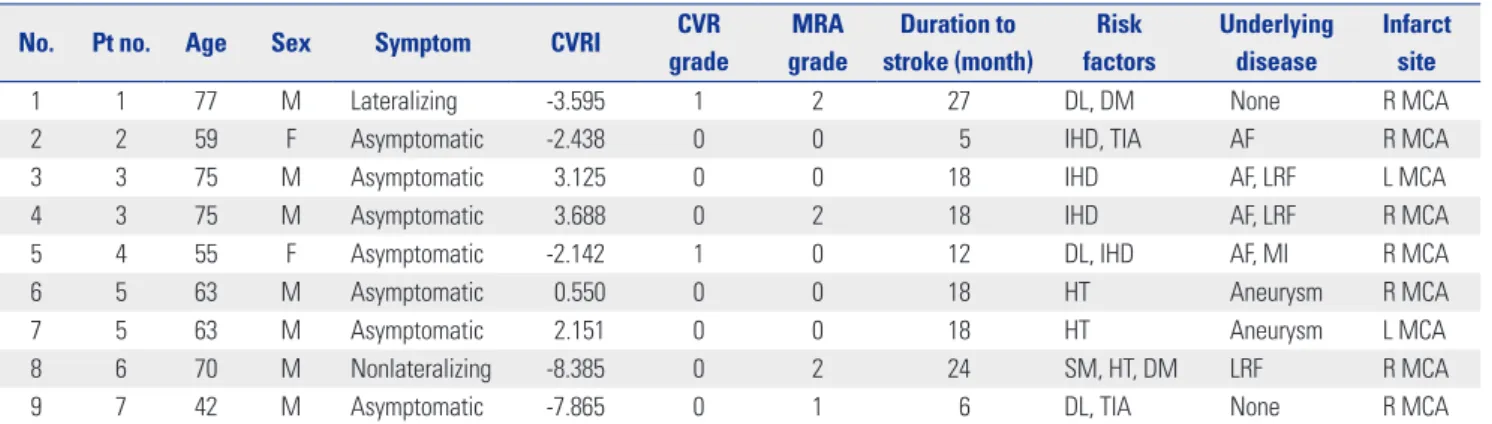
During follow-up of 18.7±9.6 months (median 20.0 months), 9 events of stroke occurred in 7 patients (Table 3). The incidence Table 1. Clinical Characteristics of 171 Patients
Parameters Values
Age (yr) 66±13 (range 18–91)
Sex (%)
Male 108 (63)
Female 63 (37)
Risk factors (%)
Hypertension 125 (73)
Hyperlipidemia 61 (36)
Diabetes 63 (37)
Ever smoker 54 (32)
Ischemic heart disease 20 (12)
Previous episodes of TIA 15 (9)
Underlying diseases (%)
Moyamoya disease 15 (9)
Ateriovenous fistula 18 (11)
Aneurysm 9 (5)
Prothrombic tendency 20 (12)
Liver or renal disease 17 (10)
Atrial fibrillation 18 (11)
Intracranial hemorrhage 12 (7)
Myocardial infarction 10 (6)
Active cancer 9 (5)
Ischemic symptoms (%)
None 48 (28)
Symptomatic MCA territories (%)
Lateralizing 76 (22)
Non-lateralizing 70 (20)
Asymptomatic MCA territories (%) 196 (57) TIA, transient ischemic attack; MCA, middle cerebral artery.
was 4.1% and the time interval between imaging studies and stroke was 5–27 months (Fig. 2). CVR grade and stenosis grade were not significantly different between patients with and with- out subsequent stroke (0.3±0.6 vs. 0.2±0.4, p=0.864 for CVR grade; 0.7±0.8 vs. 0.8±1.0, p=0.726 for stenosis grade). Of the clinical and imaging parameters tested, both stenosis and CVR grades were not significant prognostic factors, regardless of grade (Table 4). Hypertension and renal disease were signifi- cant prognostic factors for predicting subsequent stroke. Addi- tionally, aneurysm and TIA were prognostic factors with bor-
derline significance (Fig. 3).
DISCUSSION
In this study, we observed a significant correlation between ICA-MCA stenosis on MRA and CVR of MCA territory on per- fusion SPECT. However, a considerable discrepancy between stenosis and CVR also existed; 75% of grade 1 stenosis and 65%
of grade 2 stenosis were not matched with CVR impairment.
And, significant prognostic factors were different according to symptomatic and asymptomatic groups. Severe stenosis on MRA (grade 2) and combination imaging factor of decreased CVRI and MRA grade 2 were significant predictors to cause subsequent stroke in lateralizing symptomatic patients.
MRA has been performed for evaluating vascular malforma- tion,19 as well as major cerebrovascular occlusion,20,21 whereas perfusion SPECT has been used for diagnosis and prognosis prediction in cerebrovascular diseases.22-25 Acetazolamide SPECT can provide information on CVR in addition to basal cerebral perfusion. There have been several reports on the use of CVR for evaluating clinical significance of cerebrovascular stenosis. A meta-analysis by Gupta, et al.26 showed that high- grade ICA stenosis or occlusion causing CVR impairment can be a risk factor for ischemic stroke. Stenosis on MRA also has a clinical significance, and Chen, et al.27 reported that degree of stenosis on MRA was inversely correlated with cerebrovascular Table 2. CVR State of MCA Territories According to MRA Findings
MRA grade CVR grade
Proportion
0 1 2
Overall patients
0 173 11 7 56%
1 52 13 4 20%
2 53 17 12 24%
Proportion 81% 12% 7% κ=0.192
Patients with lateralizing symptoms
0 41 4 0 59%
1 7 2 2 15%
2 12 6 2 26%
Proportion 79% 16% 5% κ=0.230
Patients with non-lateralizing symptoms
0 33 1 0 49%
1 14 4 0 26%
2 10 2 6 26%
Proportion 81% 10% 9% κ=0.346
Asymptomatic patients
0 99 6 7 57%
1 31 7 2 20%
2 31 9 4 22%
Proportion 82% 11% 7% κ=0.115
CVR, cerebrovascular reserve; MCA, middle cerebral artery; MRA, magnetic resonance angiography.
Fig. 1. Proportions of decreased CVR (grade 1 and 2) according to ste- nosis grade. Decreased CVR was more frequently presented in higher stenosis grade group. CVR, cerebrovascular reserve.
40 35 30 25 20 15 10 5
0 Grade 1
Stenosis grade
Grade 0 Grade 2
Proportion of decreased CVR (%)
significant in many asymptomatic patients.
In the present study, both the stenosis and CVR were found not to be significant prognostic factors for subsequent stroke, in contrast to previous reports.11,29,30 The low event rate (9 events in 7 patients, 7/171=4.1%) in this study would have been one of the causes for the result. In addition, it could be attributed to the characteristics of our patient population. Because stroke can occur due to either thrombosis or embolism, stenosis in ICA or MCA is not the only cause for the stroke event. In a re- cent study, it was reported that approximately 47% of acute reactivity in response to increased concentration of inspired
CO2. Deteriorated autoregulation due to stenosis may cause hemodynamic impairment.
However, in the present study, only limited agreement exist- ed between stenosis and CVR, although there was a significant difference in CVR according to stenosis grade. Additionally, most of the stenosis in ICA-MCA did not cause CVR impair- ment. It is most likely due to the difference in patient popula- tion, because we enrolled consecutive patients who met the in- clusion criteria. Many patients in this study presented with no or only vague neurologic symptoms, and stenosis was inciden- tally detected in some of them. In such cases, stenosis may have been well tolerated and compensated by sufficient collateral flow. Intriguingly, in patients with any lateralizing and non-lat- eralizing neurologic symptoms, CVR impairment exhibited a fair agreement with stenosis (κ=0.230 and κ=0.346). Similarly, it has been reported that stenosis of carotid arteries is not signifi- cantly correlated with the hemispheric symptoms.28 Thus, it should be noted that stenosis in ICA-MCA may not be clinically Table 3. Clinical Characteristics of Patients Who Presented Subsequent Stroke
No. Pt no. Age Sex Symptom CVRI CVR
grade
MRA grade
Duration to stroke (month)
Risk factors
Underlying disease
Infarct site
1 1 77 M Lateralizing -3.595 1 2 27 DL, DM None R MCA
2 2 59 F Asymptomatic -2.438 0 0 5 IHD, TIA AF R MCA
3 3 75 M Asymptomatic 3.125 0 0 18 IHD AF, LRF L MCA
4 3 75 M Asymptomatic 3.688 0 2 18 IHD AF, LRF R MCA
5 4 55 F Asymptomatic -2.142 1 0 12 DL, IHD AF, MI R MCA
6 5 63 M Asymptomatic 0.550 0 0 18 HT Aneurysm R MCA
7 5 63 M Asymptomatic 2.151 0 0 18 HT Aneurysm L MCA
8 6 70 M Nonlateralizing -8.385 0 2 24 SM, HT, DM LRF R MCA
9 7 42 M Asymptomatic -7.865 0 1 6 DL, TIA None R MCA
HT, hypertension; DL, dyslipidemia; DM, diabetes mellitus; IHD, ischemic heart disease; SM, smoking; TIA, transient ischemic attack; LRF, liver or renal failure;
MI, myocardial infarction; AF, atrial fibrillation; MCA, middle cerebral artery; R MCA, right MCA; L MCA, left MCA; CVRI, cerebrovascular reserve index; CVR, cerebrovascular reserve; MRA, magnetic resonance angiography.
Fig. 2. Kaplan-Meier survival curve for subsequent stroke occurrence in overall group.
100
80
60
40
20
0
Time (month)
0 10 20 30 40
Stroke-free survival (%)
Table 4. Prognostic Factors for Subsequent Stroke
Variables Odds ratio (95% CI) p value CVR grade
0 1
1 and 2* 1.05 (0.212–5.17) 0.955
2* 0.00 (0.025–4.25) 0.394
MRA grade
0 1
1 and 2* 1.03 (0.272–3.87) 0.970
2* 1.32 (0.296–6.11) 0.701
Hypertension
(-) 1
(+) 0.198 (0.035–0.638) 0.010†
Renal disease
(-) 1
(+) 5.30 (2.27–242.3) 0.008†
Aneurysm
(-) 1
(+) 4.20 (0.993–4.20) 0.051
TIA
(-) 1
(+) 3.57 (0.725–110.8) 0.087
CVR, cerebrovascular reserve; MRA, magnetic resonance angiography; TIA, transient ischemic attack; CI, confidence interval.
*Compared with grade 0, †Statistically significant (p<0.05).
ischemic stroke had no prior occlusion within 1 month of the event and was classified as embolic occlusion.31 In the present study, 5 cases exhibited normal MRA, 6 cases exhibited normal CVR and 4 cases exhibited normal CVRI in 9 event cases, which suggests the probability of embolism as the cause for stroke.
Therefore, it is speculated that clinical factors exhibited stronger prognostic power for the subsequent stroke in this study. Our results are in line with previous reports in which obesity, smok- ing,32 end-stage renal disease,33 and cerebral aneurysm34 are sig- nificant risk factors for stroke. However, hypertension exhibited an opposite result, which may result from our classification for hypertension. Because the current state of hypertension control was not considered in this study, mild or well-controlled hyper- tension was all included in the group of hypertension. It may have been a cause for selection bias.
The results of this study suggest that the carotid or cerebral artery stenosis in asymptomatic patients should be interpreted in terms of hemodynamic significance. Even in severe stenosis group of grade 2, more than half of the stenosis did not cause CVR impairment. Thus, evaluation of CVR using brain perfu- sion SPECT or other modality would be required for re-evalua-
tion of stenotic lesions, particularly when MRA is used for screen- ing of cerebrovascular disease in asymptomatic patients. However, further study is required to determine the diagnostic roles of brain perfusion SPECT and MRA in more homogeneous group of as- ymptomatic patients or patients with high risk factors for stroke.
There are several limitations in this study. First, ICA and MCA were analyzed together as a single group on MRA, whereas CVR was analyzed for MCA territory on perfusion SPECT. Al- though both ICA and MCA stenosis can lead to CVR impair- ment in the MCA territory, the influence of specific locations of stenosis was not considered in the analysis. Second, in some patients, the time interval between MRA and perfusion SPECT was somewhat long, up to 60 days. However, the time interval was less than 2 weeks in 74% of the patients. Third, this was a retrospective study and the patient group was relatively hetero- geneous in terms of symptoms, underlying diseases, medications, and risk factors. Because we attempted to analyze the hemody- namic significance of stenosis on MRA in various conditions, we included all the consecutive patients.
In conclusion, a considerable proportion of ICA-MCA steno- sis detected on MRA did not cause CVR impairment, although Fig. 3. Kaplan-Meier survival curve for subsequent stroke occurrence according to significant risk factors (A and B) or factors with borderline signifi- cances (C and D). TIA, transient ischemic attack.
100
80
60
40
20
0
100
80
60
40
20
0
100
80
60
40
20
0
100
80
60
40
20
0 Time (months)
Time (months)
Time (months)
Time (months) p=0.010
p=0.051
p=0.008
p=0.087 0
0
0
0 10
10
10
10 20
20
20
20 30
30
30
30 40
40
40
40
Stroke-free survival (%)Stroke-free survival (%) Stroke-free survival (%)Stroke-free survival (%)
Hypertension (+) Hypertension (-)
Aneurysm (+) Aneurysm (-)
Renal disease (+) Renal disease (-)
TIA (+) TIA (-) A
C
B
D
there was a fair correlation between them. Thus, it is recom- mended that hemodynamic state should be assessed for evalu- ation of stenotic lesions, particularly in asymptomatic patients.
ACKNOWLEDGEMENTS
This study has been presented as a poster in The Korea Society of Nuclear Medicine (Oct. 25th. 2012).
This research was supported by Basic Science Research Pro- gram through the National Research Foundation of Korea (NRF) funded by the Ministry of Education (2009-0093820).
REFERENCES
1. Zhang Y, Wu S, Jia Z, Zhou Y, Liu X, Wang W, et al. The relation- ship of asymptomatic intracranial artery stenosis and Framing- ham stroke risk profile in a Northern Chinese industrial city. Neu- rol Res 2012;34:359-65.
2. Yang F, Liu L, Li M, Li M, Yin Q, Guo R, et al. Pattern of cerebrovas- cular atherosclerotic stenosis in older Chinese patients with stroke.
J Clin Neurosci 2013;20:979-83.
3. Silvestrini M, Vernieri F, Pasqualetti P, Matteis M, Passarelli F, Troisi E, et al. Impaired cerebral vasoreactivity and risk of stroke in pa- tients with asymptomatic carotid artery stenosis. JAMA 2000;283:
2122-7.
4. Gao T, Yu W, Liu C. Mechanisms of ischemic stroke in patients with intracranial atherosclerosis: a high-resolution magnetic res- onance imaging study. Exp Ther Med 2014;7:1415-9.
5. Kumar G, Kalita J, Kumar B, Bansal V, Jain SK, Misra U. Magnetic resonance angiography findings in patients with ischemic stroke from North India. J Stroke Cerebrovasc Dis 2010;19:146-52.
6. Bash S, Villablanca JP, Jahan R, Duckwiler G, Tillis M, Kidwell C, et al. Intracranial vascular stenosis and occlusive disease: evalua- tion with CT angiography, MR angiography, and digital subtrac- tion angiography. AJNR Am J Neuroradiol 2005;26:1012-21.
7. Lutsep HL, Clark WM. Association of intracranial stenosis with cortical symptoms or signs. Neurology 2000;55:716-8.
8. Man BL, Fu YP, Chan YY, Lam W, Hui CF, Leung WH, et al. Use of magnetic resonance angiography to predict long-term outcomes of ischemic stroke patients with concurrent stenoses in Hong Kong.
Cerebrovasc Dis 2009;28:112-8.
9. Lee K, Kim H, Heo JH, Bae HJ, Koh IS, Chang S. Application of mag- netic resonance imaging and magnetic resonance angiography as diagnostic measures for the first attack of suspected cerebrovascu- lar diseases in Korea. Yonsei Med J 2011;52:727-33.
10. Park SA, Park HI, Kim D, Yang CY, Zhang LQ. The prediction of gross motor outcome using cerebrovascular reserve measured by acetazolamide-challenged SPECT. NeuroRehabilitation 2012;30:
359-67.
11. Kuroda S, Houkin K, Kamiyama H, Mitsumori K, Iwasaki Y, Abe H.
Long-term prognosis of medically treated patients with internal carotid or middle cerebral artery occlusion: can acetazolamide test predict it? Stroke 2001;32:2110-6.
12. Marchal G, Bouvard G, Iglesias S, Sebastien B, Benali K, Defer G, et al. Predictive value of (99m)Tc-HMPAO-SPECT for neurological Outcome/Recovery at the acute stage of stroke. Cerebrovasc Dis 2000;10:8-17.
13. Kim JM, Jung KH, Sohn CH, Moon J, Han MH, Roh JK. Middle ce- rebral artery plaque and prediction of the infarction pattern. Arch Neurol 2012;69:1470-5.
14. Moore WS, Barnett HJ, Beebe HG, Bernstein EF, Brener BJ, Brott T, et al. Guidelines for carotid endarterectomy. A multidisciplinary consensus statement from the Ad Hoc Committee, American Heart Association. Circulation 1995;91:566-79.
15. Nederkoorn PJ, van der Graaf Y, Eikelboom BC, van der Lugt A, Bartels LW, Mali WP. Time-of-flight MR angiography of carotid artery stenosis: does a flow void represent severe stenosis? AJNR Am J Neuroradiol 2002;23:1779-84.
16. U-King-Im JM, Trivedi RA, Cross JJ, Higgins NJ, Hollingworth W, Graves M, et al. Measuring carotid stenosis on contrast-enhanced magnetic resonance angiography: diagnostic performance and re- producibility of 3 different methods. Stroke 2004;35:2083-8.
17. So Y, Lee HY, Kim SK, Lee JS, Wang KC, Cho BK, et al. Prediction of the clinical outcome of pediatric moyamoya disease with post- operative basal/acetazolamide stress brain perfusion SPECT after revascularization surgery. Stroke 2005;36:1485-9.
18. Lee HY, Paeng JC, Lee DS, Lee JS, Oh CW, Cho MJ, et al. Efficacy assessment of cerebral arterial bypass surgery using statistical parametric mapping and probabilistic brain atlas on basal/acet- azolamide brain perfusion SPECT. J Nucl Med 2004;45:202-6.
19. Raoult H, Bannier E, Maurel P, Neyton C, Ferré JC, Schmitt P, et al.
Hemodynamic quantification in brain arteriovenous malforma- tions with time-resolved spin-labeled magnetic resonance angiog- raphy. Stroke 2014;45:2461-4.
20. Kimura K, Iguchi Y, Shibazaki K, Aoki J, Uemura J. Early recanaliza- tion rate of major occluded brain arteries after intravenous tissue plasminogen activator therapy using serial magnetic resonance angiography studies. Eur Neurol 2009;62:287-92.
21. Schellinger PD, Richter G, Kohrmann M, Dorfler A. Noninvasive angiography (magnetic resonance and computed tomography) in the diagnosis of ischemic cerebrovascular disease. Techniques and clinical applications. Cerebrovasc Dis 2007;24 Suppl 1:16-23.
22. Deus-Silva L, Bonilha L, Damasceno BP, Costa AL, Yasuda CL, Costa FF, et al. Brain Perfusion Impairment in Neurologically As- ymptomatic Adult Patients with Sickle-Cell Disease Shown by Voxel-Based Analysis of SPECT Images. Front Neurol 2013;4:207.
23. Oikawa K, Ogasawara K, Saito H, Yoshida K, Saura H, Sato Y, et al.
Combined measurement of cerebral and cerebellar blood flow on preoperative brain perfusion SPECT imaging predicts devel- opment of new cerebral ischemic events after endarterectomy for symptomatic unilateral cervical carotid stenosis. Clin Nucl Med 2013;38:957-61.
24. Volkan-Salanci B, Lay Ergün E, Genc Sel Ç, Yalnizog˘lu D, Turanli G. The role of brain perfusion SPECT in Moyamoya disease. Rev Esp Med Nucl Imagen Mol 2012;31:216-8.
25. Saura H, Ogasawara K, Suzuki T, Kuroda H, Yamashita T, Ko- bayashi M, et al. Effect of combination therapy with the angioten- sin receptor blocker losartan plus hydrochlorothiazide on brain perfusion in patients with both hypertension and cerebral hemo- dynamic impairment due to symptomatic chronic major cerebral artery steno-occlusive disease: a SPECT study. Cerebrovasc Dis 2012;33:354-61.
26. Gupta A, Chazen JL, Hartman M, Delgado D, Anumula N, Shao H, et al. Cerebrovascular reserve and stroke risk in patients with ca- rotid stenosis or occlusion: a systematic review and meta-analy- sis. Stroke 2012;43:2884-91.
27. Chen J, Liu J, Xu WH, Xu R, Hou B, Cui LY, et al. Impaired dynamic cerebral autoregulation and cerebrovascular reactivity in middle cerebral artery stenosis. PLoS One 2014;9:e88232.
28. Kanber B, Hartshorne TC, Horsfield MA, Naylor AR, Robinson TG, Ramnarine KV. Wall motion in the stenotic carotid artery: as- sociation with greyscale plaque characteristics, the degree of ste- nosis and cerebrovascular symptoms. Cardiovasc Ultrasound
2013;11:37.
29. Liu M, Zhou L. Cerebrovascular reserve may be a more accurate predictor of stroke than degree of ICA or MCA stenosis. Med Sci Monit 2014;20:2082-7.
30. Kuroda S, Shiga T, Ishikawa T, Houkin K, Narita T, Katoh C, et al.
Reduced blood flow and preserved vasoreactivity characterize oxygen hypometabolism due to incomplete infarction in occlu- sive carotid artery diseases. J Nucl Med 2004;45:943-9.
31. Lee KJ, Jung KH, Byun JI, Kim JM, Roh JK. Infarct pattern and clinical outcome in acute ischemic stroke following middle cere- bral artery occlusion. Cerebrovasc Dis 2014;38:31-8.
32. Itrat A, Ahmed B, Khan M, Muhammad M, Thaver D, Khowaja Z, et al. Risk factor profiles of South Asians with cerebrovascular dis- ease. Int J Stroke 2011;6:346-8.
33. Tripepi G, Mattace-Raso F, Rapisarda F, Stancanelli B, Malatino L, Witteman J, et al. Traditional and nontraditional risk factors as predictors of cerebrovascular events in patients with end stage re- nal disease. J Hypertens 2010;28:2468-74.
34. Hokari M, Isobe M, Imai T, Chiba Y, Iwamoto N, Isu T. The impact of atherosclerotic factors on cerebral aneurysm is location depen- dent: aneurysms in stroke patients and healthy controls. J Stroke Cerebrovasc Dis 2014;23:2301-7.