Effect of 0.5 mM Dibutyryl cAMP on Meiotic Maturation during Different Incubation Time and Embryonic Development Following In Vitro Fertilization
or Parthenogenetic Activation in Porcine Oocytes
Il-jeoung Yu*
Department of Theriogenology & Reproductive Biotechnology, College of Veterinary Medicine & Bio-Safety Institute, Chonbuk National University, Jeonju 561-756, Korea
ABSTRACT
Presently, the effect of 0.5 mM dibutyryl cAMP (dbcAMP)-supplemented maturation medium during different incubation time on meiotic arrest (germinal vesicle) and resumption (metaphase II) of porcine oocytes and embryonic development of porcine oocytes following in vitro fertilization (IVF) or parthenogenetic activation (PA) was deter- mined. Porcine cumulus oocyte complexes (COCs) were cultured in 0.5 mM dbcAMP for 17, 22, 27, or 42 h, and an additional 22 h without 0.5 mM dbcAMP. The nuclear status was examined at each time point. Oocytes cultured from 39~49 h displayed more than 80% meiotic resumption. More than 85 % of meiotic arrest was presented at 17~
22 h. Oocytes were cultured for 22 h with 0.5 mM dbcAMP and additional 22 h without dbcAMP to assess develop- mental potential following IVF or PA. There were no significant differences in blastocyst rates among the dbcAMP- IVF, IVF, dbcAMP-PA, and PA groups, although cleavage rate of IVF group was significantly higher than those of dbcAMP-PA, and PA groups. In conclusion, 0.5 mM dbcAMP influenced meiotic maturation of porcine oocytes de- pending on incubation time of oocyte, although embryonic development was not improved in both IVF and PA.
(Key words : dibutyryl cAMP, porcine oocyte, meiotic arrest and resumption, embryonic development)
*Correspondence : E-mail : [email protected]
INTRODUCTION
In vitro matured oocytes have become dominant in porcine developmental technologies, replacing in vivo-derived oocytes.
Maturation of oocytes may affect embryonic development after nuclear transfer, both in parthenogenetic activation (PA) as well as in vitro-fertilized (IVF) embryos (Nagashima et al., 2003). However, the developmental competence of oocytes co- llected from various sized follicles might be different (Yoshi- da et al., 1989; Funahashi and Day, 1993; Blondin and Sirard, 1994; Funahashi et al., 1997; Wang et al., 1998).
Synchronization of oocytes before maturation in vitro is essen- tial to simultaneously complete DNA transcription and nuclear maturation (Mermillod et al., 2000; Sirad, 2001). Oocytes in arrest at the germinal vesicle (GV) during synchronization can obtain the time necessary to mature in vitro and for further develop- mental competence of embryos (Funahashi et al., 1997; Hashi- moto et al., 2002; Ponderato et al., 2002; Wu et al., 2002).
Cyclic adenosine monophosphate (cAMP) is the principal regulator of the oocyte meiotic cell cycle (Taieb et al., 1997;
Ferell, 1999; Bagg et al., 2006). In vivo, decreased cAMP con-
tent in oocytes is associated with the initiation of germinal vesicle breakdown (GVBD) (Mattioli et al., 1994). In other words, a drop of the oocyte cAMP level is involved in the re- sumption of meiosis. However, physical removal of oocytes from their follicles can induce a premature reduction of oocyte cAMP content during in vitro maturation. This adversely affects coordination of nuclear maturation and cytoplasmic maturation, and subsequently developmental potential (Bagg et al., 2006).
High intracellular levels of cAMP maintain oocyte meiotic arrest at the GV stage by addition of an analogue, dibutyryl cy- clic adenosine monophosphate (dbcAMP), into the medium for the in vitro maturation of oocytes (Cho et al., 1974; Bagg et al., 2006; Kim et al., 2008). An effective meiosis-inhibiting agent in pig oocytes is dbcAMP (Bagg et al., 2006; Kim et al., 2008). The presence of dbcAMP increases the synchrony of meiotic progression and the incidence of blastocyst formation following IVF (Funahashi et al., 1997; Somfai et al., 2003).
However, a constant concentration of 1 mM has been used to arrest porcine oocytes, although dbcAMP is linked with oocyte maturation in studies that focused on the effect of dbcAMP on embryonic development by comparing oocytes treated with
dbcAMP or dbcAMP-free oocytes (Funahashi et al., 1997;
Miyoshi et al., 2002; Bagg et al., 2006; Kim et al., 2008). I demonstrated that 0.5 mM dbcAMP could be efficient con- centration to arrest oocyte meiosis (data unpublished).
The present study was carried to determine the effect of 0.5 mM dbcAMP supplemented to maturation medium during different incubation time of oocytes on meiotic arrest and resumption of porcine oocytes and to assess the effect of 0.5 mM dbcAMP on subsequent developmental competence of embryos fertilized in vitro or activated parthenogenetically.
MATERIALS AND METHODS
1. Culture Media
Unless otherwise noted, all chemicals and reagents used were purchased from Sigma-Aldrich (St. Louis, MO, USA). The medium used for collection and washing of porcine oocyte- cumulus complexes (COCs) was modified Tyrode’s lactate- HEPES-polyvinyl alcohol (TL-HEPES-PVA) composed of 114 mM NaCl, 3.2 mM KCl, 0.4 mM Na2H2PO4, 2 mM CaCl2․ 2H2O, 0.5 mM MgCl2․6H2O, 5 mM NaHCO3, 20 mM HEPES, 16.6 mM sodium lactate (60% syrup), 0.5% (w/v) PVA, 10 IU/ml penicillin, and 10 μg/ml streptomycin. Oocyte matu- ration was accomplished in North Carolina State University 23 medium (NCSU-23; Gil et al., 2007) supplemented with 10%
porcine follicular fluid (PFF), 0.6 mM cysteine, 10 ng/ml epi- dermal growth factor (EGF), 10 IU/ml pregnant mare serum gonadotropin (PMSG), and 10 IU/ml human chorionic gona- dotropin (HCG). PFF was collected from ovarian follicles that ranged from 3~6 mm in diameter by centrifugation at 1,600
×g for 30 min at room temperature and filtration through a 1.2 μm syringe filter. The final product was stored at 20℃ until use. Fertilization medium, based on modified Tris-buffered me- dium (mTBM; 113.1 mM NaCl, 3 mM KCl, 7.5 mM CaCl2․ 2H2O, 20 mM Tris (Trizma Base), 11 mM D-glucose, and 5 mM sodium pyruvate), was supplemented with 2 mM caffeine and 0.2% bovine serum albumin (BSA). In vitro culture 1 (IVC 1) medium was composed of D-glucose-free NCSU- 23 supplemented with 0.17 mM sodium pyruvate, 2.73 mM so- dium lactate (60% syrup), and 0.4% BSA (A- 6003). In vitro culture 2 (IVC 2) medium was NCSU-23 containing 0.4% BSA.
2. In Vitro Maturation
Porcine ovaries were collected from a local abattoir and were transported to the laboratory at 34~36℃ in 0.9% saline su-
pplemented with 100 IU/ml penicillin G and 100 μg/ml strep- tomycin. COCs were aspirated through an 18 gauge needle.
Oocytes with a compact cumulus mass and a dark, evenly gra- duated cytoplasm were washed three times in TL-HEPES-PVA medium and maturation medium, respectively. Oocytes (n=20~ 25) were cultured in a 100 μl droplet of maturation medium supplemented with 10 IU/ml PMSG and 10 IU/ml HCG for 22 h, and for a further 22 h in hormone-free maturation medium.
Oocyte maturation was carried out under sterile mineral oil in a humidified atmosphere of 5 % CO2 in air at 39℃.
3. In vitro Fertilization Procedure
Extended spermatozoa supplied by Irae Yangdon (Jeonju, Korea) were maintained at 17℃ for 2 days. Percoll solutions and gradients were prepared as described previously (Yu et al., 2002). A two-layer discontinuous gradient was formed by laye- ring 1 ml of a 45% Percoll solution on top of 1 ml of a 90%
Percoll solution in a 15 ml conical tube. Aliquants of extended semen (3 ml) were layered onto Percoll gradient and centri- fuged for 20 min at 850 × g at room temperature. The pellet recovered after aspiration of the supernatant was washed two times by centrifugation with 5 ml Dulbecco’s phosphate bu- ffered saline (D-PBS) supplemented with 0.1% BSA, 10 IU/ml penicillin, and 10 μg/ml streptomycin at 350 × g for 3 min.
After the supernatant was discarded, motile spermatozoa were collected. Sperm concentration (spermatozoa/ml) was adjusted to 10 × 105 by dilution with mTBM. Oocytes with expanded cumulus cells were treated with 1 mg/ml hyaluronidase in TL-HEPES-PVA and were denuded by gentle aspiration with a small bore glass pipette. The denuded oocytes were washed three times with mTBM and oocytes were transferred to a mTBM insemination drop (45 μl) overlaid with mineral oil (20~25 oocytes per drop). A 5 μl volume of spermatozoa was added to each insemination drop to give a final concentration of 1 ×
105 spermatozoa/ml. Oocytes and spermatozoa were co-cultured for 6 h in a humidified atmosphere of 5 % CO2 in air at 39℃.
4. Pathenogenetic Procedure
Oocytes were parthenogenetically activated by incubation in IVC 1 medium containing 5 μM ionomycin for 5 min, washing twice, and incubation for 3 h in NCSU-23 medium supple- mented with 2.0 mM 6 D-methyl amino purine (6 D-MAP).
5. In Vitro Culture
Following either parthenogenic activation (PA) or in vitro
fertilization (IVF), presumptive zygotes were washed three times, transferred to 50 μl droplets (15~20 presumptive zygotes per drop) of IVC 1 medium, and incubated for 2 days at 39℃ in a humidified atmosphere of 5 % CO2 in air. After 2 days of em- bryo culture, the cleavage rate was assessed. Embryos were then washed twice, transferred to 50 μl droplets of IVC 2 me- dium, and incubated for 6 days, after which the blastocyst rate and cell numbers in blastocyst were determined.
5. Assessment of Meiotic Stage
Oocytes denuded by vigorous shaking in 3% sodium citrate solution were fixed in a 4-well culture dish (Nunc, Rochester, NY, USA) containing 500 μl of ethanol:acetic acid (3:1 v/v) for 72 h. They were then dispensed onto a slide to which a cover slip was added. Each sample was stained with aceto-or- cein [1% (w/v) orcein in 45% (v/v) acetic acid] and destained with glycerol:acetic acid:distilled water (1:1:3, v/v). Nuclear maturation of oocytes was evaluated under a light microscope at 400 × magnification. Meiotic stages were classified as pre- viously described (Romar and Funahashi, 2006) as being at ger- minal vesicle (GV), germinal vesicle breakdown (GVBD), me- taphase I (MI), anaphase I (AI), telophase I (TI), or metaphase
Ⅱ (MII).
6. Assessment of Cell Numbers in Blastocyst
Blastocysts were stained in 500 μl of Hoechst 33342 (5 μg/
ml) in TL-HEPES-PVA for 15 min at room temperature, mounted on slides, and covered with cover slips. Cell numbers in blas- tocyst were determined using a Nikon TE2000 fluorescent in- verted microscope (Nikon, Tokyo, Japan) at 400 × magnification.
7. Statistical Analysis
Each experiment was replicated 6~7 times. All percentage data was subjected to arcsine transformation. All data and data sets are presented as the mean ± SE. Maturation of oocytes and development of porcine embryos following IVF or PA were analyzed by Duncan’s multiple range test using the Statistical Analysis System ver. 8× (SAS, Cary, NC, USA). P<0.05 was considered to be statistically significant.
8. Experimental Design
Two series of experiment were used. The first experimental design assessed the effect of 0.5 mM dbcAMP-supplemented oocyte maturation medium during different incubation time on meiotic arrest and resumption of porcine oocyte. The oocytes
were cultured in maturation medium supplemented with 0.5 mM dbcAMP and hormones for 17, 22, 27, or 42 h, and then were cultured in dbcAMP- and hormone-free maturation me- dium for another 22 h. A proportion of oocytes from each group were removed, denuded, and fixed at 17, 22, 27, or 42 h; the remaining oocytes were fixed at 39, 44, 49, or 64 h after denuding cumulus cells. The meiotic stages were assessed in the populations.
The second experimental design assessed the effect of 0.5 mM dbcAMP-supplemented oocyte maturation medium on the developmental competence of embryos following PA or IVF.
Oocytes were cultured in maturation medium supplemented with 0.5 mM dbcAMP and hormones for 22 h, and were then cultured in dbcAMP- and hormone-free maturation medium for another 22 h. A portion of the oocytes were denuded and used for PA, with the remainder being denuded and used for IVF.
The rates of cleavage, and blastocyst, and the cell numbers in blastocyst were determined.
RESULTS AND DISCUSSION
1. Effect of 0.5 mM dbcAMP-supplemented Oocyte Maturation Medium during Different Incubation Time on Meiotic Arrest and Re- sumption of Porcine Oocyte
The effect of dbcAMP on reproduction in pigs has been well-studied (Funahashi et al., 1997; Miyoshi et al., 2002; Bagg et al., 2006; Kim et al., 2008). However, only 1 mM dbcAMP has been used to assess the meiosis arrest of oocytes. In pre- vious study, I sought to more decisively determine the effec- tive concentration of dbcAMP for meiotic arrest and resump- tion in porcine oocytes. I demonstrated that lower concentra- tion of 0.5 mM dbcAMP were also effective on meiotic arrest of oocytes (data unpublished).
The proportion of oocytes arrested at the GV was signifi- cantly decreased (P<0.05) in a time-dependent manner (Fig.
1a). Culture time-dependent decreases in the GV rate have been reported in porcine oocytes treated with 1 mM dbcAMP (Miyoshi et al., 2002; Bagg et al., 2006). These results are consistent with these findings that the rate of meiotic arrest de- creased as culture time increased. However, GV rate at 42 h decreased to 40% in this study was contrast with less than 10% GV rate at 40 h presented by Bagg et al (2006). On the other hand, Miyoshi et al (2002) indicated that GV rate at 42 h decreased to 40%. It is in agreement with results from our study. I suggested that 0.5 mM concentration of dbcAMP du-
ring longer culture period could be effective to arrest meiosis of porcine oocytes.
The proportions of oocytes in the MI and MII stages were significantly increased at 42 h (P<0.05). However, the propor- tion of oocytes in the GVBD stage was decreased at 42 h.
Meiotic resumption of oocytes incubated for 22 h in dbcAMP- free maturation medium was assessed (Fig. 1b). The propor- tions of MII stage oocytes at 39, 44, and 49 h were signifi- cantly higher than that of MII oocytes at 64 h (P<0.05). The proportions of GV and MI stages were significantly increased at 64 h (P<0.05). During a longer period of culture, 30% of oocytes cultured in dbcAMP for 42 h were matured to the MII
b) a)
Fig. 1. Maturation of porcine oocytes. (a) Maturation of oocytes cultured in maturation medium supplemented with 0.5 mM dbcAMP for 17, 22, 27 or 42 h. (b) Maturation of oocytes cultured in maturation medium for additional 22 h after removal of dbcAMP. Different superscripts a~c indicate sig- nificant differences in meiotic progression at a particular time point (P<0.05).
stage and 35% of the remaining oocytes progressed to the MII stage even after additional 22 h culture (a total of 64 h). These results indicate that the appropriate culture time for meiosis arrest by 0.5 mM dbcAMP is between 17 and 22 h. As to the effect of subsequent culture, the highest MII rate (90%) was shown after 22 h of culture with dbcAMP and after an additional 22 h culture without dbcAMP (a total of 44 h). This result is not different with that in other studies (Miyoshi et al., 2002; Bagg et al., 2005). However, these previous studies did not determine whether culturing for more than 44 h affected meiosis resumption of oocytes. In the present study, I demon- strated that more than 80% of oocytes proceeded to MII after 27 h culture with dbcAMP and an additional 22 h culture (a total of 49 h culture).
2. Effect of 0.5 mM dbcAMP-supplemented in Oocyte Maturation Medium on Developmental Competence of PA or IVF Embryos
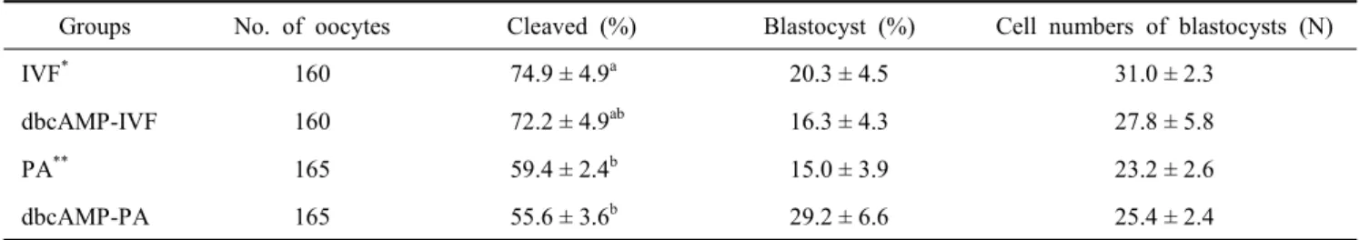
To determine whether the meiotic arrest of oocytes treated with 0.5 mM dbcAMP might contribute to improve embryo development, COCs were cultured with 0.5 mM dbcAMP for 22 h, cultured for an additional 22 h without dbcAMP, used for IVF or PA, and further cultured in vitro. The rates of cleaved embryos and blastocysts were assessed. In addition, cell num- bers in blastocyst were counted. The effect of dbcAMP on em- bryonic development was not different regardless of dbcAMP treatment, although cleavage rate of IVF embryos was signifi- cantly higher than that of parthenogenetic embryos (72~74%
vs. 55~59%, P<0.05). The blastocyst formation rate and mean cell numbers in blastocyst among groups were not significantly different, although blastocyst rate was the highest in dbcAMP- PA (29.2%; Table 1). As a result, 0.5 mM dbcAMP treatment of oocytes was not effective on improving the developmental competence of embryos following either parthenogenetic acti- vation or in vitro fertilization. This result was contrasts with studies that 1 mM dbcAMP-mediated arrest of meiotic resump- tion improved the rate of development to the blastocyst stage of in vitro matured and fertilized porcine oocytes (Funahashi et al., 1997; Kim et al., 2008). However, Miyoshi et al (2002) reported that 1 mM dbcAMP treatment of porcine oocytes did not improve the development of embryos reconstructed by nuclear transfer. On the other hand, Bagg et al (2006) reported that 1 mM dbcAMP treatment increased subsequent blastocyst rates of prepubertal oocytes following either parthenogenetic activation or in vitro fertilization, whereas blastocyst rates of adult oocytes remained unchanged. I cannot explain the diffe-
Table 1. Effect of 0.5 mM dbcAMP on development of porcine embryos following IVF or PA
Groups No. of oocytes Cleaved (%) Blastocyst (%) Cell numbers of blastocysts (N)
IVF* 160 74.9 ± 4.9a 20.3 ± 4.5 31.0 ± 2.3
dbcAMP-IVF 160 72.2 ± 4.9ab 16.3 ± 4.3 27.8 ± 5.8
PA** 165 59.4 ± 2.4b 15.0 ± 3.9 23.2 ± 2.6
dbcAMP-PA 165 55.6 ± 3.6b 29.2 ± 6.6 25.4 ± 2.4
IVF*: In vitro fertilization; PA**: Parthenogenetic activation.
rence among previous studies. However, I suggest that cytoplas- mic maturation can affect embryo development. Oocyte matura- tion includes two phenomena: nuclear maturation, involving reinitiation and completion of the first meiotic division, and cytoplasmic maturation that enables normal fertilization and further embryonic development (Marchal et al., 2001). In the future study, the assay of cytoplasmic maturation using mea- surement of glutathione contents should be considered to deter- mine the effect of dbcAMP on cytoplasmic maturation.
In conclusion, I have shown that 0.5 mM dbcAMP influenced meiotic maturation of porcine oocytes depending on incubation time of oocyte, although embryonic development including blastocyst formation was not improved in both IVF and PA.
In addition, I suggest that the appropriate culture duration to retain the GV stage of porcine oocyte in 0.5 mM-supplemented maturation medium is between 17 and 22 h and sufficient culture time to develop to MII is 39~49 h in oocytes exposed to 0.5 mM dbcAMP.
REFERENCES
Bagg MA, Nottle MB, Grupen CG and Armstrong DT. 2006.
Effect of dibutyryl cAMP on the cAMP content, meiotic pro- gression, and developmental potential of in vitro matured pre-pubertal and adult pig oocytes. Mol. Reprod. Dev. 73:
1326-1332.
Blodin P and Sirard MA. 1994. The influence of oocyte and follicular morphology on developmental competence in su- perovulated heifers. Theriogenology 41:164.
Cho WK, Stern S and Biggers JD. 1974. Inhibitory effect of dibutyryl cAMP on mouse oocyte maturation in vitro. J.
Exp. Zool. 187:383-386.
Ferell JE Jr. 1999. Xenopus oocyte maturation: new lessons from a good egg. Bioassays 21:833-842.
Funahashi H, Cantley TC and Day BN. 1997. Synchronization
of meiosis in porcine oocytes by exposure to dibutyryl cyclic adenosine monophosphate improves developmental competence following in vitro fertilization. Biol. Reprod.
57:49-53.
Funahashi H and Day BN. 1993. Effect of the duration of ex- posure to supplemental hormones on cytoplasmic matura- tion of pig oocytes in vitro. J. Reprod. Fertil. 98:179-185.
Gil MA, Alminana C, Cuello C, Parrilla I, Roca J, Vazquez JM and Martinez EA. 2007. Brief coincubation of gametes in vitro fertilization: Role of sperm:oocyte ratio and post- coincubation medium. Theriogenology 67:620-626.
Hashimoto S, Minami N, Takakura R and Imai H. 2002. Bo- vine immature oocytes acquire developmental competence during meiotic arrest in vitro. Biol. Reprod. 66:1696-1701.
Kim JS, Cho YS, Song BS, Wee G, Park JS, Cho YK, Yu K, Lee KK, Han YM and Koo DB. 2008. Exogeous dibutyryl cAMP affects meiotic maturation via protein kinase A acti- vation; it stimulates further embryonic development inclu- ding blastocyst quality in pigs. Theriogenology 69:290-301.
Marchal R, Tomanek M, Terqui M and Mermillod P. 2001. Effects of cell cycle dependent kinases inhibitor on nuclear and cytoplasmic maturation of porcine oocytes. Mol. Repod. Dev.
60:65-73.
Mattioli M, Galeati G, Barboni B and Seren E. 1994. Concen- tration of cyclic AMP during the maturation of pig oocytes in vivo and in vitro. J. Reprod. Fertil. 100:403-409.
Mermillod P, Tomanek M, Marchal R and Meijer L. 2000.
High developmental competence of cattle oocytes maintained at the germinal vesicle stage for 24 h in culture by specific inhibition of MPF kinase activity. Mol. Reprod. Dev. 55:
89-95.
Miyoshi K, Rzucidlo SJ, Pratt SL and Stice SL. 2002. Utility of rapidly matured oocytes as recipients for production of cloned embryos from somatic cells in the pig. Biol. Re- prod. 67:540-545.
Nagashima H, Fujimura T, Takahagi Y, Kurome M, Wako N, Ochiai T, Esaki R, Kano K, Saito S, Okabe M and Mura- kami H. 2003. Development of efficient strategies for the production of genetically modified pigs. Theriogenology 59:
95-106.
Ponderato N, Crotti G, Turini P, Duchi R, Galli C and Lazzari G. 2002. Embryonic and fetal development of bovine oocytes treated with a combination of butyrolactone I and rosco- vitine in an enriched medium prior to IVM and IVF. Mol.
Reprod. Dev. 62:513-518.
Romar R and Funahashi H. 2006. In vitro maturation and fertilization of porcine oocytes after a 48 h culture in ros- covitine, an inhibitor of p34cdc2/cyclin B kinase. Anim.
Reprod. Sci. 92:321-333.
Sirad MA. 2001. Resumption of meiosis: mechanism involved in meiotic progression and its relation with developmental competence. Theriogenology 55:1241-1254.
Somfai T, Kikuchi K, Onishi A, Iwamoto M, Fuchimoto D, Papp AB, Sato E and Nagai T. 2003. Meiotic arrest main- tained by cAMP during the initiation of maturation enhances meiotic potential and developmental competence and re- duces polyspermy of IVM/IVF porcine oocytes. Zygote
11:199-206.
Taieb F, Thibier C and Jessus C. 1997. On cyclins, oocytes, and eggs. Mol. Reprod. Dev. 48:397-411.
Yoshida M, Bamba K and Kojima Y. 1989. Effects of gona- dotropins and estradiol-17β on the timing of nuclear matu- ration and cumulus mass expansions in pig oocytes cul- tured in vitro. Jpn. J. Anim. Reprod. 35:86-91.
Yu I, Songsasen N, Godke RA and Leibo SP. 2002. Diffe- rences among dogs in response of their spermatozoa to cryopreservation using various cooling and warming rates.
Cryobiology 44:62-78.
Wang WH, Abeydeera LR, Prather RS and Day BN. 1998.
Morphologic comparison of ovulated and in vitro-matured porcine oocytes, with particular reference to polyspermy after in vitro fertilization. Mol. Reprod. Dev. 49:308-316.
Wu GM, Sun QY, Mao J, Lai L, McCauley TC, Park KW, Prather RS, Didion BA and Day BN. 2002. High develop- mental competence of pig oocytes after meiotic inhibition with a specific m-phase promoting factor kinase inhibitor, butylactone I. Biol. Reprod. 67:170-177.
(접수: 2011. 10. 25 / 심사: 2011. 10. 26 / 채택: 2011. 11. 15)