Requires the Lipogenic Transcription Factor Sterol Regulatory Element Binding Protein 1a
Seung-Soon Imaand Timothy F. Osborneb
Department of Physiology, Keimyung University School of Medicine, Dalseo-Gu, Daegu, South Korea,aand Metabolic Signaling and Disease Program, Diabetes and Obesity Center, Sanford-Burnham Medical Research Institute, Orlando, Florida, USAb
Sterol regulatory element binding protein (SREBP) transcription factors activate genes of lipid metabolism, but recent studies indicate they also activate genes involved in other physiologic processes, suggesting that SREPBs have evolved to connect lipid metabolism with diverse physiologic responses. There are three major mammalian SREBPs, and the 1a isoform is specifically expressed at very high levels in macrophages, where a recent study showed that it couples lipid synthesis to the proinflammatory phase of the innate immune response. In the present study, we show that loss of SREBP-1a also results in an increase in apoptosis after exposure to bacterial pore-forming toxins and we show this is a result of a selective reduction in the expression of the gene coding for the antiapoptotic factor apoptosis inhibitor 6 (Api6). Additional studies demonstrate that SREBP-1a specifically acti- vates the Api6 gene through a binding site in its proximal promoter, thus establishing the Api6 gene as a newly identified SREBP-1a target gene.
Macrophages are highly specialized cells with major functions in the mammalian innate immune response (15). They play a critical role in the first line of defense during infection and have evolved specialized mechanisms to respond to and destroy invad- ing pathogenic microbes. In this capacity, macrophages have ac- quired the ability to withstand attack by microbe-derived pore- forming toxins through activating genes required for survival.
Because the lipid-rich plasma membrane is the primary target of cellular damage mediated by the microbial toxins, the target cell needs to increase synthesis of lipid precursors to repair membrane damage caused by the actions of the microbial toxins (1). Evidence suggests that membrane expansion emanates from the endoplas- mic reticulum (ER) (4), and in a previous report, Gurcel et al. (5) provided evidence that the ER-targeted sterol regulatory element binding proteins (SREBPs) were activated after exposure to the Staphylococcus aureus pore-forming alpha-toxin. SREBPs are key transcriptional regulators of lipogenesis and cell growth, and proper regulation of their activity is critical to cellular lipid ho- meostasis. Mammals express three different SREBP isoforms:
SREBP-1a and -1c are encoded by the Srebf-1 gene and differ through alternative splicing of the SREBP-1 mRNA, whereas SREBP-2 is encoded by a distinct gene (14). The expression pat- terns for the three SREBPs vary dramatically across different tis- sues, and in liver, the SREBP-1c isoform is 10-fold more abundant than SREBP-1a. However, SREBP-1a is the more abundant iso- form in macrophages, and we showed it plays a crucial role in macrophages to coordinate cell lipid metabolism to the proin- flammatory response (8).
In this study, we demonstrate that macrophages with a selec- tive deficiency in expression of SREBP-1a (SREBP-1aDF) develop normally, but they are more susceptible to apoptosis after expo- sure to a bacterial pore-forming toxin. SREBP-1aDF macrophages express reduced levels of the antiapoptotic factor apoptosis inhib- itor 6 (Api6) (13), and we show that reintroduction of either SREBP-1a or Api6 reverses the antiapoptotic effects without re- storing lipid synthetic gene activity. These results demonstrate
that the apoptosis protection by SREBP-1a is due to its direct activation of a key antiapoptotic gene and is separate from its role in activating lipid synthesis.
MATERIALS AND METHODS
Reagents. Alpha-toxin from Staphylococcus aureus was purchased from List Biological Laboratories (Campbell, CA). Anti-mouse CD11b, anti-rat IgG2b, and the apoptosis assay kit were purchased from eBioscience. C75 was purchased from Sigma. Fetal bovine serum (FBS) was ordered from HyClone (Logan, UT).
SREBP-1aDF mice. The SREBP-1a-deficient (SREBP-1aDF) mice have been described previously (7) and have been backcrossed onto a pure C57BL/6 strain for 8 generations. We have performed all experiments described herein with backcrossed lines and have observed no significant differences in any of the parameters reported previously (7). The mice were weaned at 21 days of age, housed in a barrier facility with a 12-h- light/12-h-dark cycle, and fed Harlan rodent chow (Harlan Teklad diet, 2920X).
Isolation of BMDMs. Tissue bone marrow-derived macrophages (BMDMs) were derived from bone marrow myeloid stem cells of wild- type (WT) and SREBP-1aDF mice as described previously (8). Bone mar- row precursor cells were induced to undergo extensive proliferation and differentiation, giving rise to mature macrophages after 7 to⬃10 days in culture with L929 complement Dulbecco’s modified Eagle’s medium (DMEM) as described previously (11).
RNA isolation and RT-qPCR. Total RNA was isolated from livers of WT and SREBP-1aDF mice following the Trizol method (Life Technolo- gies). cDNA was synthesized by cDNA superscript kit (Bio-Rad) and used
Received 16 September 2011 Returned for modification 5 October 2011 Accepted 31 March 2012
Published ahead of print 9 April 2012
Address correspondence to Timothy F. Osborne, [email protected].
Supplemental material for this article may be found athttp://mcb.asm.org/.
Copyright © 2012, American Society for Microbiology. All Rights Reserved.
doi:10.1128/MCB.06294-11
on January 19, 2016 by Keimyung University Medical Libraryhttp://mcb.asm.org/Downloaded from
for real-time quantitative PCR (RT-qPCR) with an iQ5 real-time PCR detection system (Bio-Rad). mRNA levels were normalized for expression of ribosomal protein L32 mRNA as control and calculated by the compar- ative threshold cycle method. All qPCRs were repeated in triplicate. The sequences of qPCR primer pairs are as follows: Api6-Forward, 5=-GGGC TCCCTGGATGACAAC-3= and Api6-Reverse, 5=-GCTCTTCCGGGAG GAGTTCT-3=. All other primer pairs have been described previously (8).
Flow cytometry. For annexin V-propidium iodide (PI) staining, cells were incubated with annexin V-fluorescein isothiocyanate (FITC) and PI (BD BioVision) in 100l of 1⫻ annexin V binding buffer at room tempera- ture for 5 min. Next, 400loffluorescence-activatedcellsorter(FACS)buffer (0.2% bovine serum albumin [BSA], 0.2% sodium azide in Hanks’ balanced salt solution [HBSS]) was then added, and cells were measured using Cell- Quest software in a FACSCalibur (Becton, Dickinson).
For CD11b staining, cells were incubated with phycoerythrin (PE)- conjugated anti-mouse CD11b and anti-rat IgG2b-PE as a negative con- trol in FACS staining buffer (1⫻ phosphate-buffered saline [PBS] con- taining 1% BSA and 0.05% NaN3) for 30 min on ice. Cells were washed twice in ice-cold PBS and measured using CellQuest software in a FACSCalibur cytometer. Postacquisition analysis was performed using FlowJo Software (FlowJo, Inc.).
Plasmids and transient-transfection assay. The promoter regions of the Api6 gene were amplified by PCR using locus-specific primers and mouse genomic DNA and cloned into the pGL3b reporter vector (Pro- mega). The DNA sequences for all promoter constructs were confirmed before analysis. 293T cells were transfected using Lipofectamine 2000 re- agent (Invitrogen) with a luciferase reporter construct, a cytomegalovirus (CMV)–-galactosidase internal control plasmid, and a plasmid in which SREBP-1a is overexpressed as described previously (8). Luciferase activi- ties in cell extracts were measured using the Britelite Plus reporter gene assay system (PerkinElmer). Luciferase activity was normalized for trans- fection efficiency against luciferase activity with-galactosidase activities and expressed relative to the basal promoter activity of reporters in the overexpression of the control vector. The Api6 expression vector was ob- tained from Peter Tontonoz and was described before (10).
Adenovirus infection in macrophages. Macrophages were used to seed 100-mm dishes (2⫻ 106cells/dish), and recombinant adenoviruses expressing green fluorescent protein (GFP) and SREBP-1a (at a multiplic- ity of infection [MOI] of 10) were added for 6 h as described previously (8). After a further 36 h of incubation, cells were analyzed for apoptosis or harvested for RNA or protein analysis.
Electroporation. BMDMs were transiently transfected with 100 ng of GFP or Api6 expression plasmid using the Amaxa Nucleofector (Lonza) according to the manufacturer’s protocol. The presetting Y-001 program was determined to provide the highest transfection efficiency with the least amount of cell death (⬍10%). At 24 h posttransfection, cells were treated for 24 h with alpha-toxin. Control experiments with a GFP-ex- pressing vector showed 60% of macrophages were routinely transfected by this protocol (not shown).
ChIP. To fix BMDMs from WT and SREBP-1aDF mice, formaldehyde was added from a 37% stock (vol/vol) to a final concentration of 1% and samples were incubated for 10 min in the incubator at 37°C. Chromatin immunoprecipitation (ChIP) analysis was performed as described previ- ously (8). The qPCR primers used for the mouse promoters were as fol- lows: Api6-SRE forward, 5=-AACATAATTTAGACTGGAAG-3=; Api6- SRE reverse, 5=-AAGCCTAACACATAGTCTGC-3=; L32 forward, 5=-AC ATTTGCCCTGAATGTGGT; and L32 reverse, 5=-ATCCTCTTGCCCTG ACCTT-3=. The data were analyzed by Student’s t test, and unless noted otherwise in the individual figure legends, the pairwise comparisons were all significantly different at P values of⬍ 0.05.
Statistical analysis. Three to five experiments in all studies were per- formed, using triplicate replications of each sample. The data are repre- sented as means⫾ standard deviations (SD). All data sets were analyzed for statistical significance using a two-tailed unpaired Student’s t test. All
P values below 0.05 were considered significant. Statistical analysis was carried out using Microsoft Excel (Microsoft).
RESULTS
Alpha-toxin-mediated apoptosis is increased in SREBP-1aDF macrophages. We recently demonstrated that SREBP-1aDF mice have an impaired proinflammatory response in vivo and in iso- lated macrophages in culture (8). To investigate the role of SREBP-1a in other critical functions of the innate immune re- sponse, we compared the sensitivities of bone marrow-derived macrophages (BMDM) from WT and SREBP-1aDF mice to a clas- sic bacterial pore-forming alpha-toxin from S. aureus (2). We used a flow cytometry assay to monitor surface expression of an- nexin V as an indicator of both early (PI negative) and late (PI positive) apoptosis (12), and the results (Fig. 1A) reveal more surface annexin V staining in the SREBP-1aDF macrophages after FIG 1 Examination of S. aureus alpha-toxin-induced apoptosis. (A) Analysis of alpha-toxin-induced cell death by flow cytometry. Completely differenti- ated bone marrow-derived macrophages (BMDMs) (4⫻ 105cells/well) were plated in a 6-well plate. WT and SREBP-1aDF macrophages were treated with 5 ng/l alpha-toxin for 24 h with 5% CO2at 37°C; representative dot plots of annexin V (AV) and propidium iodide (PI) staining are shown. (B) Histo- grams from flow cytometry analyses for apoptosis by alpha-toxin measured using annexin V staining in BMDMs. BMDMs from WT or SREBP-1aDF macrophages were treated for 24 h with alpha-toxin, cells were stained with annexin V and propidium iodide, and apoptosis was quantified as described in Materials and Methods. WT versus SREBP-1aDF, P⫽ 0.0003. Each point represents data from a separate batch of isolated macrophages. (C) Time course for apoptosis induced by toxin treatment. Macrophages were harvested at the indicated time after toxin treatment, and the percentage of apoptosis (annexin V-positive cells) was measured. Results were from at least three dif- ferent experiments (n⫽ 3 to 5 mice per group).
SREBP-1a Protects Macrophages from Apoptosis
on January 19, 2016 by Keimyung University Medical Libraryhttp://mcb.asm.org/Downloaded from
toxin treatment, suggesting a higher degree of apoptosis in the mutant cells. Moreover, the percentages of annexin V-positive cells were counted in five different batches of macrophages, and data from each experiment are represented by a separate point in Fig. 1B. The time course in Fig. 1C demonstrates that surface annexin V staining increased over time, consistent with an effect on apoptosis (3).
Apoptosis is also increased in SREBP-1aDF macrophages treated with SLO and LPS. To evaluate whether SREBP-1a might be required to limit the response to other apoptotic mediators, we treated macrophages with both streptolysin O (SLO) as well as the bacterial cell wall lipid lipopolysaccharide (LPS). The results in Fig. 2demonstrate that apoptosis induced by these other treat- ments also was increased in the SREBP-1aDF macrophages. The immunoblot inFig. 2C also demonstrates that SREBP-1 protein
levels were induced by exposure to both alpha-toxin and strepto- lysin O in WT macrophages, and we were unable to detect any endogenous SREBP-1 in the SREBP-1aDF cells.
SREBP-1a mRNA is activated by alpha-toxin treatment. The mature nuclear SREBP-1 protein accumulates after proteolytic release of its ER membrane-bound precursor, and an increase in mature SREBP-1 can be mediated through activation of SREBP-1 gene expression or an increase in processing of the membrane- bound precursor (9). An analysis of SREBP-1a mRNA levels dem- onstrated that toxin treatment activated SREBP-1a gene expres- sion (Fig. 3A), suggesting that the increase in SREBP-1 protein is likely a result of increased SREBP-1a gene expression. The in- crease in SREBP-1a was specific because there were no changes in expression of the other two SREBPs (Fig. 3). Additionally, expres- sion of the key SREBP-responsive Fasn and AccI lipogenic target FIG 2 Apoptosis rates of BMDMs with streptolysin O (SLO) and LPS treatment detected by flow cytometry. BMDMs were treated with 4g/ml streptolysin O and 100 ng/ml LPS for 24 h, respectively. Data are shown as means⫾ standard deviations (n ⫽ 3). P ⬍ 0.05 was regarded as statistically significant. (A) Annexin V-FITC and PI staining for apoptosis detected by flow cytometry. (B) The graph shows the apoptosis rate of panel A. (C) Immunoblot analysis of SREBP-1 in WT and SREBP-1aDF macrophages. BMDMs were treated with 5 ng/l of alpha-toxin and 4 g/ml streptolysin O for 24 h and were harvested for membrane and nuclear fractions. Aliquots of 10g of nuclear protein extracts were subjected to SDS-PAGE, and immunoblot analysis was carried out with anti-SREBP-1. “P”
and “M” denote the positions of the membrane-bound precursor and soluble, mature SREBP transcription factor, respectively.
on January 19, 2016 by Keimyung University Medical Libraryhttp://mcb.asm.org/Downloaded from
genes was also significantly increased following alpha-toxin treat- ment in WT but not SREBP-1aDF macrophages (Fig. 3D and E).
These results indicate that SREBP-1a is specifically required for the induction of lipogenic gene expression in macrophages fol- lowing toxin challenge, and in its absence, macrophages are more sensitive to apoptosis.
Reduced expression of Api6 is responsible for increased apoptosis in SREBP-1aDF macrophages. Api6 expression was increased in livers of mice expressing high levels of a constitu- tively active SREBP-1a transgene (6). This observation along with the fact that Api6 protects macrophages from apoptosis following pathogen attack (10), suggested that the Api6 gene might be an SREBP-1a target gene and would be expressed at reduced levels in SREBP-1aDF macrophages. Consistent with this prediction, Api6 mRNA was induced by toxin treatment of WT macrophages, Api6 levels were lower in SREBP-1aDF mac- rophages, and the levels did not change in response to toxin treatment (Fig. 4A). However, when an adenovirus vector ex- pressing human SREBP-1a was introduced into the mutant macrophages, Api6 mRNA levels were increased and the in- crease in toxin-dependent apoptosis observed in the SREBP- 1aDF macrophages was reversed (Fig. 4B and C). Immunoblot- ting with an antibody that reacts with both mouse and human SREBP-1 showed similar SREBP-1 protein levels in both wild- type- and SREBP-1a-infected cells (Fig. 4D). SREBP-1-depen-
dent lipogenic genes were also increased by the virally encoded SREBP-1a protein, as were the other two SREBP isoforms which are known to be SREBP responsive (14) (see Fig. S1 in the supplemental material).
The Api6 gene is a direct target of SREBP-1a in the macro- phage. The reduced expression of Api6 mRNA in the SREBP- 1aDF macrophages coupled with its increase after overexpres- sion of ectopic nuclear-targeted SREBP-1a from an adenovirus vector suggested Api6 might be directly targeted by SREBP-1.
To test this prediction, we first scanned the proximal 5=-flank- ing sequence of the Api6 gene and noted potential SREBP-1 response elements, including two neighboring elements with sequence similarity to the SRE consensus sequence as well as a classic E-box, which is a known site for binding by basic helix- loop-helix leucine zipper (bHLHLZ) proteins like SREBPs (14) (Fig. 5A). To functionally evaluate these potential SREBP re- sponse elements, we first performed a series of cotransfection studies with various promoter-luciferase reporter constructs for Api6 in cultured cells (Fig. 5B and C). In this assay, expres- sion from the mouse Api6 promoter was stimulated 10-fold in 293T cells when a vector expressing the mature form of SREBP-1a was included (Fig. 4B). Truncations of the promoter confirmed that loss of SREBP-1a responsiveness resulted when the region of the promoter encompassing the putative SREBP-1 binding site(s) was deleted. To determine whether SREBP activation required one or a combination of the pro- posed SREBP binding sites, we introduced specific point mu- FIG 3 Lipogenic gene expression levels and apoptosis by alpha-toxin treat-
ment in macrophages. (A to E) qPCR for specific SREBPs, ACC1, and fatty acid synthase (FAS). BMDMs from WT or SREBP-1aDF mice were treated with 5 ng/l of alpha-toxin for 24 h, and RNA was prepared. mRNA expression was measured by qPCR. Expression levels were normalized to L32 internal control gene expression individually. Bars represent the standard deviations. Repre- sentative data from three independent experiments are shown.
FIG 4 Api6 gene expression and apoptosis by overexpression of SREBP-1a in SREBP-1aDF macrophages. (A) mRNA expression of the Api6 gene. RNA was isolated from BMDMs of WT and SREBP-1aDF mice after alpha-toxin treat- ment for 24 h, and Api6 mRNA expression was measured by qPCR. (B) Effect of SREBP-1a overexpression in SREBP-1aDF macrophages. BMDMs from SREBP-1aDF mice were infected with 6⫻ 109PFU (MOI, 10) of adenovirus expressing GFP (Ad-GFP) or adenovirus expressing SRBEP-1a (Ad-SR1a) for 24 h and treated with alpha-toxin for a further 24 h. (C) Apoptosis was mon- itored by flow cytometry. Data are presented as percentages of apoptosis⫾ standard errors of the mean (SEM) (n⫽ 3 mice per group). (D) Western blot for SREBP-1 by overexpression of SREBP-1a. BMDMs from WT and SREBP- 1aDF mice were overexpressed with adenovirus expressing GFP and SREBP-1a for 48 h, and then membrane and nuclear protein extracts were subjected to immunoblot analysis for SREBP-1a and, as a loading control,-actin. Precur- sor (p) and mature (m) forms of SREBP are noted.
SREBP-1a Protects Macrophages from Apoptosis
on January 19, 2016 by Keimyung University Medical Libraryhttp://mcb.asm.org/Downloaded from
tations into each proposed element, and mutation of the E-box decreased promoter stimulation by SREBP-1a, but mutations in the other putative response elements had minimal effect (Fig. 5D), indicating that the SREBP responsive element corre- sponds to the E-box element. To confirm that SREBP-1 binds to this proximal Api6 promoter region in macrophages, we performed a chromatin immunoprecipitation (ChIP) assay, which shows that SREBP-1a binds directly to the Api6 proxi- mal promoter in chromatin isolated from WT but not SREBP- 1aDF macrophages (Fig. 5E).
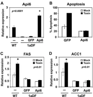
Ectopic Api6 expression protects SREBP-1aDF macrophages from apoptosis independent of changes in lipogenic gene ex- pression. The experiments presented so far demonstrate that the Api6 gene is an SREBP-1a target gene and that SREBP-1aDF mac- rophages are more susceptible to apoptosis following external challenges. However, because SREBP-1a is a transcription factor that activates genes of lipid biosynthesis as well as other pathways (9), the role of Api6 relative to other putative SREBP-1a-targeted pathways in the protection from apoptosis was unclear. To ad- dress this, we introduced an Api6 expression vector into the
SREBP-1aDF macrophages and Api6 mRNA was restored to a level similar to that observed in alpha-toxin-treated WT macro- phages (compareFig. 6A andFig. 4A). The data inFig. 6B show that the ectopic expression of Api6 was sufficient to protect the SREBP-1aDF macrophages from alpha-toxin-induced apoptosis.
Interestingly, while protection from apoptosis was restored, the level of lipogenic gene expression remained low and was not af- fected by Api6.
DISCUSSION
An earlier report in fibroblasts revealed that the SREBP pathway for lipid synthesis was increased following exposure to bacterial pore-forming toxins (5). Interference with activation of the SREBP pathway resulted in increased cell death after toxin expo- sure, and the authors suggested SREBPs were activated to provide lipid precursors to help repair the membrane damage. However, such a mechanism would only indirectly prevent cell death. In the present study, we show that the SREBP-1a isoform is specifically required in macrophages to limit cell death after exposure to a classic pore-forming bacterial alpha-toxin from S. aureus. Similar FIG 5 Api6 promoter activity and chromatin immunoprecipitation (ChIP) assay. (A) Comparison of SREBP binding element among humans and mice. A predicted SREBP binding element is shaded on the Api6 promoter between bp⫺476 and ⫺470. (B) Serial deletion constructs of the mouse Api6 promoter. The mouse Api6 promoter was amplified from mouse genomic DNA and inserted into a luciferase (Luc) reporter vector. The luciferase activity was normalized by
-galactosidase driven by a CMV promoter. (C) Deletion constructs for the Api6 promoter were analyzed for promoter activity. (D) Chromatin from BMDMs was analyzed for recruitment of SREBP-1 to the SRE region of the Api6 promoter by ChIP as described in Materials and Methods. The quantity of DNA in the precipitation with SREBP-1 antibody was normalized to input chromatin and plotted relative to the IgG background.
on January 19, 2016 by Keimyung University Medical Libraryhttp://mcb.asm.org/Downloaded from
results were obtained when we challenged macrophages with streptolysin O and LPS.
We also show that in addition to activating genes of lipid synthesis after toxin exposure, SREBP-1a specifically activates expression of Api6, a previously identified key macrophage-specific antiapoptotic factor (13). As a consequence, SREBP-1aDF macrophages express reduced levels of Api6 and are more susceptible to apoptosis follow- ing challenge with different bacterial toxins. Importantly, the increase in apoptosis is reversed when we restore expression of either SREBP-1a or Api6 in the SREBP-1aDF macrophages. Interestingly, the restoration of expression of Api6 to levels observed in WT mac- rophages protects the SREBP-1aDF cells from apoptosis without al- tering lipogenic gene expression. Thus, SREBP-1a activates genes di- rectly required for protection from apoptosis, and this function is separate from its role in activating lipogenic genes. While these two roles can be uncoupled at the molecular level, as demonstrated here, it is likely that new lipid synthesis is necessary for long-term protec- tion from the damaging effects of the pore-forming toxins on the plasma membrane. A simple model highlighting the role of SREBP-1a in activating Api6 after toxin treatment is shown inFig. 7.
In a previous report, synthetic agonists for the LXR␣/RXR het- erodimer were shown to activate Api6 and protect macrophages from apoptosis following bacterial challenge (10). This mecha- nism indicates that exogenous LXR agonists may be pharmaco- logically beneficial in preventing macrophage apoptosis in re- sponse to bacterial infection. In contrast, our results provide evidence for an endogenous host cell protective response in which Api6 is activated by SREBP-1a, which provides protection from apoptosis induced by bacterial toxins (Fig. 4A).
FIG 6 Api6 mRNA level and apoptosis level after overexpression of Api6 in SREBP-1aDF macrophages. (A) Overexpression of the Api6 mRNA level after Api6 was transfected into macrophages was confirmed by qPCR. RNA was isolated from BMDMs of WT and SREBP-1aDF mice, and Api6 mRNA ex- pression was measured by qPCR. (B) Apoptosis (annexin V-positive cells) was measured by flow cytometry. One hundred nanograms of GFP or Api6 plas- mid was transfected by electroporation, and the amounts of annexin V-posi- tive macrophages were measured by flow cytometry after 24 h. (C and D) qPCR analysis of FAS and ACC1 from BMDMs overexpressing Api6 stimu- lated for 24 h with alpha-toxin. Results are shown as the mean of triplicate samples⫾ SEM relative to unstimulated WT samples (n ⫽ 3 mice per group).
FIG 7 Scheme of the antiapoptotic role of SREBP-1a through the Api6 gene in macrophages. A model for how SREBP-1a regulates bacterial-toxin-induced apoptosis of the macrophage through direct activation of the antiapoptotic factor Api6 gene is shown. Our results suggest that bacterial alpha-toxin-induced SREBP-1a activates not only lipogenesis but also the antiapoptotic factor Api6 gene to limit apoptosis following bacterial infection of macrophages.
SREBP-1a Protects Macrophages from Apoptosis
on January 19, 2016 by Keimyung University Medical Libraryhttp://mcb.asm.org/Downloaded from
We showed in previous studies with the SREBP-1aDF mouse that SREBP-1a activates the initial proinflammatory phase of the innate immune response by regulating Nlrp1a gene expression, which is a key scaffold component of the caspase 1-activating in- flammasome (8). The activation of both Api6 and Nlrp1a by SREBP-1a provides a mechanism to coordinate antiapoptotic and inflammation pathways, which are two key adaptive processes that unite to protect cells against pathogen attack. However, be- cause SREBP-1a activates genes directly required for both pro- cesses, we cannot rule out that the protection from apoptosis is partly due to the inflammasome pathway and vice versa. Regard- less, the identification of key genes for both pathways that are direct targets of SREBP-1a further highlights the multifunctional roles of SREBP-1a in cellular physiology and further supports our overarching hypothesis that SREBPs have evolved to link diverse physiologic processes together with lipid metabolism (9).
SREBPs are conserved regulators of lipid metabolism throughout the eukaryotic kingdom, and along with the current studies, other recent reports suggest SREBPs regulate additional aspects of cellular physiology that directly or indirectly couple a wide variety of seem- ingly disparate cell-environment interactions with lipid metabolism (8,14,16). Thus, we propose that SREBPs have evolved as “metabolic integrators” that effectively bridge broader physiological processes with basic intermediary lipid metabolism (9).
ACKNOWLEDGMENTS
We thank our laboratory members for helpful discussions. We also thank Craig Walsh for reagents and, along with Manuela Raffatellu, Judith Behnsen, Andrea Tenner, and Debra Fraser, for helpful discussions and experimental advice. Peter Tontonoz, UCLA, kindly provided the Api6 plasmid.
This work was supported in part by a grant from the NIH to T.F.O.
(HL48044).
REFERENCES
1. Aderem A, Underhill DM. 1999. Mechanisms of phagocytosis in macro- phages. Annu. Rev. Immunol. 17:593– 623.
2. Bhakdi S, Tranum-Jensen J. 1991. Alpha-toxin of Staphylococcus aureus.
Microbiol. Rev. 55:733–751.
3. Earnshaw WC, Martins LM, Kaufmann SH. 1999. Mammalian caspases:
structure, activation, substrates, and functions during apoptosis. Annu.
Rev. Biochem. 68:383– 424.
4. Gagnon E, et al. 2002. Endoplasmic reticulum-mediated phagocytosis is a mechanism of entry into macrophages. Cell 110:119 –131.
5. Gurcel L, Abrami L, Girardin S, Tschopp J, van der Goot FG. 2006.
Caspase-1 activation of lipid metabolic pathways in response to bacterial pore-forming toxins promotes cell survival. Cell 126:1135–1145.
6. Horton JD, et al. 2003. Combined analysis of oligonucleotide microarray data from transgenic and knockout mice identifies direct SREBP target genes. Proc. Natl. Acad. Sci. U. S. A. 100:12027–12032.
7. Im SS, et al. 2009. Sterol regulatory element binding protein 1a regulates hepatic fatty acid partitioning by activating acetyl coenzyme A carboxylase 2. Mol. Cell. Biol. 29:4864 – 4872.
8. Im SS, et al. 2011. Linking lipid metabolism to the innate immune re- sponse in macrophages through sterol regulatory element binding pro- tein-1a. Cell Metab. 13:540 –549.
9. Jeon TI, Osborne TF. 2012. SREBPs: metabolic integrators in physiology and metabolism. Trends Endocrinol. Metab. 23:65–72.
10. Joseph SB, et al. 2004. LXR-dependent gene expression is important for macrophage survival and the innate immune response. Cell 119:299 –309.
11. Joshi AD, Raymond T, Coelho AL, Kunkel SL, Hogaboam CM. 2008. A systemic granulomatous response to Schistosoma mansoni eggs alters re- sponsiveness of bone-marrow-derived macrophages to Toll-like receptor agonists. J. Leukoc. Biol. 83:314 –324.
12. Koopman G, et al. 1994. Annexin V for flow cytometric detection of phosphatidylserine expression on B cells undergoing apoptosis. Blood 84:
1415–1420.
13. Miyazaki T, Hirokami Y, Matsuhashi N, Takatsuka H, Naito M. 1999.
Increased susceptibility of thymocytes to apoptosis in mice lacking AIM, a novel murine macrophage-derived soluble factor belonging to the scav- enger receptor cysteine-rich domain superfamily. J. Exp. Med. 189:413–
422.
14. Osborne TF, Espenshade PJ. 2009. Evolutionary conservation and adap- tation in the mechanism that regulates SREBP action: what a long, strange tRIP it’s been. Genes Dev. 23:2578 –2591.
15. Pluddemann A, Mukhopadhyay S, Gordon S. 2011. Innate immunity to intracellular pathogens: macrophage receptors and responses to microbial entry. Immunol. Rev. 240:11–24.
16. Seo YK, et al. 2011. Genome-wide localization of SREBP-2 in hepatic chromatin predicts a role in autophagy. Cell Metab. 13:367–375.
on January 19, 2016 by Keimyung University Medical Libraryhttp://mcb.asm.org/Downloaded from