MINI-FOCUS ON FRACTIONAL FLOW RESERVE Clinical Research
Clinical and Physiological Outcomes of
Fractional Flow Reserve-Guided Percutaneous Coronary Intervention in Patients With
Serial Stenoses Within One Coronary Artery
Hack-Lyoung Kim, MD,* Bon-Kwon Koo, MD, P
HD,* Chang-Wook Nam, MD, P
HD,†
Joon-Hyung Doh, MD, P
HD,‡ Ji-Hyun Kim, MD,§ Han-Mo Yang, MD, P
HD,*
Kyung-Woo Park, MD, P
HD,* Hae-Young Lee, MD, P
HD,* Hyun-Jae Kang, MD, P
HD,*
Young-Seok Cho, MD, P
HD,储 Tae-Jin Youn, MD, P
HD,储 Sang-Hyun Kim, MD, P
HD,¶
In-Ho Chae, MD, P
HD,储 Dong-Ju Choi, MD, P
HD,储 Hyo-Soo Kim, MD, P
HD,*
Byung-Hee Oh, MD, P
HD,* Young-Bae Park, MD, P
HD*
Seoul, Daegu, Koyang, and Seongnam, Korea
Objectives This study was performed to evaluate the physiological and clinical outcomes of frac- tional flow reserve (FFR)-guided revascularization strategy with drug-eluting stents in serial stenoses within the same coronary artery.
Background Identifying a functionally significant stenosis is difficult when several stenoses exist within 1 coronary artery.
Methods A total of 131 patients (141 vessels and 298 lesions) with multiple intermediate stenoses within the same coronary artery were assessed by FFR with pullback pressure tracings. In vessels with an FFR ⬍0.8, the stenosis that caused the largest pressure step-up was stented first. Major ad- verse cardiac events were assessed during follow-up.
Results FFR was measured 239 times and there were no procedure-related complications. There was a weak negative correlation between FFR and angiographic percent diameter stenosis (r ⫽ ⫺0.282, p ⬍ 0.001). In total, 116 stents were implanted and revascularization was deferred in 61.1% (182 of 298) of lesions. When the vessels with an initial FFR ⬍0.8 were divided into 2 groups according to FFR after first stenting (FFR ⱖ0.8 vs. FFR ⬍0.8), there were no differences in baseline angiographic and physiological parameters between the 2 groups. During the mean follow-up of 501 ⫾ 311 days, there was only 1 target vessel revascularization due to in-stent restenosis. There were no events related to deferred lesions.
Conclusions FFR-guided revascularization strategy using pullback pressure tracing in serial stenoses was safe and effective. This strategy can reduce unnecessary intervention and maximize the benefit of percu- taneous coronary intervention with drug-eluting stents in patients with multiple stenoses within 1 coronary artery. (J Am Coll Cardiol Intv 2012;5:1013–8) © 2012 by the American College of Cardiology Foundation
From the *Cardiovascular Center, Seoul National University Hospital, Seoul, Korea; †Keimyung University Dongsan Medical Center, Daegu, Korea; ‡Inje University Ilsan Paik Hospital, Koyang, Korea; §Dongguk University Ilsan Hospital, Koyang, Korea;
储Cardiovascular Center, Seoul National University Bundang Hospital, Seongnam, Korea; and the ¶Cardiovascular Center, Seoul National University Boramae Hospital, Seoul, Korea. This study was supported by a grant from the Seoul National University Hospital (A062260), Innovative Research Institute for Cell Therapy, the Clinical Research Center for Ischemic Heart Disease (0412-CR02-0704-0001) sponsored by the Ministry of Health and Welfare, Republic of Korea, and the SNUH Research Fund (03-2010-0270). The authors have reported that they have no relationships relevant to the contents of this paper to disclose.
Manuscript received April 9, 2012; revised manuscript received June 14, 2012, accepted June 21, 2012.
The presence of myocardial ischemia is a major prognostic factor in patients with symptomatic coronary artery disease (1,2) and the decision to perform revascularization should be guided based on the presence of myocardial ischemia. Frac- tional flow reserve (FFR) is a reliable physiological parameter to determine the functional significance of coronary stenosis (3,4). FFR-guided revascularization strategy was reported to be safe and effective in patients with various lesion subsets (5–8).
However, identification of the culprit lesion, which causes myocardial ischemia and warrants revascularization, is chal- lenging in patients with diffuse disease or multiple sequential stenoses with intermediate severity.
See page 1019
In patients with multiple stenoses of intermediate severity in 1 coronary artery, FFR measurements with pullback pressure recording can be helpful to identify the lesion that has functional significance (9,10). Although more and more patients with complex coronary lesions are now treated with drug-eluting stents (DES), the outcomes of this strategy in the era of DES have not yet been fully evaluated.
We performed this study to eval- uate the physiological and clinical outcomes of FFR-guided revascu- larization strategy with DES in pa- tients with serial stenoses within 1 coronary artery.
Methods
Study subjects. Between March 2009 and December 2011, patients who underwent elective coronary angiography and had multiple intermediate steno- ses in the same epicardial coronary artery (vessel size ⬎2 mm in diameter) were prospectively enrolled from 2 Korean centers. An intermediate stenosis was defined as 40% to 70% diameter stenosis by visual assessment. To be included, each lesion should be separated by an angiographically normal-looking segment of at least 20 mm. Patients were excluded if any of the following were present: in-stent restenosis, acute ST-segment elevation myocardial infarc- tion, regional wall motion abnormalities of a target vessel segment, left ventricular ejection fraction ⬍40%, primary myocardial or valvular disease, contraindication to adeno- sine, or angiographically visible thrombus at a target lesion.
In patients with acute coronary syndrome, only the noncul- prit vessels were included. The study protocol was approved by the Institutional Review Board of each participating hospital and informed consent was obtained from every study participant.
Procedures. Target vessel engagement was performed via radial or femoral approach using 5-F to 7-F guide catheters.
Angiographic images were acquired after intracoronary nitroglycerin (100 to 200 g) administration. FFR was measured using a 0.014-inch pressure guidewire (St. Jude Medical, Minneapolis, Minnesota) as previously described (4) and hyperemia was induced by the continuous intrave- nous infusion of adenosine (140 g/kg/min). The pressure wire was initially positioned distal to the most distal lesion, and FFR was measured. FFR was calculated as the mean distal coronary pressure divided by the mean aortic pressure during maximal hyperemia and functional significance was defined with the threshold of FFR ⬍0.8 (11). In vessels with an FFR ⬍0.8, the pressure wire was slowly pulled back to the ostium of the coronary artery under steady-state hyperemia and the stenosis that caused the largest pressure step-up (primary target lesion) during pressure wire pull- back was treated first. Percutaneous coronary intervention (PCI) of other stenoses was determined by FFR measured after stenting of the primary target lesion. In this study, apparent and true FFR of nonprimary target lesion were calculated. Apparent FFR was defined as the initial ratio of proximal and distal pressures across the nonprimary target lesion, and true FFR was defined as the ratio of pressures across that stenosis after the stenosis of a primary target lesion was eliminated by PCI (9,10) (Fig. 1). All pressure tracings were recorded on the RadiAnalyzer Xpress (St. Jude Medical) for offline analysis. All PCI procedures were performed using DES.
Quantitative coronary angiography. Quantitative coronary an- giography was performed by an independent core laboratory at Seoul National University Cardiovascular Center. Quantitative coronary angiography was performed by a single experienced observer who was unaware of FFR findings. Using the guide catheter for calibration and an edge detection system (CAAS, version 5.7, QCA System, Pie Medical, Maastricht, the Nether- lands), the reference diameter, minimum lumen diameter, and lesion length were measured, and the percentage of diameter stenosis was calculated.
Follow-up. Patients were recommended to visit the hospital for follow-up at 1, 6, and 12 months after initial angiogra- phy. Information relating to major adverse cardiac events, including cardiac death, target vessel-related myocardial infarction, and revascularization, was collected. Telephone contact was performed if necessary. There was no loss to follow-up in this study.
Statistical analysis. Data were expressed as mean ⫾ SD for continuous variables and percentages for categorical vari- ables. Comparison of continuous variables was performed using the Student t test or paired t test. Analysis of discrete variables was performed using the chi-square test. Pearson correlation was used to calculate the association between angiographic stenosis and FFR as well as between apparent and true FFR. The value of p ⬍ 0.05 was considered as significant. All statistical analyses were performed using SPSS (version 16.0, SPSS Inc., Chicago, Illinois).
Abbreviations and Acronyms
DESⴝ drug-eluting stent(s) FFRⴝ fractional flow reserve
PCIⴝ percutaneous coronary intervention
Results
Among 161 eligible coronary arteries with 2 or more intermediate stenoses, 20 were excluded (10 protocol viola- tions, 6 pressure tracing artifacts, 3 bypass surgeries, and 1 balloon angioplasty), and 141 coronary arteries (131 patients
and 298 lesions) were finally analyzed in this study. Sixteen vessels had 3 stenoses and the other 125 vessels had 2 stenoses. Baseline clinical characteristics of study patients and angiographic findings were summarized in Table 1. The most commonly involved artery was the left anterior de- scending coronary artery (95 vessels, 67.4%). The mean
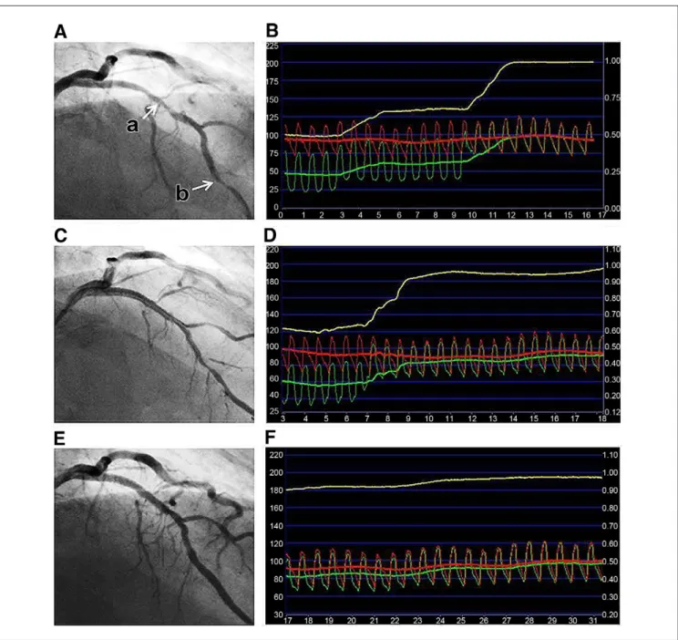
Figure 1.Representative Case of FFR with Pullback Pressure Tracing-Guided PCI
(A, B) Two consecutive intermediate stenoses (labeled a and b with arrows) were observed in the left anterior descending artery. As the fractional flow reserve (FFR) was 0.48, pullback pressure tracing was performed while simultaneously monitoring the intracoronary pressure (green line), aortic pressure (red line), and FFR (yellow line). Two step-ups of intracoronary pressure were observed during pullback pressure tracing under maximal hyperemia (B). Apparent FFR of lesions a and b were 0.67 (ratio of pressures across lesion a⫽ 60/90) and 0.75 (ratio of pressures across lesion b ⫽ 45/60), respectively. As the larger pressure step-up was observed across lesion a (30 mm Hg) than lesion b (16 mm Hg), the proximal stenosis was regarded as the primary target lesion and stenting was per- formed. (C, D) After stenting lesion a (C), pullback pressure tracings (D) were performed again. FFR was 0.59 and intracoronary pressure step-up across lesion b was 20 mm Hg. Therefore, stenting to the distal stenosis followed. True FFR of lesion b was 0.73 (55/75 mm Hg). (E, F) After stenting both proximal and distal lesions, FFR was 0.85 and no significant pressure step-up was found across lesion a or lesion b. PCI⫽ percutaneous coronary intervention.
diameter stenosis was 51.6 ⫾ 13.2% and 167 (56%) of 298 lesions had ⱖ50% stenosis.
FFR was measured 239 times in total, and there were no procedure-related complications. The association between FFR and angiographic stenosis of the most severe lesion in a target vessel is shown in Figure 2. There was a weak negative correlation between FFR and angiographic percent
diameter stenosis (r ⫽ ⫺0.282, p ⬍ 0.001) and there was no difference in FFR between the vessels with ⱖ50% and
⬍50% stenosis (0.73 ⫾ 0.11 vs. 0.76 ⫾ 0.12, p ⫽ 0.231).
Mean distal FFR before PCI was 0.74 ⫾ 0.11 in all vessels.
In total, 116 stents (70 proximal and 46 distal) were implanted and revascularization was deferred in 61.1% (182 of 298) of lesions. Two or more stents were implanted in only 26 vessels (18.4%). In 89 vessels with an FFR ⬍0.8, baseline FFR was 0.67 ⫾ 0.09 and increased to 0.84 ⫾ 0.07 after stenting (p ⬍ 0.001).
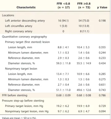
The pressure step-up of the primary target lesion was 19.7 ⫾ 6.6 mm Hg, and all primary target lesions had step-up of more than 10 mm Hg. When the primary target lesion was a proximal lesion, FFR was increased from 0.69 ⫾ 0.08 to 0.84 ⫾ 0.08 (21.7% increment) after stenting. In cases of distal lesions, stenting increased FFR from 0.66 ⫾ 0.10 to 0.87 ⫾ 0.05 (31.8% increment). After stenting the primary target lesion, pressure step-up of a nonprimary target lesion was increased from 7.7 ⫾ 5.9 mm Hg to 10.9 ⫾ 7.8 mm Hg (p ⫽ 0.013). Figure 3 shows the association between apparent and true FFR of nonprimary target lesions. When the vessels with an initial FFR ⬍0.8 was divided into 2 groups according to the FFR after first stenting (FFR ⱖ0.8 vs. FFR ⬍0.8), there were no differ- ences in baseline angiographic and physiological parameters between the 2 groups (Table 2). There was a trend toward a higher pressure step-up of nonprimary target lesion(s)
Table 1.Clinical and Angiographic Characteristics
Age, yrs 63.8⫾ 9.2
Male 88 (66.2)
Previous history
Diabetes 47 (35.9)
Hypertension 82 (62.6)
Hypercholesterolemia 52 (39.7)
Current smoking 24 (18.3)
Stable angina 64 (48.9)
Unstable angina 29 (22.1)
Multivessel disease 88 (66.1)
Left ventricular ejection fraction 61.4⫾ 7.6
Angiographic characteristics Involved arteries, 141 vessels
Left anterior descending artery 95 (67.4)
Left circumflex artery 21 (14.9)
Right coronary artery 25 (17.7)
Quantitative coronary angiography, 298 lesions
Lesion length, mm 11.1⫾ 6.9
Minimum lumen diameter, mm 0.9⫾ 0.4
Reference diameter, mm 2.6⫾ 0.6
Diameter stenosis, % 51.6⫾ 13.2
Values are mean⫾ SD or n (%).
Figure 2.Relationship Between FFR and Maximal Angiographic Stenosis Obtained by QCA
FFR⫽ fractional flow reserve; QCA ⫽ quantitative coronary angiography;
r⫽ correlation coefficient.
Figure 3.Relationship Between Apparent and True FFR of Nonprimary Target Lesions
Proximal lesion is the primary target lesion and distal lesion is a nonpri- mary target lesion (open circles); distal lesion is the primary target lesion and proximal lesion is a nonprimary target lesion (solid circles). Abbrevia- tions as inFigure 2.
before intervention in patients with an FFR ⬍0.8 after stenting the primary target lesion (9.7 ⫾ 6.2 mm Hg vs. 6.9 ⫾ 4.7 mm Hg, p ⫽ 0.094).
During the mean follow-up of 501 ⫾ 311 (median 509) days, there was only 1 target vessel revascularization that occurred due to in-stent restenosis. There were no events related to deferred lesions. One noncardiac death (due to acute subdural hemorrhage) and 1 nontarget vessel-related myocardial infarction occurred during follow-up (Table 3).
Discussion
The present study demonstrated that FFR measurement with repetitive pullback pressure recordings is safe and useful to determine the proper target lesions for revascular- ization with DES and can reduce unnecessary intervention in patients with serial stenoses in 1 coronary artery.
FFR is a well-established physiologic parameter for the assessment of the hemodynamic significance of coronary stenosis (7,12–14). However, clinical application of FFR in vessels with multiple stenoses is not easy. In cases of multiple serial stenoses, 1 stenosis influences the FFR of the others, which complicates the determination of FFR of each individual stenosis (9,10). In this situation, pullback pres- sure recordings under maximal hyperemia have been known to be a useful and practical method to identify the stenosis that has hemodynamic significance requiring revasculariza-
tion (9,10). In our study of 141 vessels with 298 interme- diate lesions, 239 FFR measurements were required to perform the FFR-guided revascularization strategy. How- ever, there were no procedure-related complications, dem- onstrating that this approach can provide a safe and feasible physiological assessment for patients with serial stenoses in a real-world practice.
When multiple stenoses exist in 1 vessel, the functional significance of each stenosis can be underestimated due to hemodynamic interaction among the lesions (9,10). There- fore, the functional significance of a nonprimary target lesion should be reassessed after the treatment of a primary target lesion. Like previous reports (9,10), our study showed that true FFR was lower than apparent FFR in both proximal and distal stenoses and the pressure step-up of a nonprimary target lesion was increased from 7.7 ⫾ 5.9 mm Hg to 10.9 ⫾ 7.8 mm Hg after stenting the primary target lesion. In addition, there was no difference in baseline angiographic and physiological characteristics that can pre- dict the FFR ⬎0.8 after first stenting. All of these findings emphasize the importance of a repeated measurement of FFR after stenting the primary target lesion to accurately assess the functional significance of nonprimary target lesions. Otherwise, functionally significant lesions can be left untreated. The increment of pressure gradient across the nonprimary target lesion after PCI of primary target lesion was less than that from the previous study by Pijls et al. (10).
This difference seems to be due to the differences in lesion characteristics and severity of stenosis between the 2 studies.
It is well known that stent placement should be per- formed only for functionally significant stenosis (15,16). In this study, PCI was deferred in 182 lesions of 298 lesions (61.1%) based on the FFR value and only 26 vessels (18.4%) required more than 2 stents. During mean follow-up of 501 ⫾ 311 days, there was no clinical event related to deferred lesions. Considering the excellent clinical outcomes and the number of stents saved in our study, FFR measure- ment with pullback pressure tracing in patients with serial stenoses can maximize the benefit of PCI with DES, reduce the number of implanted stents, and minimize stent-related complications. This study’s results are in line with the results of the FAME (Fractional Flow Reserve Versus Angiography for Multivessel Evaluation) study (11) and
Table 3.Clinical Outcomes of FFR-Guided Revascularization in Patients With Serial Stenoses
Cardiac death 0
Target vessel-related MI 0
Nontarget vessel-related MI 1
TLR of stented lesion 1
TLR of deferred lesion 0
FFR⫽ fractional flow reserve; MI ⫽ myocardial infarction; TLR ⫽ target lesion revascularization.
Table 2.Comparison of Angiographic and Physiological Characteristics Between 2 Groups Divided by FFR After Stenting the Primary Target Lesion
Characteristic
FFR <0.8
(nⴝ 17) FFR >0.8
(nⴝ 72) p Value Locations
Left anterior descending artery 16 (94.1) 54 (75.0) 0.198
Left circumflex artery 1 (5.9) 10 (13.9)
Right coronary artery 0 8 (11.1)
Quantitative coronary angiography Primary target (first stented) lesion
Lesion length, mm 8.8⫾ 4.1 10.4⫾ 5.3 0.355
Minimum lumen diameter, mm 1.1⫾ 0.3 1.4⫾ 0.6 0.244 Reference diameter, mm 2.9⫾ 0.3 2.6⫾ 0.6 0.233 Diameter stenosis, % 59.3⫾ 11.8 55.5⫾ 14.9 0.434 Nonprimary target lesion
Lesion length, mm 13.4⫾ 7.1 10.9⫾ 6.6 0.285
Minimum lumen diameter, mm 1.3⫾ 0.3 1.5⫾ 0.6 0.275 Reference diameter, mm 2.7⫾ 0.4 2.6⫾ 0.6 0.651 Diameter stenosis, % 51.1⫾ 11.0 49.6⫾ 12.6 0.743
FFR before stenting 0.68⫾ 0.09 0.68⫾ 0.08 0.786
Pressure step-up before stenting
Primary target lesion, mm Hg 19.2⫾ 6.2 19.9⫾ 6.9 0.729 Nonprimary target lesion, mm Hg 9.7⫾ 6.2 6.9⫾ 4.7 0.094 Values are mean⫾ SD or n (%).
FFR⫽ fractional flow reserve.
show the clinical and economic benefit of FFR measure- ment in patients with multiple lesions.
Study limitations. First, the number of patients was rela- tively small, and there was no control group. Second, accurate calculation of FFR of each stenosis could not be performed in our study, as we did not measure coronary wedge pressure (9,10). However, as balloon occlusion is required for the calculation of FFR of each stenosis, it was not clinically applicable, as about 40% of the lesions did not require coronary intervention.
Conclusions
FFR-guided revascularization strategy using pullback pres- sure tracing method in serial stenoses was safe and effective.
This strategy can reduce unnecessary intervention and maximize the benefit of PCI with DES in patients with multiple stenoses within 1 coronary artery.
Reprint requests and correspondence: Dr. Bon-Kwon Koo, Division of Cardiology, Department of Internal Medicine, Seoul National University College of Medicine, Cardiovascular Center, Seoul National University Hospital, 101 Daehak-ro, Jongno-gu, Seoul 110 –744, Korea. E-mail: [email protected].
REFERENCES