Copyright 2006 by the Korean Society for Clinical Neurophysiology 91 대한임상신경생리학회지 8(1):91~93, 2006 ISSN 1229-6414
Uremic encephalopathy is usually associated with cortical involvement in patients with uremia, as evidenced by frequent seizures and abnormal findings on electroencephalography.1,2 Involuntary movements in uremic encephalopathy typically consist of asterixis and myoclonus, which reflect cortical involvement.1,2Other symptoms of abnor- mal movement disorder including parkinsonism and evidence of basal ganglia pathology as a con- sequence of uremia have been reported very rarely.3 A case of acute parkinsonism with bilat- eral basal ganglia lesions in a patient with uremia
is examined here.
Case report
A 47-year-male visited to our hospital in June 2005 because of general weakness and gait dis- turbance. he had hypertension and diabetes mel- litus for 7 years. The diabetes had been treated with oral hypoglycemic agents. Uremia was diag- nosed in January 2005, and hemodialysis started in March.
Neurologic examination revealed mild bradyki- nesia, generalized rigidity, dysarthria, dysphagia and masked face. His gait was short-stepped and shuffling with mild postural impairment. His cra- nial nerve and cerebellar functions were normal.
The deep tendon reflexes were normoactive and pathologic reflexes were absent. Blood chemistry showed elevated BUN (56 mg/dL) and creatinine (6.1 mg/dL) levels. The blood glucose level was 136 mg/dL and electrolyte levels were within normal
요독증 환자의 양측 기저핵 병변에 의해 발생된 급성 파킨슨증 1예
인제대학교 의과대학 부산백병원 신경과학교실
박강민∙김상진
Acute Parkinsonism with Bilateral Basal Ganglia Lesions in A Patient with Uremia
Kang-Min Park, M.D., Sang-Jin Kim, M.D.
Department of Neurology, Busan Paik Hospital, Inje University College of Medicine
A 47-year-old male who had hypertension and diabetes mellitus for 7 years suddenly developed bradykinesia, gener- alized limb muscular rigidity, dysarthria and dysphagia. Uremia developed 5 months prior to this and he had been on hemodialysis. A T2-weighted brain MRI showed extensive hyperintensity over the bilateral basal ganglia, extending to the adjacent periventricular white matter. In T1-weighted images the lesions were hypointense. Supportive treatments were given and his symptoms improved. Exacerbation of glucose utilization failure or vasogenic edema is suggested as the etiology of basal ganglia lesions, but the exact underlying pathophysiology is unknown.
Key Words: Uremia, Basal ganglia, Parkinsonis
Address for correspondence Sang-Jin Kim, M.D.
Department of Neurology, Busan Paik Hospital, Inje University College of Medicine, 633-165 Gaegeum-dong Busanjin-gu, 614-735, Busan, Korea
Tel: +82-51-890-6130 Fax: +82-51-895-6367 E-mail: [email protected]
� This study was financially supported by research fund from inje University in 2006.
박강민∙김상진
limits.
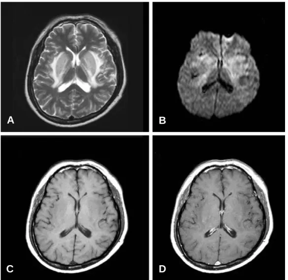
A T2-weighted brain MRI performed 5 days after symptoms onset showed extensive hyperin- tensity over the bilateral basal ganglia, extending to the adjacent periventricular white matter. In T1-weighted images the lesions were hypointense (Fig. 1).
Supportive treatments and hemodialysis were given, His symptoms gradually improved. One month after symptoms onset, he had mild dysarthria but was otherwise perfectly well.
Discussion
Acute movement disorders and bilateral basal ganglia lesions in patients with uremia are a rare
distinct clinical syndrome. The lesions are sym- metrical and have unique imaging characteristics.
They are high signal intensity on T2-weighted images and low signal intensity on T1-weighted images with mass effect and perifocal edema.3.4 Our patient had uremia and suffered from acute parkinsonism associated with symmetrical bilat- eral basal ganglia lesions. Our case also exhibited high signal intensity on T2-weighted images and low signal intensity on T1-weighted images.
Wang et al. first reported similar findings in 1998 and 2003.3.4 All of their patients had both uremia and diabetes mellitus. They proposed the following as a possible underlying pathophysiolo- gy. The basal ganglia cellular functions had already been compromised by long-term diabetes
92 J Korean Society for Clinical Neurophysiology / Volume 8 / June, 2006
Figure 1. (A) T2 weighted MRI images show extensive hyperintensity over basal ganglia bilaterally, extending to the adjacent periventricular white matter. (B) On diffusion MRI images the lesions are slightly hyperintense. (C) T1 weighted MRI images show hypointensity on bilateral basal ganglia. (D) Gadolinium-enhanced T1 weighted images show slight enhancement of lesions.
A B
C D
경요독증 환자의 양측 기저핵 병변에 의해 발생된 급성 파킨슨증 1예
J Korean Society for Clinical Neurophysiology / Volume 8 / June, 2006 93
mellitus through microangiopathic changes and energy utilization failure. When they were further exposed to markedly elevated levels of uremic toxins, their regional cellular metabolism and/or vascular autoregulation collaped, and tissue was damaged.3.4Wang et al. also reported a FDG-PET study.5 The two cases showed markedly reduced glucose metabolism in the basal ganglia, espe- cially in the bilateral putamens where glucose uptake was nealy absent. They described that focal deficit of glucose utilization in the bilateral basal ganglia regions could explain these findings of FDG-PET study.5
But Lee et al. suggested vasogenic edema as the pathophysiology when examining diffusion weighted images and apparent diffusion coeffi- cient maps in their case with uremia only.6 The exact etiology underlying thess lesions is still unknown. Probably, pathophysiology of basal ganglia lesions is multifactorial including exacer- bation of glucose utilization failure and vasogenic edema.
The prognosis for our patient was good, but most patients of Wang et al. died or required pro- longed intensive care due to complications.
Therefore, acute parkinsonism in uremic patients may require more careful observation and follow-
up. The definitive treatment of these patients is uncertain, but it is advisible to remove uremic toxins as soon as possible because of the proposed pathogenesis.
In conclusion. We have reported a rare distinct clinical syndrome of acute parkinsonism with bilateral basal ganglia lesions in a patient with uremia.
REFERENCES
01. Raskin NH. Neurological complication of renal failure. In:
Aminoff MJ. Neurology and general medicine. 3rd. New York: Churchill Livingstone 2001:293-306.
02. Raskin NH, Fishman RA. Neurological disorders in renal failure. N Engl J Med 1976;294:143-148.
03. Wang HC, Brown P, Lees AJ. Acute movement disorders with bilateral basal ganglia lesions in uremia. Movement Disorders 1998;13(6):952-957.
04. Wang HC, Cheng SJ. The syndrome of acute bilateral basal ganglia lesions in diabetic uremic patients. J Neurol 2003;250:948-955.
05. Wang HC, Hsu JL, Shen YY. Acute bilateral basal ganglia lesions in patients with diabetic uremia: an FDG-PET study. Clin Nucl Med 2004;29:475-478.
06. Lee PH, Shin DH, Kim JW, Song YS, Kim HS.
Parkinsonism with basal ganglia lesions in patient with uremia: evidence of vasogenic edema. Parkinsonism and Related Disorders 2006;12:93-96.