Effect of Cymbidium Root Extracts on Oxidative Stress-induced Myoblasts Damage
Wan Joong Kim
1, Han-Sung Kim
2, Joerg Opitz
3, Kazuya Kabayama
4and Tack-Joong Kim
1*
1
Division of Biological Science and Technology, Yonsei-Fraunhofer Medical Device Lab, College of Science and Technology, Yonsei University, Wonju 220-710, Korea
2
Department of Biomedical Engineering, Yonsei-Fraunhofer Medical Device Lab, College of Health Science, Yonsei University, Wonju 220-710, Korea
3
Fraunhofer Institute for Ceramic Technologies and Systems-Material Diagnostics, Dresden, Germany
4
Department of Chemistry, Graduate School of Science, Osaka University, Osaka, Japan
Received July 21, 2014 /Revised August 9, 2014 /Accepted August 12, 2014Skeletal muscle atrophy can be defined as a decrease in or a disease of the muscle tissue, or as a disorder of the nerves that control the muscle, through injury or lack of use. This condition is asso- ciated with reactive oxygen species (ROS), resulting in various muscular disorders. Exposure to ROS induces muscle atrophy through several biological factors, such as SOD1 and HSP70. We found that cymbidium root extract reduced the H
2O
2-induced viability loss in C2C12 myoblasts and inhibited apoptosis. In addition, we showed that the cymbidium root extract increased the expression of HSP70 and decreased the expression of SOD1 in the H
2O
2-induced C2C12 myoblasts. These results suggest that cymbidium root extract might have therapeutic value in reducing ROS-induced muscle atrophy.
Key words : Cymbidium, heat shock protein 70, reactive oxygen species
*Corresponding author
*Tel : +82-33-760-2242, Fax : +82-33-760-2183
*E-mail : [email protected]
This is an Open-Access article distributed under the terms of the Creative Commons Attribution Non-Commercial License (http://creativecommons.org/licenses/by-nc/3.0) which permits unrestricted non-commercial use, distribution, and reproduction in any medium, provided the original work is properly cited.
Journal of Life Science 2014 Vol. 24. No. 9. 1019~1024 DOI : http://dx.doi.org/10.5352/JLS.2014.24.9.1019
서 론
근위축 (atrophy)은 근육을 사용하지 않음으로써 발생하는 근육 조직의 손실 또는 근육을 지배하는 신경의 손상으로 정 의할 수 있으나 , 정확한 진단이 어려워 한국 및 서양에서도 정확한 통계를 산출하기 어렵다 [1].
근위축은 움직이지 못하는 상태에서의 근육사용 감소 , 장 기적인 요양 , 신경제거, 우주탐사 등의 결과로 나타날 수 있으 며 [20], 근위축이 발병하면 완전한 치료가 이루어지지 않아 진행을 늦추어 주는 약물효과만의 연구가 진행되고 있다 . 근 위축은 산화적인 환경의 근육에서 전자이동과 산소흐름이 갑 자기 많아질 때 활성산소에 의하여 발생하기도 하며 심한 운 동을 하면 활성산소 생산의 증가로 인해 근육손상이 유도된 다 [6, 11, 20]. 이러한 기전은 활성산소와 관련된 물질대사와 매개되어 세포 내 높은 라디칼 농도를 유지시켜 단백질 또는 DNA에 의해 세포사를 유도한다[5, 21]. 즉, 과다한 활성산소 에 의해 높은 라디칼이 형성되고 이는 세포막 , DNA, 그 외의 모든 세포 구조를 손상시키며 손상의 범위에 따라 세포가 기 능을 잃거나 변질된다 . 이와 같이 과산화수소가 세포 내에서
높은 수준의 농도를 유지할 수 있는 이유는 막 투과성을 이용 한 확산 등으로 OH- 또는 자유라디칼을 세포 내로 유입시키 기 때문으로 보고되고 있다 [14].
세포 내 세포사의 관련된 신호전달 경로 단백질 중 하나인 열충격단백질 (heat shock protein: HSP)은 샤페론과 같이 세 포간에 다른 단백질의 제대로 된 구조형성을 도와주며 , 기능 적인 부류에 관련되는 단백질이다 . 이 단백질은 열손상, 감염, 염증 , 격렬한 운동을 포함한 다양한 스트레스와 세포의 독성, 기아 , 저산소증, 알코올, 아미노산 유사체 등의 스트레스 유발 인자에 노출 시 발현된다 [9]. 특히, HSP70의 발현은 tumor necrosis factor (TNF)-α로 유도된 세포독성물질과 화학적 손 상물질의 공격으로부터 방어하기 위해 유도되며 산화스트레 스 , 세라마이드, 방사선과 같은 세포 내 세포사 자극신호들로 부터 세포를 보호하는 역할을 한다 [9, 10, 24].
한편 , 심비디움은 난초과에 속하는 식물로 동남아시아, 중
국 , 일본, 오스트레일리아 북부 등에 분포한다. 다수의 연구결
과에 따르면 심비디움의 뿌리에서 추출된 phenanthrene과
phenylpropanoid는 항균력 실험에서 고초균, 폐렴간균, 적색
백선균에 대해 항균기능을 가지고 [22], carrageenin으로 유도
된 쥐의 부종에 대한 항염증 실험에서 효과를 나타내었으며
[7], 착향 및 화장품 성분으로 이용된다고 알려진 linalool과
4-methyl-phenol은 심비디움 뿌리에서 색층분석법을 통해 분
석되었다 [4]. 특히, 심비디움에서 추출된 유효성분인 ar-
omatic glucosides는 과산화물제거 실험에서 항산화능 효과
를 확인하였다 [23]. 하지만, 근육세포에서의 심비디움 추출물
에 대한 효과 및 기전은 현재까지 보고되지 않았다 . 따라서,
- Note -
본 연구에서는 근육세포의 산화적 손상에 대한 심비디움 뿌 리 추출물의 효과와 분자기전을 연구하였다 .
재료 및 방법
실험재료
실험에 사용된 세포배양 재료는 Gibco-BRL (Gaithersburg, MD, USA)로부터 구입하였으며, HSP70 antibody는 Enzo Life Science (Switzerland)로부터 Beta-actin (β-actin)는 Cell Signaling Technology (Danvers, MA, USA)로부터 구입하였 다 . 비교군으로 사용되는 N-acetyl-L-cytein (NAC)는 Sigma- Aldrich (St. Louis, MO, USA)에서, 세포 염색시 사용하는 4’,6’-diamidine-2’-phenylindole dihydrochloride (DAPI)는 Vector Laboratories (Burlingame, CA, USA)에서 구입하였 다 . 심비디움 뿌리추출물은 농촌진흥청에서 제공받아 고체추 출물 100 mg을 물 1 ml에 녹인 sample을 희석하여 사용하였다.
자유라디컬 소거율 측정
2,2-Diphenyl-1-picrylhydrazyl (DPPH) 용액은 항산화 기 작 중 Pronton-radical scavaenger에 의해 전자를 내어주면서 라디칼이 소거되며 가시적으로는 보라색으보부터 노란색으 로 색의 변화가 관찰되는데 본 연구에서는 심비디움 뿌리추 출물과 DPPH의 두 물질을 암상태에서 30분간 반응시켜 추출 물의 각 농도에서 자유라디칼 소거율을 517 nm 파장영역에 서 packard EL800 microplate reader (BioTek Instruments, Winooski, VT, USA)로 측정하였다[2].
세포배양
본 실험에 사용된 C2C12 근육세포는 10% heat-inactivated fetal bovine serum (FBS), 100 units/ml penicillin, 100 μg/ml streptomycin과 L-glutamine을 첨가한 dulbecco’s modified eagle medium (DMEM) 배지를 이용하여37℃ 습윤한 CO
2배 양기 (5% CO
2/ 95% air)에서 배양하였다. 세포는 실험을 위해 80% 채워서 사용하였고 계대 배양을 하기 전에 phosphate buffered saline (PBS, pH7.4)로 세척한 후에 0.25% tryp- sin-EDTA를 처리하여 계대 배양을 하였다.
세포생존율 측정
24 well plate에 C2C12 근육세포를 1×10
5cells/well이 되 도록 분주하고 , 24시간 37℃ 습윤한 CO
2배양기 (5% CO
2/ 95% air)에서 배양하였다. 이 후, 배지를 serum free DMEM으 로 바꾸어주고 각각의 농도로 심비디움 추출물을 첨가하여 24시간 배양한 후에 각각 0, 1 mM의 H
2O
2가 포함된 serum free DMEM으로 배지를 교체하였다. Control 세포들은 DMEM에 H
2O
2를 넣지 않은 것으로 정하고 MTT assay를 이 용하여 550 nm의 흡광도에서 packard EL800 microplate
reader로 측정하였다. 시험물질을 첨가하지 않은 대조군 세포 수를 100%로 하여 각 시험물질의 상대적인 생존율을 구하였 다 .
세포사 확인
C2C12 근육세포는 2.5×10
5cell/cm
2로 커버글라스 위에 23시간 배양한 후, 배지를 serum free DMEM으로 바꾸어주 고 각각의 농도로 심비디움 추출물을 첨가하여 24시간 배양 하였다 . 이후 각각 0, 1 mM의 H
2O
2가 포함된 serum free DMEM으로 배지를 교체하였다. 1시간 후 C2C12 근육세포는 4% paraformaldehyde로 고정하였다. 상온에서 PBS에 0.1%
Triton X-100을 넣어준 것에 30분 동안 노출시켜서 DAPI 용 액이 세포막에 잘 투과될 수 있도록 하였다 . 죽은 세포는 형광 현미경으로 염색된 세포에서 형태적인 변화로 확인하였다 . 각각 3번의 독립된 100개 세포를 세어 확인하였고, 핵의 형태 가 깨어진 세포로 진행된 것을 눈으로 확인하여 세포사가 진 행되었다고 예상되는 세포를 전체 세포수의 퍼센트로 표시하 였다 . 핵의 형태가 응축되거나 단편화 된 것은 세포사가 진행 된 것으로 간주하여 측정한 세포는 전체 세포수의 퍼센트로 표시하였다 .
단백질 분리 및 Western blot 분석
전기영동을 위한 단백질 시료의 추출을 위해 H
2O
2와 각 농 도별로 심비디움 추출물을 처리한 세포를 PBS로 1회 세척한 후 , PRO-PREP 단백질 추출 키트 (iNtRON, Korea)를 사용하 여 각 well에 처리하고 얼음 위에서 20분 반응시켰다. 13,000 rpm, 4℃에서 5분간 원심분리 시킨 후, 단백질 정량은 Bradford protein assay kit를 사용하여 595 nm에서 흡광도를 측정하였다 . 단백질의 농도 비율을 균일하게 맞춰준 뒤 so- dium dodecyl sulfate (SDS)-polyacrylamide gel 전기영동으 로 분리시킨 후 , PVDF membrane (Bio-Rad, Hercules, CA, USA)에 전이시켰다. 이 membrane을 항체의 비특이적 결합 을 차단하기 위하여 blocking buffer (5% skim milk와 0.1%
Tween 20을 함유한 Tris-buffered saline)에서 1시간 동안 반 응시켰다 . 그 후 각 검증 단백질에 대한 항체를 BSA/Tris- buffered saline에 희석하여 가한 후 4℃에서 overnight 반응 시켰다 . 이어서 0.1% Tween 20을 함유한 Tris-buffered saline (TBS-T)로 membrane을 5분간 3회 세척하고, 이차 항체를 상 온에서 1시간 반응시킨 후, 다시 세척하고 enhanced chem- iluminescence (ECL) detect kit (GE healthcare, Buckingham- shire, UK)를 처리하여 ImageQuant LAS4000 (GE health- care, Buckinghamshire, UK)를 통해 단백질 발현을 확인하였 다 .
통계처리
본 실험적 결과는 mean ± standard error로 표시하였고,
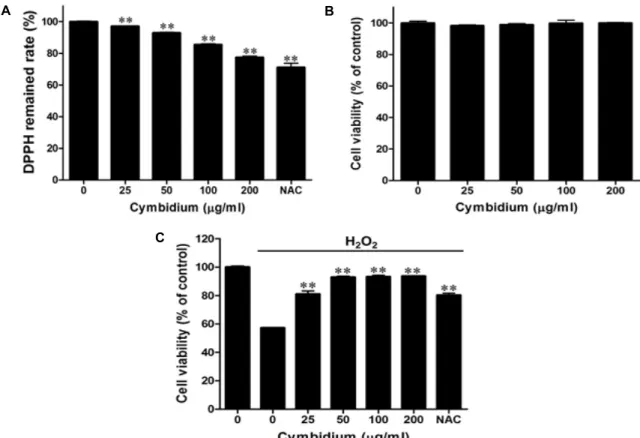
A B
C
Fig. 1. Effect of cymbidium extract on H2O2-induced oxidative stress in C2C12 myoblasts (A) Scavaenging of cymbidium extract by DPPH assay. Extract was treated in 96 well plate with ethanol and DPPH solution. Then sample was incubated for 30 min without light. Optical density was determined at 517 nm. DPPH remained rate was calculated by the following equation. DPPH remained rate (%) = [(absorbance of sample / absorbance of control) ×100]. (B) Cytotoxicity of cymbidium extract
.
C2C12 myoblasts were cultured in 96-well plates until confluent, and the medium was replaced with serum-free medium with or without cymbidium extract (0-200 μg/ml) for 24 hr. The EZ-Cytox reagent was added to the medium, and C2C12 myoblasts were incubated for 1 hr. Optical density was determined at 450 nm using a microplate reader. (C) Effect of cymbidium extract on H2O2-induced oxidative stress in C2C12 myoblasts. C2C12 myoblasts were cultured in 96-well plates until confluent and the medium was then replaced with a serum-free medium with cymbidium extract (0-200 μg/ml).After pre-incubating for 23 hr, 1 mM H2O2 was added for 1 hr. The EZ-Cytox reagent was added to the medium, and C2C12 myoblasts were incubated for an additional 1 hr. The optical density was set at 450 nm by using a microplate reader.
The cell viability was calculated using the following equation: Cell viability (%) = [(absorbance of the H2O2-treated sam- ple/absorbance of the H2O2-untreated control) ×100]. Each value represents the mean (± SD) from three experiments in which each experiment was performed in triplicate. NAC: N-acetyl cysteine. **
p
<0.01 versus H2O2 alone.통계처리는 student’s t-test에 의해 p<0.05인 경우 유의한 것 으로 간주하였다 .
결과 및 고찰
심비디움 추출물의 자유라디칼 소거 효과와 H
2O
2로 유도된 C2C12 근육세포 손상에서 심비디움 추출물의 영향
산화스트레스에 의한 C2C12 근육세포 손상에서 심비디움 추출물의 예방효과를 확인하기 전에 심비디움 추출물의 양에 따른 항산화 효과를 확인하였다 . 산화제인 DPPH로 산화를 개시시킨 후 0-200 μg/ml 심비디움 추출물에 의한 라디칼 소 거 능력을 측정하였다 . 심비디움 추출물을 농도별(0-200 μg/
ml)로 처리한 결과, 자유라디컬이 농도 의존적으로 소거되었 다 . 특히, 200 μg/ml의 심비디움 추출물은 Control군에 비해 약 23%정도 감소한 것을 확인하였다(Fig. 1A).
또한 , C2C12 근육세포에서 심비디움 추출물의 농도에 따 른 세포 내 미치는 영향과 그 유효농도를 설정하기 위하여 세포독성을 확인하였다 . 심비디움 추출물을 농도별(0-200 μ g/ml)로 24시간 동안 처리한 결과 Fig. 1B와 같이 200 μg/ml 의 농도범위 내에서 C2C12 근육세포에 대한 심비디움 추출 물의 세포독성은 확인되지 않았다 .
C2C12 근육세포를 H
2O
2에 노출시켜 산화스트레스에 대한
반응에서 심비디움 추출물의 예방효과를 측정하였다 . Control
군은 심비디움 추출물을 처리하지 않았고 , 1 mM의 H
2O
2처
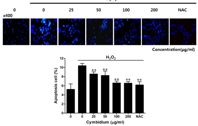
Fig. 2. Effect of cymbidium extract on cell death by H2O2-induced oxidative stress in C2C12 myoblasts. C2C12 myoblasts were cultured on a cover glass until confluent, and then the medium was replaced with a serum-free medium with NAC (2 mM) and cymbidium extract (0-200 μg/ml). After pre-incubating for 23 hr, C2C12 myoblasts were treated with 1 mM H2O2 for 1 hr. C2C12 myoblasts on a cover glass were mounted with DAPI-mounting media, and then examined by fluorescence microscopy. The apoptotic cells were estimated by direct counting after staining. Each value represents the mean (± SD) from three experiments, each performed in triplicate. NAC:
N
-acetyl cysteine, DAPI: 4, 6-diamino-2-phenylindole. **p
<0.01 versus H2O2 alone.리에 따라 세포생존율을 약 43% 감소시켰다. 이와 대조적으 로 심비디움 추출물의 전처리는 H
2O
2처리에 의한 C2C12 근 육세포 생존의 손실을 농도 의존적으로 감소시켰다 . 특히, 심 비디움 추출물의 농도 200 μg/ml에서는 세포손상을 93%까 지 보호하였다 (Fig. 1C). 이 결과들은 산화 스트레스에 의해 유도된 근육세포의 손상을 심비디움 추출물이 보호하는 효과 가 있음을 확인할 수 있었다 . 즉, 근위축은 근육세포의 전자전 달과 일반적인 산소접합에 민감한 산화적 손상을 유발하는데 산화적 스트레스는 격렬한 운동으로도 증가될 수도 있다 [3, 11, 15]. 근위축을 포함한 근육질환의 환원상태는 다른 기관의 환원상태보다 더 산화적이며 , 세포수준의 항산화 방어기전의 활성 및 본 실험에서의 심비디움 추출물은 기본적으로 근육 의 산화적 손상과 관계된 질병 조건의 조절과정 효율성을 높 일 것으로 사료된다 .
H
2O
2로 유도된 C2C12 근육세포에서 심비디움 추출물이 세포사에 미치는 영향
H
2O
2로 유도되는 세포사에서 심비디움 추출물의 효과를 확인하고자 DAPI 고정염색을 통해 염색질 응축 및 단편화
형태 변화를 통해 확인하였다 . H
2O
2를 처리한 표본에서는 처 리하지 않은 Control에 비해 염색질 응축 및 단편화 형태의 세포수가 높게 관찰되었고 , 심비디움 추출물의 0-200 μg/ml 농도별로 측정한 염색질 응축 및 단편화 형태의 세포수는 농 도 의존적으로 감소되는 것을 확인하였다 (Fig. 2). H
2O
2로 유 도된 근육세포의 경우 세포 내 높은 라디칼 농도를 유지하여 단백질 또는 DNA에 손상을 주게 되며 이는 세포 외막, 세포 내 구조 단백질 , 핵안 DNA 등에서 산화를 시켜 구조적 변화 로 인한 손상을 유발함으로써 세포사를 유도한다 [16]. 본 연 구에서는 심비디움 추출물을 이용하여 근육세포의 산화적 스 트레스로 유도된 세포사 및 증가된 라디칼을 잠정적으로 소 거함으로써 산화적 손상인 세포사 및 세포의 구조적 변화에 대항할 것이라 사료된다 .
H
2O
2로 유도된 C2C12 근육세포에서 HSP70, SOD1 발현에 대한 심비디움 추출물의 영향
근육 손상에서 HSPs의 종류 중 하나인 HSP70은 근육을
보호할 수 있는 활성을 나타낸다는 이미 알려진 사실을 토대
로 심비디움 추출물이 근육세포의 산화적 스트레스에 대한
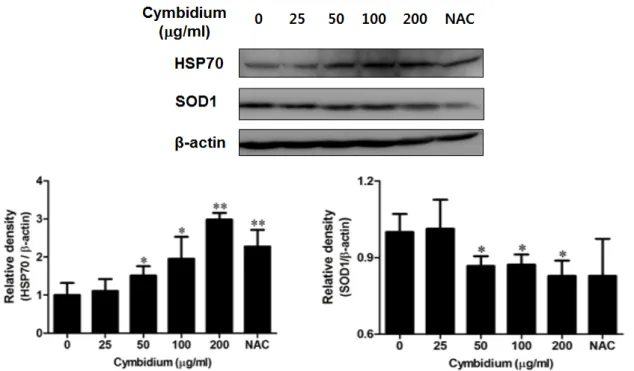
Fig. 3. Effect of cymbidium extract on HSP70 and SOD1 protein expression by H2O2-induced oxidative stress in C2C12 myoblasts.
C2C12 myoblasts were cultured in 6 well plates until confluent, and then the medium was replaced with a serum-free me- dium with cymbidium extract (0-200 μg/ml). After pre-incubating for 23 hr, C2C12 myoblasts were treated with 1 mM H2O2for 1 hr and then C2C12 myoblasts were lysed using PRO-PREP. Whole lysates were analyzed using SDS-PAGE. Each protein was analyzed by Western blotting using specific antibodies. Expression of HSP70, SOD1 and β-actin are shown.
Western blots were analyzed by densitometry and the values are given as the density of control. The results are an average of 3 similar experiments, expressed by mean ± SD. The inserts display representative blots of 3 similar independent experi- ments, respectively. *
p
<0.05, **p
<0.01 versus H2O2 alone.반응에서 HSP70 신호의 세포보호 효과를 확인하였다[17].
H
2O
2를 처리한 근육세포가 단백질 수준인 HSP70의 발현에 서 심비디움 추출물의 효과를 보면 , C2C12 근육세포에 추출 물의 농도 0-200 μg/ml로 24시간 동안 처리하여 배양하고, 그 후 , 1시간 동안 1 mM의 H
2O
2로 처리하였다 . 그 결과, HSP70 단백질은 심비디움 추출물을 처리한 세포에서 농도 의존적으 로 증가하고 대조군인 NAC보다 더 증가함을 볼 수 있었다.
즉 , HSP70의 발현을 통하여 근육세포의 산화적 스트레스에 대한 세포보호 효과를 나타냄을 알 수 있었다 . 또한, SOD1은 세포질에 존재하는 단백질로 초과산화이온을 산소와 과산화 수소로 변환시켜 줌으로써 활성산소로부터 세포를 보호하는 역할을 하는데 , 이는 활성산소 존재 시 발현이 증가한다고 알 려져 있다 [18]. 동일한 조건에서 Western blot을 통해 확인한 결과 , 심비디움 추출물을 처리한 세포에서 농도 의존적으로 감소하는 것을 확인하였다 (Fig. 3). 이러한 결과는 산화적 스 트레스인 활성산소의 자유라디칼을 심비디움 추출물이 감소 시켜 활성산소에 대해 나타나는 SOD1의 발현을 억제한 것으 로 사료된다 . 현재 산화적 근육 손상 연구와 관련된 다양한 신호전달체계 연구에서 단백질 및 메커니즘 , 대사적 작용 등 이 보고되고 있다 [8, 9, 12, 13]. 본 연구는 HSP70, SOD1 두가 지 단백질에 대한 항산화적 방어기능에 연관되어 집중했지
만 , 자유라디칼을 만드는 세포의 기질들을 포함하는 다른 신 호체계에 관계된 것을 토대로 연구의 범위를 확대할 수 있을 것으로 사료된다 .
감사의 글
이 논문은 정부 (미래창조과학부)의 재원으로 한국연구재 단의 지원을 받아 수행된 해외우수연구기관유치사업 연구 (2010-00757)와 MOU근거 한일협력사업 연구(NRF- 2012K2A2A4016850)임.
Reference
1. Booth, F. W. 1982. Effect of limb immobilization on skeletal muscle.
J Appl Physiol Respir Environ Exerc Physiol
52, 1113-1118.2. Brand-Williams, W., Cuvelier, M. E. and Berset, C. 1995.
Use of a free radical method to evaluate antioxidant activity.
J Food Sci Technol
28, 25-30.3. Chan, K. M. and Decker, E. A. 1994. Endogenous skeletal muscle antioxidants.
Crit Rev Food Sci Nutr
34, 403-426.4. Gaytan, V. G., Mendoza, M. N. R., Hernandez, M. S., Trejo-Tellez, L. I., Santos, M. P. and Ponce, G. V. 2013.
초록:산화스트레스에 의해 유도된 근세포 손상에서 심비디움 뿌리추출물의 효과 김완중
1․김한성
2․오피츠 요크
3․가바야마 카즈야
4․김택중
1*
(
1연세대학교 과학기술대학 생명과학기술학부 ,
2연세대학교 보건과학대학 의공학부 ,
3독일 프라운호퍼연구 소 ,
4일본 오사카대학교 과학대학원 화학과 )
근위축은 근육을 사용하지 않음으로써 발생하는 근육 조직의 손실 또는 근육을 지배하는 신경의 손상으로 정 의할 수 있다 . 이 상태는 다양한 근육질환에 관여하는 활성산소종이 관여 한다. 우리는 심비디움 뿌리 추출물이 과산화수소에 의해 유도된 C2C12 근육세포 생존율 손실과 세포사를 억제한다는 것을 찾았다. 또한 심비디움 뿌리 추출물이 HSP70 단백질 발현증가와 SOD1 단백질 발현감소를 확인하였다. 이들의 결과는 심비디움 뿌리 추출물 이 근위축에서 활성산소종을 환원하는 치료약물로서 작용이 가능할 것으로 사료된다 .
Volatile Components in the Flower, Pedicellate Ovary and Aqueous Residue of Cymbidium sp. (ORCHIDACEAE).
J Anal Sci Meth Instrum
3, 212-218.5. Giorgio, M., Trinei, M., Milgliaccio, E. and Pelicci, P. G.
2007. Hydrogen peroxide: a metabolic by-product or a com- mon mediator of ageing signals?
Nat Rev Mol Cell Biol
8, 722-728.6. Halter, B., Gonzales de Aquilar, J. L., Rene, F., Petri, S., Fricker, B., Echaniz-Laguna, A., Dupuis, L., Larmet, Y. and Loeffler, J. P. 2010. Oxidative stress in skeletal muscle stim- ulates early expression of Rad in a mouse model of amyo- trophic lateral sclerosis.
Free Radic Biol Med
48, 915-923.7. Howlader, M. A., Alam, M., Ahmed, Kh. T., Khatun, F. and Apu, A. S. 2011. Antinociceptive and anti-inflammatory ac- tivity of the ethanolic extract of Cymbidium aloifolium (L.).
Pak J Biol Sci
19, 909-911.8. Kolesnick, R. N., Haimovitz-Friedman, A. and Fuks, Z.
1994. The sphingomyelin signal transduction pathway me- diates apoptosis for tumor necrosis factor, Fas, and ionizing radiation.
Biochem Cell Biol
72, 471-474.9. Li, C. Y., Lee, J. S., Ko, Y. G., Kim, J. I. and Seo, J. S. 2000.
Heat shock protein 70 inhibits apoptosis downstream of cy- tochrome c release and upstream of caspase-3 activation.
J Biol Chem
275, 25665-25671.10. Mitsuhashi, M., Yamaguchi, M., Kojima, T., Nakajima, R.
and Kasai, K. 2011. Effects of HSP70 on the compression force-induced TNF-α and RANKL expression in human pe- riodontal ligament cells.
Inflamm Res
60, 187-194.11. Nishida, H., Ichikawa, H. and Konishi, T. 2007. Shengmai- san enhances antioxidant potential in C2C12 myoblasts through the induction of intracellular glutathione peroxidase.
J Pharmacol Sci
105, 342-352.12. Obeid, L. M., Linardic, C. M., Karolak, L. A. and Hannun, Y. A. 1993. Programmed cell death induced by ceramide.
Science
259, 1769-1771.13. Powers, S. K., Kavazis, A. N. and McClung, J. M. 2007.
Oxidative stress and disuse muscle atrophy
. J Appl Physiol
102, 2389-2397.14. Stone, J. R. and Yang, S. 2006. Hydrogen peroxide: a signal-
ing messenger.
Antioxid Redox Signal
8, 243-270.15. Strobel, N. A., Peake, J. M., Matsumoto, A., Marsh, S. A., Coombes, J. S. and Wadley, G. D. 2011. Antioxidant supple- mentation reduces skeletal muscle mitochondrial biogenesis.
Med Sci Sports Exerc
43, 1017-1024.16. Seifried, H. E., Anderson, D. E., Fisher, E. I. and Milner, J. A. 2007. A review of the interaction among dietary anti- oxidants and reactive oxygen species.
J Nutr Biochem
18, 567-579.17. Senf, S. M., Dodd, S. L., McClung, J. M. and Judge, A. R.
2008. Hsp70 overexpression inhibits NF-kappaB and Foxo3a transcriptional activities and prevents skeletal mus- cle atrophy.
FASEB J
22, 3836-3845.18. Smietana, M. J., Arruda, E. M., Faulkner, J. A., Brooks, S.
V. and Larkin, L. M. 2010. Reactive oxygen species on bone mineral density and mechanics in Cu,Zn superoxide dis- mutase (Sod1) knockout mice.
Biochem Biophys Res Commun
403, 149-153.19. Thomason, D. B., Biggs, R. B. and Booth, F. W. 1989.
Protein metabolism and beta-myosin heavy-chain mRNA in unweighted soleus muscle.
Am J Physiol
257, R300-305.20. Walker, P. M. 1991. Ischemia/reperfusion injury in skeletal muscle.
Ann Vasc Surg
5, 399-402.21. Yen, Y. P., Tsai, K. S., Chen, Y. W., Huang, C. F., Yang, R. S. and Liu, S. H. 2012. Arsenic induces apoptosis in my- oblasts through a reactive oxygen species-induced endo- plasmic reticulum stress and mitochondrial dysfunction pathway.
Arch Toxicol
86, 923-933.22. Yoshikawa, K., Baba, C., Iseki, K., Ito, T., Asakawa, Y., Kawano, S. and Hashimoto, T. 2014. Phenanthrene and phenylpropanoid constituents from the roots of Cymbi- dium Great Flower 'Marylaurencin' and their antimicrobial activity.
J Nat Med
68,
743-747.23. Yoshikawa, L., Otsu, M., Ito, T., Asakawa, Y., Kawano, S.
and Hashimoto, T. 2013. Aromatic constituents of Cymbidium Great Flower Marie Laurencin and their anti- oxidative activity
. J Nat Med
67, 217-221.24. Zylicz, M. and Wawrzynow, A. 2001. Insights into the function of Hsp70 chaperones.