Development of Broad-range and Specific 16S rRNA PCR for Use in Routine Diagnostic Clinical Microbiology
Hyun-Chul Kim
1,2, Yun-Tae Kim
1, Hyogyeong Kim
1, Sanghoo Lee
1, Kyoung-Ryul Lee
1and Young-Jin Kim
1*1
Department of Molecular Biology, Seoul Medical Science Institute/Seoul Clinical Laboratories, Seoul 152-766, Korea
2
Department of Biomaterials Science and Engineering, Yonsei University, Seoul 120-749, Korea
Received January 27, 2014 /Revised March 6, 2014 /Accepted March 6, 2014Broad-range and specific 16S rRNA gene PCR is used for detection and identification of bacterial pathogens in clinical specimens from patients with a high suspicion for infection. We describe the de- velopment of a broad-range and specific PCR primer, based on bacterial 16S rRNA, for use in routine diagnostic clinical microbiology services. The primers were designed by using conservative regions of 16S rRNA sequences from 10 strains. Ninety-eight clinical strains were isolated from clinical patient specimens. A total of 98 strains of bacteria were identified by phenotypic methods; PCR with newly designed primers and universal primers. All purified PCR products were sequenced using both for- ward and reverse primers on an automated DNA analyzer. In this study, we evaluated the usefulness of the newly designed primers and the universal primers for the detection of bacteria, and both these techniques were compared with phenotypic methods for bacteria detection. When we also tested 98 strains of clinical isolates with newly designed primers, about 778 bp DNA fragments were amplified and identified from all strains. Of the 98 strains, 94 strains (95.9%) correspond in comparison with phenotypic methods. The newly designed primers showed that the identities of 98 (100%) strains were the same as those obtained by universal PCR primers. The overall agreement between the newly de- signed primers and universal primers was 100%. The primer set was designed for rapid, accurate, and cheap identification of bacterial pathogens. We think the newly designed primer set is useful for the identification of pathogenic bacteria.
Key words : 16S rRNA, clinical isolates, the newly designed primers, universal primers
*Corresponding author
*Tel : +82-2-330-2011, Fax : +82-2-858-2814
*E-mail : [email protected]
This is an Open-Access article distributed under the terms of the Creative Commons Attribution Non-Commercial License (http://creativecommons.org/licenses/by-nc/3.0) which permits unrestricted non-commercial use, distribution, and reproduction in any medium, provided the original work is properly cited.
Journal of Life Science 2014 Vol. 24. No. 4. 361~369 DOI : http://dx.doi.org/10.5352/JLS.2014.24.4.361
Introduction
Identification of bacteria in clinical microbiology is tradi- tionally performed by isolation of the organisms and study of their phenotypic characteristics, including gram staining, morphology, culture requirements, and biochemical re- actions. However, these methods of bacterial identification have major weakness [10]. First, they cannot be used for non-cultivable organisms. Second, we are occasionally faced with organisms exhibiting biochemical characteristics that do not fit into patterns of any known genus and species.
Third, identification of slow-growing organisms would be extremely slow and difficult [10]. Especially, conventional methods often cannot fully characterize bacterial or fungal
isolates, and laboratories are now relying on broad-range DNA sequencing for microorganism identification. There- fore, CLSI (Clinical and Laboratory Standards Institute) guideline suggests 16S rRNA for bacteria and ITS (Internal transcribed spacer) regions for fungi [8]. 16S rRNA gene se- quences to study bacterial phylogeny and taxonomy have been by far the most common housekeeping genetic marker used for a number of reasons. These reasons include (i) its presence in almost all bacteria, often existing as a multigene family, or operons; (ii) the function of the 16S rRNA gene over time has not changed, suggesting that random sequence changes are a more accurate measure of time (evolution);
and (iii) the 16S rRNA gene (approximately 1,5 kb) is large enough for informatic purposes [7]. Despite its accuracy, 16S rRNA gene sequence analysis lacks widespread use beyond the large and reference laboratories because of cost and tech- nical considerations. We describe the development of a broad-range PCR primer set, based on bacterial 16S rRNA, for use in the routine diagnostic clinical microbiology service.
The purpose of this study was to evaluate the utility of
newly designed 16S rRNA primers in a clinical microbiology
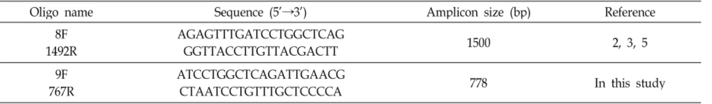
Table 1. Synthetic oligonucleotide used as primer for PCR
Oligo name Sequence (5’→3’) Amplicon size (bp) Reference
8F
1492R AGAGTTTGATCCTGGCTCAG
GGTTACCTTGTTACGACTT 1500 2, 3, 5
9F
767R ATCCTGGCTCAGATTGAACG
CTAATCCTGTTTGCTCCCCA 778 In this study
Bacterial strains and DNA isolation
For the specificity of conventional PCR assay with de- signed primer, it was verified using strains of both American Type Culture Collection (ATCC; Manassas, VA, USA) refer- ence and clinical origins. 20 strains obtained from the ATCC, were used in this study. And 98 clinical (50 gram-negative and 48 gram-positive) strains were isolated from patient specimens. Bacterial strains were cultured on sheep blood or chocolate agar depending upon the specific growth re- quirement of each species. For identification, Vitek system (bioMerieux Vitek, NC, USA) and several conventional bio- chemical methods were used for identification of the bacte- rial strains [11]. Bacterial DNA was prepared from collected bacteria samples using the G-spin
TMfor Bacteria Genomic DNA Extraction Kit according to the manufacturer’s in- structions (Intron Biotechnology, Seoul, Korea).
Conventional PCR assay
The sequences of the PCR primers that were evaluated in this study to detect broad bacteria species are all shown in Table 1. Used primer pairs are 2 primer sets. One primer set was 9F (5’-ATCCTGGCTCAGATTGAACG-3’), 767R (5’-CTAATCCTGTTTGCTCCCCA-3’). These were designed with highly conserved regions from the 16S rRNA gene se- quences of variable species. Conserved regions were con- firmed by CLUSTALW2 software. And the other one used universal primer pair: F (5’-AGAGTTTGATCCTGGCTCAG- 3’), R (5’- GGTTACCTTGTTACGACTT-3’) [7, 8, 10]. All oli- gonucleotides were provided from the Bioneer (Daejeon, Korea). DNA was amplified in 20 μl reaction mixture consist- ing of 2 μl of 10× PCR buffer, 1 μl of 2 mM dNTP, 1 μl of 10 μM forward primer, 1 μl of 10 μM reverse primer, 13 μ l of DW, and 0.25 U of Super-Therm Gold Taq DNA poly- merase (JMR Holdings, Kent, UK). The conditions for ampli-
cles of amplification with denaturation at 95°C for 30 sec, annealing at 53°C for 40 sec, and extension at 72°C for 1 min, followed by a final elongation step at 72°C for 5 min.
After amplification, 6 μl aliquot of each amplication product was analyzed using electrophoresis on 1% agarose gels cast and ran in 0.5× TAE buffer. A 100 bp maker (Cosmogene- tech, Seoul, Korea) was included in the gel. Gel was stained with ethidium bromide and visualized using transmitted ul- traviolet illumination and photographed using gel doc- umentation system.
DNA sequencing and analysis
The PCR products were directly sequenced and analyzed using an ABI 3130XL DNA sequencers (Applied Biosystems, USA) and ABI sequencing Analysis Software. To eliminate errors caused by amplification artifacts, the forward and re- verse sequences of each 16S rRNA gene sequences were de- termined for products from at least two independent PCRs.
The sequences of the PCR products were compared with known 16S rRNA gene sequences in NCBI GenBank (http://www.ncbi.nlm.nih.gov/genbank/).
Results
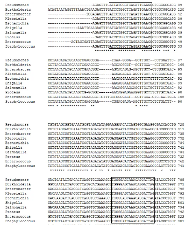
The 9F/767R bacterial primer set was designed from a conserved region of the 16S rRNA gene, which is located between the universal bacterial primer set (Fig. 1, Table 1).
Bacterial 16S rRNA sequences were amplified from type strians and clinical isolates. We describe new primer set (9F/767R) and universal primer set (8F/1492R) that may be used to amplify DNA from many different species of micro- organism, and use this DNA to identify the organisms.
For each sample, two different PCR products, using 9F/
767R and 8F/1492R primers, were independently processed.
Fig. 1. Alignment of sequences in the region of the 16S rRNA genes for
P. aeruginosa
(EF079669),B. cepacia
(GQ359110),E. cloacae
(GU191924),K. pneumoniae
(HQ200406),E. coli
(FJ823386),S. flexneri
(EU009189),S. typhi
(Z47544),P. mirabilis
(HM771658),E. faecalis
(AY692453),S. aureus
(AB634830), (lines 1 to 10, respectively). Square boxes represent primers newly designed from 16S rRNA sequences.Amplification using the universal primer pair generated a single DNA fragment of approximately 1,492 bp (Fig. 2A).
Amplification using the new primer pair generated DNA bands at about 778 bp (Fig. 2B). This pairs were used to 16S rRNA amplification from the 20 species type strains (Acinetobacter lwoffii ATCC 17925, Bacillus subtilis ATCC 6633,
Brevibacilus brevis ATCC 8246, Enterobacter cloacae ATCC
700323, Enterobacter sakazakii ATCC 29544, Enterococcus casse-
liflavus ATCC 700327, Enterococcus faecalis ATCC 51299,
Enterococcus faecalis ATCC 29212, Escherichia coli ATCC 25922,
Escherichia coli O157:H7 ATCC 43888, Klebsiella pneumoniae
ATCC 13883, Proteus mirabilis ATCC 7005, Providencia alcalifa-
B
Fig. 2. PCR products in a 1% agarose gel for the 20 bacterial species tested with universal primer and the newly designed primers.
(A) Universal PCR with primers 8F and 1492R (amplicon size: 1,492 bp), (B) PCR with newly designed primers 9F and 767R (amplicon size: 778 bp), Lanes: M, DNA size markers; 1,
Acinetobacter lwoffii
ATCC 17925; 2,Bacillus subtilis
ATCC 6633; 3,Brevibacilus brevis
ATCC 8246; 4,Enterobacter cloacae
ATCC 700323; 5,Enterobacter sakazakii
ATCC 29544; 6,Enterococcus casseliflavus
ATCC 700327; 7,Enterococcus faecalis
ATCC 51299; 8,Enterococcus faecalis
ATCC 29212; 9,Escherichia coli
ATCC 25922; 10,Escherichia coli
O157:H7 ATCC 43888; 11,Klebsiella pneumoniae
ATCC 13883;12,Proteus mirabilis
ATCC 7005; 13,Providencia alcalifaciens
ATCC 51902; 14,Pseudomonas aeruginosa
ATCC 9027; 15, Pseudomonas aeruginosa
ATCC 27853; 16,Salmonella typhimurium
ATCC 1925; 17,Staphylococcus aureus
ATCC 25923; 18, Streptococcus pneumoniae
ATCC 49619; 19,Streptococcus pyogenes
ATCC 19615; 20,Vibrio parahaemolyticus
NCCP 10511.ciens ATCC 51902, Pseudomonas aeruginosa ATCC 9027, Pseudomonas aeruginosa ATCC 27853, Salmonella typhimurium ATCC 1925, Staphylococcus aureus ATCC 25923, Streptococcus pneumoniae ATCC 49619, Streptococcus pyogenes ATCC 19615, Vibrio parahaemolyticus NCCP 10511), all of which were am- plified successfully and showed no additional bands. The newly designed primers showed about 778 bp size DNA band (PCR product) in the 98 clinical isolated strains.
Using universal primers, the 16S rRNA gene PCR amplifi- cation of the 98 isolates showed about 1,492 bp. Using new primers, the 16S rRNA gene PCR amplification of the 98 iso- lates was successful, yielding target bands at about 778 bp.
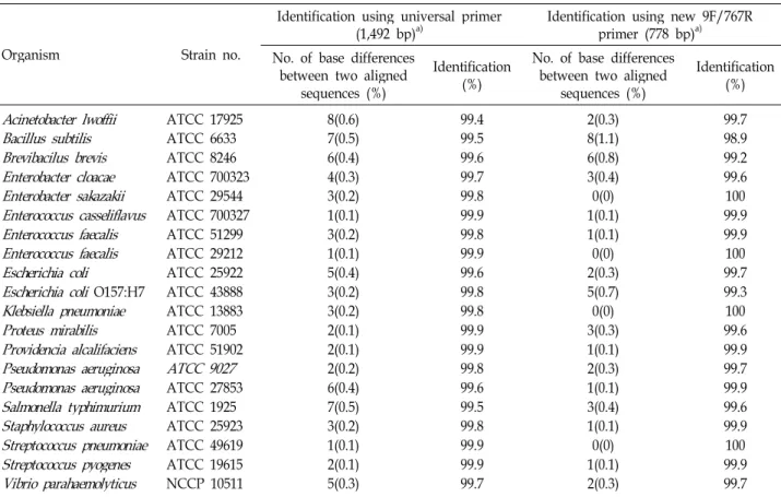
Furthermore, DNA sequencing of the corresponding PCR products using the same oligonucleotides as sequencing pri- mers posed no problems. Purified PCR products were di- rectly sequenced in both forward and reverse directions us- ing the same primers as for PCR. Table 1 shows bacteria identification using either the universal primer or the newly designed 16S rRNA primers. The 16s rRNA sequence of type strains were compared with the universal primers and new- ly designed 16S rRNA primers. The nucleotide similarity values were calculated, and similarity values were de- termined using the BLAST program at NCBI website. An observed percentage difference is the number of base mis- matches between two aligned sequences, as determined.
Obtained full sequences showed approximately average
99.74% (99.4~99.8%) identification with the database se- quences using universal primers, indicating that correct am- plification had been achieved. Also, average bacteria identi- fication by new primers were 99.72% (Table 2). All strains, except Bacillus subtilis, analysed sequences revealed identi- fication above 99% (99.2~100%), Bacillus subtilis is identified as having a high rate of 98.9%. The identities of 19 type strains were the same as those obtained by universal primers and the rate of identification is above 99%. For all 20 type strains, there was <1.1% difference between the 16S rRNA gene sequences of the type strains and the most closely matched sequence in the GenBank (Table 2).
Analysis of the 20 strains sequences using the newly de-
signed primers by 16S rRNA-based bacterial identification
database showed that the identities of 20 strains were the
same as those obtained by universal 16S rRNA gene
sequencing. In other words, twenty of 20 strains were identi-
fied as the same species by newly designed primers, in
agreement with the naming by the ATCC or NCCP. Then,
we evaluated the usefulness of this primer in the identi-
fication of isolates clinically significant bacterial strains that
showed phenotypic identification methods. A total of 98 bac-
terial strains were tested by molecular method including the
newly designed primers with universal primers and pheno-
typic methods. The identification of 98 strains analyzed and
the alignment tool used can also affect the comparison of
Table 2. Analysis of DNA sequences of type strains identified using 16S rRNA bacteria database
Organism Strain no.
Identification using universal primer
(1,492 bp)a) Identification using new 9F/767R primer (778 bp)a) No. of base differences
between two aligned sequences (%)
Identification (%)
No. of base differences between two aligned
sequences (%)
Identification (%)
Acinetobacter lwoffii
Bacillus subtilis Brevibacilus brevis Enterobacter cloacae Enterobacter sakazakii Enterococcus casseliflavus Enterococcus faecalis Enterococcus faecalis Escherichia coli
Escherichia coli
O157:H7Klebsiella pneumoniae Proteus mirabilis Providencia alcalifaciens Pseudomonas aeruginosa Pseudomonas aeruginosa Salmonella typhimurium Staphylococcus aureus Streptococcus pneumoniae Streptococcus pyogenes Vibrio parahaemolyticus
ATCC 17925 ATCC 6633 ATCC 8246 ATCC 700323 ATCC 29544 ATCC 700327 ATCC 51299 ATCC 29212 ATCC 25922 ATCC 43888 ATCC 13883 ATCC 7005 ATCC 51902
ATCC 9027
ATCC 27853 ATCC 1925 ATCC 25923 ATCC 49619 ATCC 19615 NCCP 105118(0.6) 7(0.5) 6(0.4) 4(0.3) 3(0.2) 1(0.1) 3(0.2) 1(0.1) 5(0.4) 3(0.2) 3(0.2) 2(0.1) 2(0.1) 2(0.2) 6(0.4) 7(0.5) 3(0.2) 1(0.1) 2(0.1) 5(0.3)
99.499.5 99.6 99.799.8 99.999.8 99.999.6 99.899.8 99.9 99.999.8 99.699.5 99.899.9 99.9 99.7
2(0.3) 8(1.1) 6(0.8) 3(0.4) 0(0) 1(0.1) 1(0.1) 2(0.3)0(0) 5(0.7) 0(0) 3(0.3) 1(0.1) 2(0.3) 1(0.1) 3(0.4) 1(0.1) 0(0) 1(0.1) 2(0.3)
99.798.9 99.2 99.6100 99.999.9 99.7100 99.3100 99.6 99.999.7 99.999.6 99.9100 99.9 99.7
a)Analysis using database of GenBank
sequences. An observed percentage is the number of match- es or mismatches between blasted sequences, as determined.
Four of 98 isolated strains showed a considerable number of mismatches with clinical isolates in phenotypic method.
Newly designed PCR primers and universal PCR primers analysis of the isolates revealed same results for 98 clinical isolates, while in 4 isolates, phenotypic method were incon- sistent (Table 3). It reaffirms the 16S rRNA genes amplifica- tion of the 4 isolates showed bands at about 1,492 bp and 778 bp, respectively. Four strains of incorrect identity (one of Escherichia coli, one of Stapylococcus aureus, one of Corynebacterium striatum and one of Streptococcus sp.) were studied to assess and compare newly designed PCR primers and universal PCR primers by 16S rRNA-based identi- fication of bacteria. The 4 strains (E. coli, S. aureus, C. striatum, Streptococcus sp.) has been identified as a strain of Citrobacter freundii, Stapylococcus saprophyticus, Corynebacterium con- fusum, Arthrobacter cumminsii, respectively (Table 3). After all, identification of phenotypic method of 4 isolates was not discriminative enough. However, new primers and univer- sal primers of these 98 isolates were compared to the known 16S rRNA gene sequences in the GenBank, yielded the cor-
rect identity, with good discrimination. The overall agree- ment between the newly designed primers and universal primers were 100%. Therefore, the newly designed primers of identification have the advantage over phenotypic meth- ods of identification, the newly designed primers 16S rRNA-based, is investigation and approach focused on iden- tifying to solved the problem of phenotypic method.
In this case, in fact, bacteria identification can be detected by the use of the newly designed PCR primers, instead of the "universal" primers 8F/1492R.
Discussion
Clinically significant strains should be identified at the species level by a reliable and reproducible method to pro- vide a better understanding of pathogenic potential of vari- ous species. Phenotypic methods for identification of strains appear to be unreliable. The purpose of the present study was to compare commonly used phenotypic methods, the Vitek system and PCR amplicon-sequencing based methods targeting the 16S rRNA gene.
In this study, we evaluated the usefulness of the newly
E. coli K. pneumoniae Serratia marcescens
Morganella morganii ssp morganii Proteus mirabilis
Providencia rettgeri Citrobacter braakii
Acinetobactor baumanii complex Stapylococcus aureus
Enterococcus faecalis Streptococcus agalactiae Stapylococcus haemolyticus Enterococcus faecium Corynebacterium striatum Staphylococcus pneumoniae Staphylococcus lugdunensis Stapylococcus hominis
Stapylococcus capitis ssp. capitis Stapylococcus epidermidis
Streptococcus dysgalacitae ssp. equisimilis Streptococcus pyogenes
Streptococcus
sp.Streptococcus mitis Streptococcus anginosus
Corynebacterium pseudodiphtherigoicum Corynebacterium urealyticum
Staphylococcus caprae Streptococcus gordonii
28 76 2 4 1 11 10
8 8 33 2 1 1 11 1 1 1 11 1 1 1 11
27 (96.4) 7 (100) 6 (100) 2 (100) 4 (100) 1 (100) 1 (100) 1 (100) 9 ( 90) 8 (100) 8 (100) 3 (100) 3 (100) 1 ( 50) 1 (100) 1 (100) 1 (100) 1 (100) 1 (100) 1 (100) 1 (100) 1 (100) 1 (100) 1 (100) 1 (100) 1 (100) 1 (100)
1 (3.6)
1c) (10)
1d) (50)
1e) (100)
27 (96.4) 7 (100) 6 (100) 2 (100) 4 (100) 1 (100) 1 (100) 1 (100) 9 ( 90) 8 (100) 8 (100) 3 (100) 3 (100) 1 ( 50) 1 (100) 1 (100) 1 (100) 1 (100) 1 (100) 1 (100) 1 (100) 1 (100) 1 (100) 1 (100) 1 (100) 1 (100) 1 (100)
1 (3.6)
1c) (10)
1d) (50)
1e) (100)
a)Vitek system (bioMerieux Vitek) was used for identification of 50 clinical isolates of 50 gram-negative and 30 clinical isolates of 48 gram-positive (except clinical isolates of 10
Staphylococcus aureus
and 8Streptococcus agalactiae
) 10S. aureus
clinical isolates were identified using the biochmical test (catalase, coagulase test, DNase, Mannitol salt agar), and 8S. agalactiae
clinical isolates were identified using the Pastorex Strep test (Bio-Rad, Hercules, CA,. USA).b)
Citrobacter freundii
c)
Stapylococcus saprophyticus
d)
Corynebacterium confusum
e)
Arthrobacter cumminsii
designed primers and of universal primers (8F and 1492R) for identification of bacteria, and both these techniques were compared with phenotypic methods (Vitek, biochemical test, and streptex) for bacteria identification. Ninety-eight clinical strains (50 gram-negative and 48 gram-positive) were iso- lated from clinical patient specimens. A total 98 strains of bacteria were identified by molecular method (PCR) with newly designed primers and universal primers and pheno- typic methods.
Nucleotide sequences of PCR new primers (9F and 767R)
designed from the universally conserved amino acid se- quences of the 16S rRNA gene from 10 type strains (Acineto- bacter lwoffii, Bacillus subtilis, Brevibacilus brevis, Enterobacter cloacae, Enterobacter sakazakii, Enterococcus casseliflavus, Enter- ococcus faecalis, Enterococcus faecalis, Escherichia coli, Escherichia coli O157, Klebsiella pneumoniae, Proteus mirabilis, Providencia alcalifaciens, Pseudomonas aeruginosa, Pseudomonas aeruginosa, Salmonella typhimurium, Staphylococcus aureus, Streptococcus pneumoniae, Streptococcus pyogenes, Vibrio parahaemolyticus).
Each strain sequences were aligned using ClustalW. And
then the specific PCR primers for type strains were designed using the Primer3Plus (Fig. 1, Table 1).
DNA obtained from ATCC standard control strains was examined by the universal primers and newly designed pri- mers (Fig. 2). All of these DNA samples generated the uni- versal PCR product of the expected 1,492 bp size (Fig. 2A).
The newly designed primers PCR band of approximately 778 bp was also produced (Fig. 2B).
Table 2 shows identification using either the universal pri- mers or the newly designed 16S rRNA primers. We used BLAST to compare the sequence of PCR product with known nucleotide sequences of type strains in the database.
Observed percentage difference is the number of base mis- matches between two aligned sequences. Rates of correct identification by the universal primers and new primers were 99.74(99.4~99.9%) and 99.72(98.9~100%), respectively (Table 2). When all type strains were considered, that new primer have very similar rate to universal primer. We found that all of the type strain sequences they generated had an identical perfect matching sequence when analyzed by new primers. In other words, twenty of 20 strains were identified as the same species by newly designed primer, in agreement with the naming by the ATCC or NCCP.
In this study, we evaluated the utility of newly designed primer as a means to identify 98 isolates obtained from clin- ical sources. Surveys looked at the feasibility of identifying medically important bacteria using newly designed primers.
When we also tested 98 strains of clinical isolates with newly designed primers and universal primers, DNA fragment was amplified and was identified from all strains. Phenotypic identification including Vitek system, biochemical test, and strep test were performed in the 98 isolates. 16S rRNA genes using two primer sets were amplified by PCR, sequencing and then the sequence was put in GenBank BLAST to com- pare with database in NCBI.
Of the total 98 strains, 94 strains (95.9%) correspond in comparison with phenotypic methods. 4(4.1%) isolates were misidentified at phenotypic methods. The newly designed primers showed that the identities of 98(100%) strains were the same as those obtained by universal PCR primers (Table 3). The overall agreement between the newly designed pri- mers and universal primers were 100%. Newly designed PCR primers and universal PCR primers analysis of the clin- ical isolates revealed same results for 50 gram negative iso- lates, while phenotypic result of 1 isolate was inconsistent.
One Citrobacter freundii strain was incorrectly identified as
E. coli with the phenotypic methods (Table 3).
Even if 16S rRNA gene sequencing is highly useful in re- gards to bacterial identification, it has low phylogenetic power at the species level and poor discriminatory power for some genera [1, 6, 9], and DNA relatedness studies are necessary to provide absolute resolution to these taxonomic problems. Previously, it was reported that the type strains of Edwardsiella species exhibit 99.35 to 99.81% similarity to each other, and yet these three species are clearly dis- tinguishable biochemically and by DNA homology (28 to 50% relatedness) [4].
The misidentification rate was higher with the Vitek than with the sequence. These misidentifications did lead to er- rors in interpretation of clinical isolates. However, in this study, Newly designed PCR primers and universal PCR pri- mers for K. pneumoniae, Serratia marcescens, Morganella morga- nii ssp morganii, Proteus mirabilis, Citrobacter braakii and Acinetobactor baumanii complex showed good agreement with phenotypic methods. With the exception of 3 isolates (S. aur- eus, Corynebacterium striatum, Streptococcus sp.), 45 isolates showed good agreement with phenotypic methods. A higher percentage of species identifications were obtained using newly designed primers and universal primers results than phenotypic methods.
All strains except 8 E. faecalis, 8 Streptococcus agalactiae, 3 S. haemolyticus, 3 E. faecium, 1 S. pneumoniae, 1 S. lugdunensis, 1 S. hominis, 1 Stapylococcus capitis ssp. Capitis, 1 Stapylococcus epidermidis, 1 Streptococcus dysgalacitae ssp. Equisimilis, 1 Streptococcus pyogenes, 1 Streptococcus mitis (viridans strep), 1 Streptococcus anginosus, 1 Corynebacterium pseudodiphther- igoicum, 1 Corynebacterium urealyticum, 1 Staphylococcus cap- rae, and 1 Streptococcus gordonii, phenotypic results matched molecular methods (New primer and universal primer).
Four strains of incorrect identity (one of Escherichia coli, one of Stapylococcus aureus, one of Corynebacterium striatum and one of Streptococcus sp.) were studied to assess and com- pare newly designed PCR primers and universal PCR pri- mers by 16S rRNA-based identification of bacteria. 4 strains, E. coli, S. aureus, C. striatum, Streptococcus sp., has been identi- fied as a strain of Citrobacter freundii, Stapylococcus saprophyti- cus, Corynebacterium confusum, Arthrobacter cumminsii, re- spectively. The overall agreement between the newly de- signed primers and universal primers were 100%.
Therefore, the newly designed primers for identification
have the advantage over phenotypic methods, the newly de-
signed primers 16S rRNA-based, is investigation and ap-
genic bacteria. In this case, in fact, bacteria identification can be detected by the use of the newly designed PCR primers, instead of the "universal" primers 8F/1492R.
Some bacteria are difficult to identify with phenotypic identification schemes commonly used outside reference laboratories. 16S rRNA-based identification of bacteria po- tentially offers a useful alternative when phenotypic charac- terization methods fail. However, as yet, the usefulness of 16S rRNA sequence analysis in the identification of conven- tionally unidentifiable isolates has not been evaluated with a large collection of isolates. In this study, we evaluated the utility of 16S rRNA sequencing as a means to identify a col- lection of isolates obtained from clinical sources.
The sensitivity and specificity for pathogens identification were compared among different methods. 98 clinical speci- mens from different sources underwent phenotypic identi- fication and molecular identification using 16S rRNA univer- sal PCR primers and the newly designed primers. As results, our new primers have the usefulness.
Acknowledgment
This work was supported by Seoul Medical Science Institute in 2013.
References
1. Bosshard, P. P., Zbinden, R., Abels, S., Bo¨ddinghaus, B., Altwegg, M. and Bo¨ttger, E. C. 2006. 16S rRNA gene se- quencing versus the API 20 NE system and the Vitek 2 ID-GNB card for identification of nonfermenting gram- negative bacteria in the clinical laboratory.
J Clin Microbiol
Microbial diversity in marine sediments from Sagami Bay and Tokyo Bay, Japan, as determined by 16S rRNA gene analysis.
Microbiology
145, 3305-3315.4. John, M. J. and Sharon, L. A. 2007. 16S rRNA gene sequenc- ing for bacterial identification in the diagnostic laboratory:
pluses, perils, and pitfalls.
J Clin Microbiol
45, 2761-2764.5. Les, D. and Thomas, M. S. 2007. Performance of the transla- tional apparatus varies with the ecological strategies of bacteria.
J Bacteriol
189, 3237-3245.6. Mignard, S. and Flandrois, J. P. 2006. 16S rRNA sequencing in routine bacterial identification: a 30-month experiment
. J Microbiol Methods
67, 574-581.7. Patel, J. B. 2001. 16S rRNA gene sequencing for bacterial pathogen identification in the clinical laboratory.
Mol Diagn
6, 313-321.8. Petti, C. A., Bosshard, P. P., Brandt, M. E., Clarridge, J. E.
III., Feldblyum, T. V., Foxall, P., Furtado, M. R., Pace, N.
and Procop, G. 2008. Interpretive criteria for identification of bacteria and fungi by DNA target sequencing, approved guideline document MM18-A, 28. Wayne, PA: Clinical and Laboratory Standards Institute.
9. Rudolf, I. A., Wolfgang, L. and Karl-heinz, S. 1995.
Phylogenetic identification and in situ detection of in- dividual microbial cells without cultivation.
Microbiol Rev
59, 143-169.10. Woo, P. C., Ng, K. H., Lau, S. K., Yip, K. T., Fung, A. M., Leung, K. W., Tam, D. M., Que, T. L. and Yuen, K. Y. 2003.
Usefulness of the microSeq 500 16S ribosomal DNA-based bacterial identification system for identification of clinically significant bacterial isolates with ambiguous biochemical profiles.
J Clin Microbiol
41, 1996-2001.11. Woo, P. C., To, A. P., Lau, S. K., Fung, A. M. and Yuen, K. Y. 2004. Phenotypic and molecular characterization of er- ythromycin resistance in four isolates of