Extracts from Gracilaria vermiculophylla Prevent Cellular Senescence and Improve Differentiation Potential in Replicatively Senescent Human Bone Marrow Mesenchymal Stem Cells
Sin-Gu Jeong1,2, Tae Oh Cho1 and Goang-Won Cho1,2*
1Department of Biology, College of Natural Science, Chosun University, Gwangju 501-759, Korea
2Department of Life Science, BK21-Plus Research Team for Bioactive Control Technology, Chosun University, Gwangju 501-759, Korea Received May 18, 2018 /Revised July 3, 2018 /Accepted July 3, 2018
The red algae Gracilaria vermiculophylla is widespread on seashores worldwide and has been used as food in Asian countries. Previous studies have reported that extracts of Gracilaria red algae have bene- ficial anti-oxidant and anti-inflammatory effects. The present study examined the anti-senescence ef- fects of Gracilaria vermiculophylla extracts (GV-Ex) in replicatively senescent human bone marrow mes- enchymal stem cells (hBM-MSCs). GV-Ex pretreatment improved the cellular viability of hBM-MSCs that had been injured by oxidative stress. These effects of GV-Ex were confirmed by MTT assay and immunoblot analysis using the apoptotic proteins p53 and cleaved caspase-3. The reactive oxygen spe- cies (ROS) levels were examined in long-term cultured Passages 17 (P-17) mesenchymal stem cells (MSC) and compared to P-7 MSC. The ROS accumulation was greater in the P-17 than in the P-7.
However, these increased ROS levels in the P-17 were decreased significantly after treatment with GV-Ex, and restoration of the levels of the anti-oxidant enzymes SOD1, SOD2, and CAT was also ob- served under these conditions. In addition, P-17 hBM-MSC treated with GV-Ex had decreased levels of the senescence proteins p53, p21, and p16. The results show that the ability of P-17 hBM-MSC to differentiate into osteocytes and adipocytes was improved by GV-Ex treatment, suggesting that GV-Ex ameliorates the functional decline of senescent stem cells.
Key words : Anti-oxidant, Gracilaria vermiculophylla, mesenchymal stem cells, reactive oxygen species, replicative senescence
*Corresponding author
*Tel : +82-62-230-6641, Fax : +82-62-230-6650
*E-mail : [email protected]
This is an Open-Access article distributed under the terms of the Creative Commons Attribution Non-Commercial License (http://creativecommons.org/licenses/by-nc/3.0) which permits unrestricted non-commercial use, distribution, and reproduction in any medium, provided the original work is properly cited.
Journal of Life Science 2018 Vol. 28. No. 9. 1042~1047 DOI : https://doi.org/10.5352/JLS.2018.28.9.1042
서 론
인체의 노화는 이를 구성하는 각각의 기관(organ)과 조직 (tissue)의 노화(ageing)로 야기되는데, 그 중심 원인에 조직 항상성(tissue homeostasis)이 있다[18]. 조직의 항상성이란 개 개의 조직을 구성하는 세포들의 사멸과 증식의 균형유지를 의미한다[2]. 이 균형이 깨어질 때 노화와 퇴행성질환과 같은 질병을 앓게 되는데, 성체줄기세포(adult stem cells)의 기능 저하와 직접적으로 관련이 있다[7, 11, 21]. 즉, 성체줄기세포는 각 조직에 필요한 세포를 공급하는 역할을 수행하는데, 줄기 세포의 기능 저하는 기관에 노화된 세포의 축적을 야기하여 문제를 일으킨다. 이러한 질병들을 미연에 방지하기 위한 일 환으로 줄기세포의 노화를 제어하는 것이 필요하다.
성체 줄기세포는 계대배양을 통해 빠르게 노화되어 가는데,
telomere가 짧아지는 것, 활성산소(reactive oxygen species;
ROS)의 증가, 손상된 세포내 거대 분자들의 축척, 손상된 소기 관들의 축척, 세포내 손상의 축척 등 여러가지 요인이 제시되 었다[1, 4, 13, 17]. 이러한 노화의 원인들 중에 근본적인 요인으 로 ROS의 증가가 중요시 되고 있다. 세포는 정상적인 에너지 대사과정에서 ROS가 생성되는데 이 ROS는 반응성이 뛰어나 세포내 분자들, 단백질, 지질, 핵산과 반응하여 손상을 초래한 다[19, 23, 24]. 세포는 이러한 ROS를 제거하기 위한 항산화 효소인 SOD1, SOD2, CAT가 있고, 손상을 복구하기 위한 re- pair system을 가지고 있다[6, 8]. 하지만 세포배양 과정에서 ROS는 점점 더 축척되어지고 그만큼 손상도 축적되면서 세포 는 점진적으로 정상적인 생리학적 기능을 상실하게 된다. 이 러한 손상은 간혹 세포의 형질전환으로 나타나 세포의 암화를 초래하기도 하는데, 세포는 이를 억제하기 위해 빠르게 노화 를 유도하여 자폭하게 한다[22]. 이상과 같이 ROS의 축척은 세포의 비정상적인 상태로 되거나, 기능 상실로 이어진다. 이 러한 이유로 세포내 축척되는 ROS를 제거하는 것은 매우 중 요하다.
홍조류인 꼬물꼬시래기(Gracilaria vermiculophylla)는 전 세 계의 해변 지역에 널리 퍼져 있으며 아시아 국가에서 식량 자원으로도 이용되어왔다[15]. 선행 연구에 따르면, Gracilaria
속 홍조류 추출물에서 항산화 및 항염증 효과가 보고되었다[3, 25]. 본 연구에서는 이러한 효과를 지닌 홍조류에서 항노화 물질을 추출하기위해 홍조류에 속하는 다양한 해조류를 채집 하여 분석하였고, 꼬물꼬시래기(Gracilaria vermiculophylla)에 서 효과를 관찰하였다. 또 그 효과를 검증하기위해 노화된 인 간의 골수 유래 중간엽 줄기세포(hBM-MSCs)를 이용하여 G.
vermiculophylla 추출물(GV-Ex)의 항노화 효과를 검증하였다.
결과적으로, GV-Ex가 세포내 ROS를 감소시키고, 줄기세포 기 능을 개선함을 관찰하였다.
재료 및 방법
Cell culture
인간 골수 유래 중간엽줄기세포(Human bone marrow mesenchymal stem cells; hBM-MSCs)는 Cell Engineering for Origin (CEFO, Seoul, Korea)로부터 구입하여 이용하였다.
hBM-MSCs는 T-75 flasks (Becton Dickinson, San Jose, CA, USA)에서 판매사가 제공하는 매뉴얼에 따라 배양하였다. 배 양액은 DMEM (Gibco, Grand Island, NY, USA)에 mesen- chymal cell growth supplements와 L-glutamine, penicillin, streptomycin등을 첨가하여 이용하였고, 95% air와 5% CO2를 함유하는 37°C 항습배양기에 유지되었다. MSCs는 5일 단위로 계대 배양하였고, 매 3일마다 배양액을 교체하였다. 모든 실험 은 7 혹은 17번 계대 배양한(P-7, P-17) 세포를 이용하였다.
Preparation of Gracilaria vermiculophylla extract G. vermiculophylla는 대한민국 부산 바다에서 채집하여 (TC17055; deposited in the Marine Brown Algae Resources Bank, Chosun University, Gwangju, Korea), 이물질과 염분을 수돗물로 제거한 후에 상온에서 건조하였다. 이를 분말로 만 들어 80% ethanol (20× volume)에 담궈 7일 동안 추출한 후, 150 mm 지름의 Qualitative Filter Paper (Hyundai Micro, Gyeonggi-do, Korea)로 필터 하였다. Rotary Evaporator (Eyela, Tokyo, Japan)를 이용하여 에탄올을 제거하여 -70°C에 하루 동안 저장하였다. 최종 건조된 추출 물질은 freeze dryer (Ilshin Lab. Co. Ltd., Gyeonggi-do, Korea)로 3일 동안 동결건 조하여 얻었다. 실험을 위해 동결 건조된 20 mg 시료를 1 ml의 DMSO/에탄올(GV-crude; GV-Cr), 증류수(GV-H2O; GV- H), 아세톤(GV-acetone; GV-Ac)를 이용하여 녹여 이용하였다.
MTT assay
MSCs에서 GV-Ex에의한 보호효과를 관찰하기 위해 3-(4,5- dimethylthiazol-2-yl)-2,5-diphenyltetrazolium bromide (MTT) assay (Sigma-Aldrich)를 수행하였다. 먼저, hBM-MSCs를 96- well plates에 8×10³ cells/well로 접종하여 하루 동안 배양하 였다. 1 μg/ml GV-Ex 24시간 처리한 후에, 0-3 mM H2O2를
1시간 처리하여 세포독성을 제작사 매뉴얼에 따라 분석하였 다.
Detection of intracellular ROS
2‘,7’-dichlorofluorescein diacetate (DCFH-DA; Sigma- Aldrich, St. Louis, MO, USA)를 이용하여 세포내의 ROS량을 분석하였다. 24-well plates에 4×10⁴ cells/well와 96-well plates에 8×10³ cells/well의 MSCs를 접종하여 하루 동안 안정 화 하였다. 1 μg/ml GV-Ex로 24시간 전 처리한 후에 20 μM DCFH-DA를 첨가하여 37°C에서 1시간 배양하였다. Phos- phate-buffered saline (PBS)로 두 번 세척한 후, 1 mM hydro- gen peroxide (H2O2)를 1시간 동안 처리하였다. PBS 세척 후, 세포는 4% paraformaldehyde (PFA)로 10분간 고정하여, 4‘,6- diamidino-2-phenylindole (DAPI)를 포함하는 mounting sol- ution (Molecular Probes, Eugene, OR, USA)으로 마운팅하였 다. Fluorescence microscopy (Nikon Eclipse 80Ti microscope;
Nikon, Tokyo, Japan)로 관찰하였고, ELISA plate reader (Molecular Devices, Sunnyvale, CA, USA)로 정량 하였다. 세 포 사진은 DS-Rl1 digital camera (Nikon)를 이용하여 촬영하 였다.
Immunoblot analysis
처리된 세포는 400 μl RIPA buffer (2 mM phenylmethyl- sulfonyl fluoride와 1 mM sodium orthovanadate, protease inhibitor cocktail을 포함; Santa Cruz Biotechnology, Dallas, TX, USA)를 이용하여 추출하고, 16 000× g 20분간 원심 분리 하였다. 전체 단백질(50 μg)은 10% sodium dodecyl sul- fate-polyacrylamide gel로 전기영동하여 분리하였고, poly- vinylidene difluoride (PVDF) membranes (AGFA, Mortsel, Belgium)에 blot하였다. 5% skim milk로 1시간 blocking, 1차 항체를 첨가하여 4℃에서 16시간, 세척 후에 2차 항체를 첨가 하여 상온에서 1시간 반응하였다. Amersham ECL reagents (GE Healthcare, Buckinghamshire, UK)를 이용하여 단백질의 발현을 식별하였다. 1차 항체로는 SOD1 혹은 CAT, p53, p21, p16 (Santa Cruz Biotechnology, Inc., Dallas, TX, USA; 1:500 dilution), SOD2 (Abcam, Cambridge, UK; 1:5000 dilution), cleaved caspase-3 (Merck Millipore, Darmstadt, Germany;
1:500 dilution), 그리고 β-actin (Sigma-Aldrich; 1:5,000 dilu- tion)을 이용하였고, 2차 항체는 1차 항체와 조화되는 horse- radish peroxidase-conjugated secondary antibodies (Jackson ImmunoResearch Laboratories, West Grove, PA, USA)를 이 용하였다.
Osteocyte and adipocyte differentiation
처리된 MSCs는 선행연구에서 제시된 방법에 따라 뼈세포 (osteocytes)와 지방세포(adipocytes)로 분화 유도되었다[9,
A
B
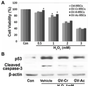
Fig. 1. Protective effects of G. vermiculophylla extracts against oxi- dative stress in hBM-MSCs. (A) hBM-MSCs were treated with 1 μg/ml GV-Cr, GV-H, or GV–Ac for 24 hr, then treated with 0.5, 1, 2, or 3 mM H2O2 for 1 hr. The cell viabilities were determined using an MTT assay. The graph shows the viability of cells under each treatment condition as a percentage of the viability of untreated cells (Con; Control), and * indicates a p-value of less than 0.01 in a paired t-test against the control hBM-MSCs (no GV extract; mean ± SD, n=4) from the same H2O2 treat- ment condition. Statistically significant protective effects were observed when GV-Cr or GV-Ac treatment pre- ceded treatment with 1 mM H2O2. (B) hBM-MSCs were incubated with 1 μg/ml GV-Cr or GV-Ac, and then treat- ed with 1 mM H2O2 for 1 hr. Total protein extracts from the cells were then analyzed by Western blot with anti- bodies against the apoptotic proteins p53 and cleaved caspase-3. A β-actin antibody was used as a loading control. Vehicle indicates a mixture of equal parts DMSO and ethanol.
20]. 뼈세포로 분화는, hBM-MSCs를 osteogenic medium에서 14일 동안 배양하여 분화하였다. 분화배양액은 DMEM (Gibco) 에 10% fetal bovine serum (FBS; Hyclone, Logan, Utah, USA) 와 100 nM dexamethasone (Cayman Chemical, Ann Arbor, Michigan, USA), 100 μM L-ascorbic acid (Sigma-Aldrich), 10 mM β-glycerophosphate (Sigma-Aldrich)를 혼합하여 제작하 였고, 매 3일마다 새 배양액으로 교환하였다. 분화된 세포는 alizarin red S 염색을 통해 식별하였다.
지방세포로 분화를 위해, MSCs는 24-well plates에서 adi- pogenic medium에서 14일간 분화 유도하였다. 분화 배양액 은 DMEM에 10% FBS와 500 μM 3-isobutyl-1-methylxanthine (Cayman Chemical), 1 μM dexamethasone, 100 μM indome- thacin (Cayman Chemical), 10 μg/ml insulin (Tocris Biosci- ence, Bristol, UK) 등을 혼합하여 제작하였다. 분화된 세포는 oil red O staining solution (200 ml of 100% isopropanol에 0.7 g oil red O powder)로 염색하여 식별하였다. 모든 염색한 세포의 관찰은 Nikon Eclipse TS100 microscope를 이용하였 고, Canon i-Solution IMTcam3 digital camera (Canon, Tokyo, Japan)를 이용하여 촬영하였다.
Statistical analysis
실험결과는 4번 이상 반복하여 평균을 내어 ± S.D. (stan- dard deviation)로 표기하였고, paired t-test법으로 통계 처리 하였다. p<0.01 일 때 통계적 의미를 부여하였다.
결과 및 고찰
G. vermiculophylla 추출물을 이용한 MSC의 보호 효과 Gracilaria 속의 홍조류에는 polyhenols계의 몇몇 생리활성 천연 물질이 분리되었지만[3, 10, 25], 같은 속에 속하는 G. ver- miculophylla에서는 항산화물질이 분리되지않았다. 본 연구에 서는 G. vermiculophylla에 항산화물질의 존재여부를 확인하고 검출하기 위하여 연구되었다. 먼저, DMSO/에탄올(GV-Cr), 물(GV-H), 아세톤(GV-Ac) 등의 용매를 이용하여 추출 물질이 함유하는 다양한 화합물질을 추출하였다. 배양한 hBM-MSC 에 GV-Cr, GV-H 또는 GV-Ac를 각각 1 μg/ml로 24시간 처리 한 후, 0~3 mM H2O2를 1시간 동안 처리하였다. 세포 독성은 H2O2 농도의존적으로 증가하였고, 1 μg/ml GV-Cr 또는 GV-Ac로 전처리 한 세포에서 보호효과가 관찰되었다(Fig.
1A). GV-H로 처리 한 결과 보호 효과가 관찰되지 않았는데, 해당 화합 물질이 수용성 용매에는 추출되지 않고, 유기 용매 에 용해된다는 것을 암시한다. 보호효과를 분자적 수준에서 검증하기 위해, hBM-MSCs에 1 μg/ml GV-Cr 또는 GV-Ac를 각각 전 처리한 후 세포사멸 관련 단백질인 p53과 cleaved cas- pase-3에 대한 immunoblot분석을 수행하였다. 그 결과 p53과 cleaved caspase-3의 수준이 감소되었다(Fig. 1B). 이 결과는
GV 추출물이 산화 스트레스에 의해 유발되는 세포 손상을 억제하고 세포 사멸을 예방할 수 있음을 암시한다.
GV-Ex에 의한 노화줄기세포에 ROS의 완화
줄기세포는 배양접시에서 여러 번 계대배양하면 점진적으 로 노화가 일어난다. 여러가지 노화관련 현상들이 나타나는데 그 중에 하나가 세포 내에 내재하는 ROS가 증가와 항산화 효소의 활성 감소이다[5, 12, 16]. 항산화 물질은 이러한 ROS를 효과적으로 제거하여 세포의 손상을 억제하고 노화를 지연하 다. 이를 확인하기 위해 MSCs를 장기간 배양(passage 17;
P-17)하여 짧게 배양한(P-7)과 비교하였다. P-17에서 ROS는 증가되었고 GV-Cr와 GV-Ac를 처리하면 의미있게 감소됨을 형광현미경(Fig. 2A)과 형광분광기(ELISA; Fig. 2B)를 이용하 여 확인하였다. 이와 같이, GV-Ex는 노화된 세포에서 ROS를 감소하여 항산화효과를 나타낸다.
A
B
Fig. 2. Reduction of iROS by GV-Ex in replicative senescence hBM-MSCs. (A) Intracellular ROS levels were visualized by fluorescence microscopy following DCFH-Da treat- ment in hBM-MSCs at P-7, or under various pre-treat- ment conditions at P-17. (B) Fluorescence intensities were quantified using an ELISA plate reader. The graph shows the relative DCF fluorescence compared to ve- hicle treatment, and an * indicates a p-value of less than 0.01 in a paired t-test against the vehicle treated cells (mean ± SD, n=4).
A B
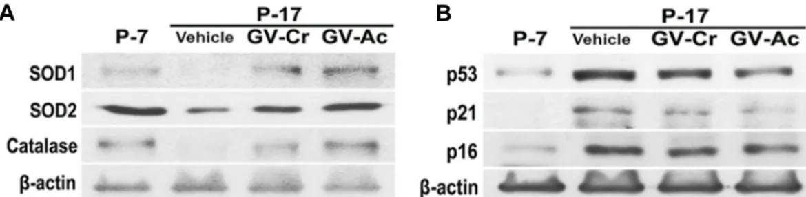
Fig. 3. Restoration of antioxidant enzymes and senescent proteins by GV-Ex in replicative senescence hBM-MSCs. (A) Total soluble-ly- sates from P-7, P-17, and GV-extract treated P-17 hBM-MSCs were examined by immunoblot for SOD1, SOD2, and CAT protein levels. β-Actin was used as a loading control. (B) The levels of senescence proteins such as p53, p21, and p16 were examined by immunoblot analysis. β-actin is used as a loading control. Vehicle indicates a mixture of equal parts DMSO and ethanol.
Fig. 4. GV-Ex restores differentiation potential in replicative sen- escence hBM-MSCs. (A) hBM-MSCs (P-7, P-17, and GV- extract treated P-17) were induced to differentiate into osteocytes (upper panels) or adipocytes (lower panels).
Differentiation capacities were measured by staining with alizarin red S (osteocytes) and oil red O (adipocytes).
GV-Ex에 의한 항산화 효소 및 노화 단백질의 회복 줄기세포의 장기간 배양은 ROS의 축적으로 이어지고 세포 는 점진적으로 노화되어 항산화 효소의 감소를 수반한다[5, 12, 16]. GV 추출물은 과잉의 ROS 생성을 제어되는데, 이것이 hBM-MSC에서 항산화 효소(SOD1, SOD2 및 CAT)의 발현을 회복에 따른 결과인지 실험하였다. MSC(P-17)에서 SOD1와 SOD2, CAT의 발현이 감소되었고, GV 추출물 처리하였을 때 발현이 개선됨을 관찰하였다(Fig. 3A). 노화 단백질인 p53와 p21, p16 역시 P-17에서 증가되었고, GV-Cr 또는 GV-Ac 처리 에 따라 부분적으로 감소됨을 관찰하였다(Fig. 3B). 이러한 결 과는 GV 추출물이 장기간 배양을 통해 노화된 hBM-MSC에서
축적된 ROS 수준을 완화하여 세포 노화를 예방할 수 있음을 나타낸다.
GV-EX의 줄기세포의 분화능력을 개선
건강한 BM-MSC는 골 세포와 지방 세포로 분화하는 능력 에 따라 정의된다[20]. 이 분화 능력은 종종 MSC의 기능적 활동을 결정하는 기준으로 사용된다[5, 14]. 본 연구에서는, GV 추출물이 노화된 hBM-MSC에서 ROS 수준을 감소시키고 줄기세포의 노화를 예방하거나 역전시킬 수 있음을 보였다.
이러한 효과가 줄기세포의 분화능력의 개선으로 나타나는지 확인하였다. P-17 줄기세포에서 급격한 분화능력 감소가 관찰 되었고, GV 추출물을 처리 한 P-17는 처리하지 않은 세포보다 더 효과적으로 골 세포 또는 지방 세포로 분화하였다(Fig. 4).
이러한 결과는 노화 된 hBM-MSC (P-17)의 분화능력 감소가 GV 추출물 처리에 의해 부분적으로 반전되거나 방지됨을 시 사한다.
본 연구에서는 hBM-MSC를 이용하여 홍조류 G. vermic- ulophylla 추출물에 대한 노화 방지 효과를 조사하였다. GV 추출물을 처리하면 노화된 줄기세포에 과잉으로 축적된 ROS 수준을 조절하였는데, 이는 항산화 효소인 SOD1, SOD2 및
CAT의 발현을 증가 시켜 성취된다. 그 결과로 세포의 노화 단백질 p53, p21 및 p16의 수준을 감소 시키고, 한편으로는 분화능력을 회복시킴을 관찰하였다. 이와 같이 GV 추출물질 은 줄기세포의 노화를 억제하며, 줄기세포 치료시에 이를 병 용하여 이용한다면 치료효과를 향상시킬 수 있을 것으로 생각 된다.
감사의 글
이 논문은 해양 수산부의 Marine Biotechnology Program (PJT200669) 지원을 받아 연구되었음. 이 연구는 조선대학교 대학원(2017.2.)에 "The mechanism of cellular senescence and neuronal differentiation in human bone marrow mesen- chymal stem cells"라는 제목의 1저자 박사학위 논문을 토대로 추가 연구됨.
References
1. Arai, Y., Martin-Ruiz, C. M., Takayama, M., Abe, Y., Take- bayashi, T., Koyasu, S., Suematsu, M., Hirose, N. and von Zglinicki, T. 2015. Inflammation, but not telomere length, predicts successful ageing at extreme old age: a longitu- dinal study of semi-supercentenarians. EBioMedicine 2, 1549-1558.
2. Biteau, B., Hochmuth, C. E. and Jasper, H. 2011. Maintaining tissue homeostasis: dynamic control of somatic stem cell activity. Cell Stem Cell 9, 402-411.
3. de Almeida, C. L., Falcao Hde, S., Lima, G. R., Montenegro Cde, A., Lira, N. S., de Athayde-Filho, P. F., Rodrigues, L.
C., de Souza Mde, F., Barbosa-Filho, J. M. and Batista, L.
M. 2011. Bioactivities from marine algae of the genus Gracilaria. Int. J. Mol. Sci. 12, 4550-4573.
4. Fang, C., Gu, L., Smerin, D., Mao, S. and Xiong, X. 2017.
The interrelation between reactive oxygen species and au- tophagy in neurological disorders. Oxid. Med. Cell. Longev.
2017, 8495160.
5. Jeong, S. G. and Cho, G. W. 2015. Endogenous ROS levels are increased in replicative senescence in human bone mar- row mesenchymal stromal cells. Biochem. Biophys. Res.
Commun. 460, 971-976.
6. Jeong, S. G. and Cho, G. W. 2015. Trichostatin A modulates intracellular reactive oxygen species through SOD2 and FOXO1 in human bone marrow-mesenchymal stem cells.
Cell Biochem. Funct. 33, 37-43.
7. Jeong, S. G. and Cho, G. W. 2016. Accumulation of apopto- sis-insensitive human bone marrow-mesenchymal stromal cells after long-term expansion. Cell Biochem. Funct. 34, 310- 316.
8. Johnson, F. and Giulivi, C. 2005. Superoxide dismutases and their impact upon human health. Mol. Aspects Med. 26, 340-352.
9. Kim, E. K., Lim, S., Park, J. M., Seo, J. K., Kim, J. H., Kim,
K. T., Ryu, S. H. and Suh, P. G. 2012. Human mesenchymal stem cell differentiation to the osteogenic or adipogenic line- age is regulated by AMP-activated protein kinase. J. cell.
Physi. 227, 1680-1687.
10. Kim, M. H., Hong, H. N., Hong, J. P., Park, C. J., Kwon, S. W., Kim, S. H., Kang, G. and Kim, M. 2010. The effect of VEGF on the myogenic differentiation of adipose tissue derived stem cells within thermosensitive hydrogel matrices.
Biomaterials 31, 1213-1218.
11. Liu, L. and Rando, T. A. 2011. Manifestations and mecha- nisms of stem cell aging. J. Cell Biol. 193, 257-266.
12. Lu, T. and Finkel, T. 2008. Free radicals and senescence. Exp.
Cell Res. 314, 1918-1922.
13. Luceri, C., Bigagli, E., Femia, A. P., Caderni, G., Giovannelli, L. and Lodovici, M. 2018. Aging related changes in circulat- ing reactive oxygen species (ROS) and protein carbonyls are indicative of liver oxidative injury. Toxicol. Rep. 5, 141-145.
14. Marcus, A. J., Coyne, T. M., Rauch, J., Woodbury, D. and Black, I. B. 2008. Isolation, characterization, and differ- entiation of stem cells derived from the rat amniotic mem- brane. Differentiation 76, 130-144.
15. Mazarrasa, I., Olsen, Y. S., Mayol, E., Marba, N. and Duarte, C. M. 2014. Global unbalance in seaweed production, re- search effort and biotechnology markets. Biotechnol. Adv. 32, 1028-1036.
16. Nguyen, A., Leblond, F., Mamarbachi, M., Geoffroy, S. and Thorin, E. 2016. Age-dependent demethylation of Sod2 pro- moter in the mouse femoral artery. Oxid. Med. Cell. Longev.
2016, 8627384.
17. Okuda, K., Bardeguez, A., Gardner, J. P., Rodriguez, P., Ganesh, V., Kimura, M., Skurnick, J., Awad, G. and Aviv, A. 2002. Telomere length in the newborn. Pediatr. Res. 52, 377-381.
18. Pellettieri, J. and Sanchez Alvarado, A. 2007. Cell turnover and adult tissue homeostasis: from humans to planarians.
Annu. Rev. Genet. 41, 83-105.
19. Piazza, N., Hayes, M., Martin, I., Duttaroy, A., Grotewiel, M. and Wessells, R. 2009. Multiple measures of functionality exhibit progressive decline in a parallel, stochastic fashion in Drosophila Sod2 null mutants. Biogerontology 10, 637-648.
20. Pittenger, M. F., Mackay, A. M., Beck, S. C., Jaiswal, R. K., Douglas, R., Mosca, J. D., Moorman, M. A., Simonetti, D.
W., Craig, S. and Marshak, D. R. 1999. Multilineage poten- tial of adult human mesenchymal stem cells. Science 284, 143-147.
21. Rossi, D. J., Jamieson, C. H. and Weissman, I. L. 2008. Stems cells and the pathways to aging and cancer. Cell 132, 681- 696.
22. Strasser, A., O'Connor, L. and Dixit, V. M. 2000. Apoptosis signaling. Annu. Rev. Biochem. 69, 217-245.
23. Vehvilainen, P., Koistinaho, J. and Gundars, G. 2014. Me- chanisms of mutant SOD1 induced mitochondrial toxicity in amyotrophic lateral sclerosis. Fron. Cell. Neurosci. 8, 126.
24. Watanabe, K., Shibuya, S., Ozawa, Y., Nojiri, H., Izuo, N., Yokote, K. and Shimizu, T. 2014. Superoxide dismutase 1 loss disturbs intracellular redox signaling, resulting in glob-
초록:홍조류인 Gracilaria vermiculophylla 추출물에 의한 노화 골수유래 중간엽줄기세포의 항노화 및 분화능력 개선 효과
정신구1,2․조태오1․조광원1,2*
(1조선대학교 자연과학대학 생명과학과, 2조선대학교 대학원 생명과학과, BK21-플러스 생리활성제어 연구팀, 3조선
대학교 자연과학대학 식품영양학과)
홍조류인 꼬물꼬시래기(Gracilaria vermiculophylla)는 전 세계의 해변 지역에 널리 퍼져 있으며 아시아 국가에서 식량 자원으로 이용되어왔다. 이전 연구에 따르면, Gracilaria 속 홍조류 추출물에서 항산화 및 항염증 효과가 보고 되었다. 본 연구에서는 노화된 인간의 골수 유래 중간엽 줄기세포(hBM-MSCs)를 이용하여 Gracilaria vermic- ulophylla 추출물(GV-Ex)의 항노화 효과를 조사하였다. MTT 분석와 immunoblot 분석(apoptotic protein p53과 cleaved caspase-3)을 이용하여, GV-Ex 전처리는 산화적 스트레스에 의해 손상된 hBM-MSCs의 세포생존력을 향 상시킴을 확인하였다. 또, 세포내 생성된 ROS는 장기간 배양 된 MSCs (Passages 17; P-17)와 P-7 MSC에서 측정하 여 서로 비교하였는데, P-17 MSC에서 증가되었고, GV-Ex 처리하면(GV-Ex treated P-17 MSCs) 유의하게 감소되 었다. 또한, 항산화 효소인 SOD1와 SOD2, CAT의 발현 역시 GV-Ex 처리함에 따라 복원됨을 관찰하였다. 노화 표지단백질인 p53와 p21, p16 등의 발현 또한 GV-Ex를 처리한 P-17 MSC에서 감소되었다. 줄기세포의 골세포 (osteocytes) 혹은 지방세포(adipocytes)로 분화하는 능력 역시 GV-Ex를 처리한 P-17 MSCs에서 개선되었다. 이상 과 같은 결과를 통해, GV 추출물은 노화된 줄기세포의 기능을 개선함을 시사한다.
al age-related pathological changes. BioMed Res. Internat.
2014, 140165.
25. Yang, J. I., Yeh, C. C., Lee, J. C., Yi, S. C., Huang, H. W., Tseng, C. N. and Chang, H. W. 2012. Aqueous extracts of
the edible Gracilaria tenuistipitata are protective against H(2)O(2)-induced DNA damage, growth inhibition, and cell cycle arrest. Molecules 17, 7241-7254.