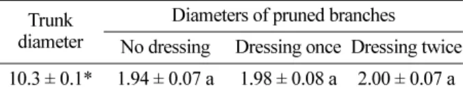
Effects of Wound Dressing with Thiophanate-Methyl Paste on Compartmentalization of Pruning Wounds
6
0
0
전체 글
(2)
(3)
(4)
(5)
(6)
수치

관련 문서
Salty taste intensity scores for chicken rice porridge of salt solution and various level of soy sauce solution ··· 26 Fig.. Photograph of Salad dressing
Environment Council Decision (EU) 2015/146 of 26 January 2015 on the signing, on behalf of the European Union, of the agreement between the European Union and its Member
Instead of having a bricklayer bend over and pick up a brick from a pile of bricks on a relatively unadjustable brickstand, rotate the brick to find the best side, and then
It considers the energy use of the different components that are involved in the distribution and viewing of video content: data centres and content delivery networks
2) Serum Cortisol, ACTH 3) Serum TSH and Free T4 4) Serum Prolactin and GH 5) Serum 25OHD and PTH.. What is the most common laboratory. abnormality expected in this
After first field tests, we expect electric passenger drones or eVTOL aircraft (short for electric vertical take-off and landing) to start providing commercial mobility
1 John Owen, Justification by Faith Alone, in The Works of John Owen, ed. John Bolt, trans. Scott Clark, "Do This and Live: Christ's Active Obedience as the
손질한 원료를 미리 조리(또는 훈제)한 후 통조림에 넣고 식물성 오일을 넣어 만들어 진 캔 저장 식품3. 2 양념류 수산 통조림 canned
