✝Division of General Education, Pyeongtaek University, Gyeonggi-do 405-701, Korea
*Biosensor Research Institute, Seoul National University of Technology
(Received November 19, 2014; Revised December 11, 2014; Accepted December 11, 2014)
Abstract : An in-vivo diagnosis of trace Mg(II) ion was performed using a low-cost and environment-friendly voltammetric method, using a graphite counter and reference electrodes and a fluorine-immobilized graphite working electrode, and clean deep seawater was used as an electrolyte solution. Under optimum conditions, the analytical working ranges attained microgram ranges, and a detection limit of 80.6 ugL-¹ was obtained using stripping voltammety with 60 sec accumulation time. Ex-vivo application was performed on fish liver and mice droppings. The developed techniques can be applicable to tumor cell analysis.
Keywords : Magnesium, seawater, liver, mice, droppings, voltammetry
1. Introduction
Magnesium (Mg(II)) has been shown to be one of the most important and common elements in an organism. It is present in several regions of the biological human-body systems because of its role in enzymatic and some complexation reactions [1, 2]. It is also the most important element for controlling the tumor cell growth rate and regulatory ions in the physiological systems inside or outside the cells [3]. Mg affects many cellular functions, including the transport of potassium and calcium ions, and it modulates signal transduction, energy metabolism, and cell proliferation [4]. It also plays an important role in human-cell growth. On the other
✝Corresponding author (E-mail: [email protected])
hand, it has been reported that Mg can cause pleiotropic, often diverging effects on tumor growth, vascularization, and metastatization.
Thus, both favorable and unfavorable effects of Mg have been identified [3]. For this reason, under in-vivo or ex-vivo conditions, the droppings cell detection methods have been highly connected to cancer growth. Here, many methods for the determination of trace Mg ion have been developed, such as thermal analyses [5, 6], microstructure analysis [7], derivative spectrophotometry [8], suppressed-ion chromatography [9], graphite furnace atomic absorption [10], quantitative capillary electrophoretic analysis [11], atomic-absorption spectrometry [12], electrospray mass spectrometry [13], high-performance liquid chromatography [14, 15], and cost-effective sequential-injection analysis [16]. These photometric and separation techniques,
(A) (B)
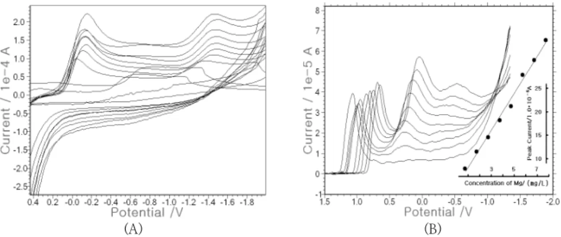
Fig. 1. (A) Cyclic voltammetric scan within the 10-90 mgL-¹ range, using a graphite working electrode in clean deep seawater. (B) Square-wave cathodic stripping voltammetry within the 10-90 mgL-¹ varying ranges using optimum parameters.
(A) (B) (C)
Fig. 2. SW optimization. (A): 0.025~0.2 V amplitude variation. (B): 5~50 Hz SW frequency variation. (C): 0.005~0.04 V increment potential variation, using 80 mg/L Mg(II) constant. The other parameters were 1.5 V initial potential and 60 sec accumulation time
however, are time-consuming, complicated systems and are not applicable for diagnostics assay. Simpler and more sensitive voltammetric detection methods than previously known have been developed, such as those involving the use of glassy carbon electrode [17], gold electrode modified with cysteine [18], and mercury-film-plated carbon paste electrode [19]. These techniques, however, are not used for cell detection as they are complicated and are too expensive to modify. Recently, a method involving the use of graphite pencil electrode (GE), which is simple and inexpensive, was developed, such as for the
determination of voltammetric behavior and the square-wave voltammetric determination of trepibutone [20], that involving the use of an inexpensive and renewable pencil electrode [21], and in a study on pencil-lead bismuth-film electrodes [22]. In this study, stripping voltammetric GE was used for trace analysis. Moreover, a fluorine-immobilized graphite working electrode [23] and deep seawater were used as electrolyte solutions instead of the expensive acid or base standard solutions, and a sensitive working range was attained. The results of the use of the developed method can be applied to fish liver
voltammetry
Analytical systems were used with the bioelectronics-2 system, which was constructed at the authors’ institute. Graphite pencil leads (HB, H, and 3H; 0.3 mm diameter; Korea) were used as working, reference, and counter electrodes. An Mg(II) standard solution (1000 mg/L) was obtained from Merck. All the chemicals that were used were of standard-grade purity. Highly purified water was prepared via three-time distillation, using 18 Mcm-1 Milli-Q Ultra-Pure Water System (Millipore, Bedford). HF immobilization was performed via a 10-cycle scan in an HF standard electrolyte solution, with a 1.0 V initial potential, a -1.0 V switching potential, and a 0.5 V/s scan rate, with a GE working electrode. All the experiments were performed at room temperature, without removing oxygen. Electrolytes from the deep seawater obtained from the east coast of Korea were used. First, cyclic voltammetry (CV) and stripping voltammetry (SV) were performed using GE, then non-treated clean deep seawater was used as an electrolyte solution instead of acid or base solutions. Fig. 1(A) shows the CV effects within the 10, 20, 30, 40, 50, 60, 70, 80, and 90 mg/L Mg(II) addition. In the blank solution, no signal was obtained, then the peak current increased from 3.225 to 15.02x10⁻⁴ A within the 10-70 mg/L Mg(II) variations. After 80 and 90 mg/L Mg(II) was spiked, it decreased from 12.83 to 11.31x10⁻⁴ A. Under these conditions, only a reduction peak was obtained. Fig. 1(B) shows the effects of cathodic stripping on SV concentration. In the blank solution consisting of deep seawater, the signal was clear and
3. Results
3.1. Square-wave (SW) stripping voltammetry
The optimization of the SW parameters was studied by maintaining the 80 mg/L Mg(II) spike in the 10-mL deep seawater. Fig. 2(A) shows the 0.025, 0.05, 0.075, 0.1, 0.125, 0.15, 0.175, and 0.2 V SW amplitude variation. The peak current increased from 1.812 to 6.512x10⁻⁵ A within the 0.025-0.125 V amplitude range. After 0.15 V, it decreased from 6.326 to 4.565x10⁻⁵ A. As the maximum peak current was obtained at 0.0125 V amplitude, such amplitude was chosen as an optimum condition. Under this condition, Fig.
2(B) shows the frequency variation range of 5, 10, 15, 20, 25, 30, 35, 40, 45, and 50 Hz.
The peak current increased from 2.642 to 10.29x10⁻⁵ A within the 5-45 Hz range. At 50 Hz frequency, it decreased to 10.11x10⁻⁵ A. The maximum current was thus obtained at 45 Hz, which was chosen as an optimum condition. Fig. 2(C) shows the voltammograms of the SW increment potential within the 0.005, 0.01, 0.015, 0.02, 0.025, 0.03, 0.035, and 0.04 V increment potential. The signal of the peak current increased from 8.579 to 29.15x10⁻⁵ A and decreased slowly to 28.73x10⁻⁵ A. It reached its maximum level at the 0.035 V increment potential, which was thus chosen as an optimum condition. After identifying the optimized conditions, the analytical working ranges were applied to fish liver and mice droppings pellet.
(A) (B) (C)
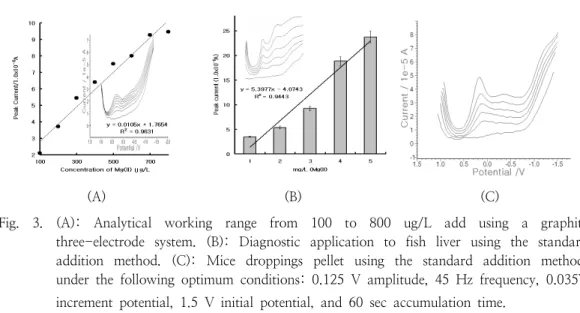
Fig. 3. (A): Analytical working range from 100 to 800 ug/L add using a graphite three-electrode system. (B): Diagnostic application to fish liver using the standard addition method. (C): Mice droppings pellet using the standard addition method, under the following optimum conditions: 0.125 V amplitude, 45 Hz frequency, 0.035V increment potential, 1.5 V initial potential, and 60 sec accumulation time.
Under the optimized conditions, the analytical working ranges were examined using GE in the deep-seawater electrolyte. Fig. 3(A) shows the SW result in micro ranges. The 100, 200, 300, 400, 500, 600, 700, and 800 ug/L Mg(II) variations were spiked using deep seawater under optimum conditions. The peak current increased from 2.104 to 9.465x10⁻⁶ A.
The peak width was sharp and sensitive. The linear equation was y=0.0105x+1.7654, and the precision was R²=0.9631, which can attain microranges and can be used for ex-vivo or in-vivo applications. Analytical application was then made on fish liver cell and mice droppings. Fig. 3(B) shows the tissue application to fish liver using the standard addition method. The bottom curve represents the blank solution containing seawater, which did not obtain any current. The next curve represents the fish liver sample, which was prepared by cutting 0.1-g tissue from a fish and dissolving it in 10-ml conc nitrate. This unknown 0.1-ml sample was spiked in seawater electrolyte. The 5.365x10⁻⁶ A peak current was obtained. After that, 0.1 mg/L of the 100-ppm Mg(II) standard was spiked three times, and 9.22, 18.8, and 23.74x10⁻⁶ A peak currents were obtained, respectively. The peak
current increased linearly, and 1.91 mg/L Mg(II) contents were obtained. Fig. 3(C) shows the standard addition methods for the mice dropping sample, which was prepared by collecting 0.0168 g mice dropping, dissolving it in 1-ml conc nitrate, and diluting it with 100-ml water. Using the standard addition method, no signal was obtained from the blank solution. The unknown sample was spiked, and a 13.07x10⁻⁶ A peak current was obtained. After that, 0.1-mL Mg(II) standard (100 mg/L) was spiked three times, and 16.89, 21.33, and 31.28x10⁻⁶ A peak currents were obtained. These results show that 55.1 mg Mg(II) exists not only in fish liver but also in mice droppings.
4. Discussion
Even though a low-cost electrode and a deep-seawater electrolyte solution were used in this study, the following analytical optimum conditions were identified for the detection of low Mg(II) concentrations, using stripping voltammetry: 0.125 V amplitude, 45 Hz frequency, 0.035 V increment potential, 1.5 V initial potential, and 60 sec accumulation time.
References
1. M. Benamor, N. Aguerssif imultaneous determination of calcium and magnesium by derivative spectrophotometry in pharmaceutical products", Spectrochimica Acta Part A 69; 676-681 (2008).
2. Sema Ba˘gdat Yasar,Seref Güçer Fractionation analysis of magnesium in olive products by atomic absorption spectrometry", Analytica Chimica Acta 505;
43–49 (2004).
3. Federica I. Wolf, Achille R.M. Cittadini, Jeanette A.M. Maier Magnesium and tumors: Ally or foe, Cancer Treatment Reviews 35; 378-382 (2009).
4. Nils-Erik L. Saris, Eero Mervaala, Heikki Karppanen,Jahangir A. Khawaja, Andrzei Lewenstam Magnesium An update on physiological, clinical and analytical aspects", Clinica Chimica Acta 294; 1–26 (2000).
5. Pierre Bracconi, Cyrille Andr`es, Augustin N’iaye, Yvette Pourcelot Thermal analyses of commercial magnesium stearate pseudopolymorphs", Thermochimica Acta 429; 43–51 (2005).
6. Yukitaka kato, Fu-uta Takahashi,Akihiko watanabe, Yoshio Yoshizawa Thermal analysis of a magnesium oxide/water chemical heat pump for congeneration", Applied Thermal Engineering 21;
1067-1081 (2001).
7. S.Y. Liu, J.D. Hu, Y. Yang, Z.X. Guo , H.Y. Wang Microstructure analysis of magnesium alloy melted by laser irradiation, Applied Surface Science 252 ; 1723–1731 (2005).
sodium–atrix waters" , Analytica Chimica Acta 410; 11–23 (2000).
10. Yueying Zhen, Kay B. Franz, Steven W.
Graves novel assay of cell rubidium uptake using graphite furnace atomic absorption: Application to rats on a magnesium-deficient diet", Journal of Nutritional Biochemistry 16; 291–296 (2005).
11. S. Motellier, S. Petit, P. Decambox Quantitative capillary electrophoretic analysis for calcium and magnesium in sodium–atrix waters", Analytica Chimica Acta 410; 11–3 (2000).
12. Sema Bagdat Ya¸sar, Seref Güçer Fractionation analysis of magnesium in olive products by atomic absorption spectrometry", Analytica Chimica Acta 505;
43–49 (2004).
13. Shi Ouyang, Murthy A. Vairavamurthy Effect of magnesium ion in the analysis of
carboxylated compounds by
electrospray-mass spectrometry", Analytica Chimica Acta 422; 101–112 (2000).
14. Brett Paull*, Miroslav Macka, Paul R.
Haddad Determination of calcium and magnesium in water samples by high-performance liquid chromatography on a graphitic stationary phase with a mobile phase containing o-cresolphthalein complexone", Journal of Chromatography A, 789; 329–337 (2009).
15. Toyohide Takeuchi, Shiro Inoue, Mamie Yamamoto, Miharu Tsuji, Tomoo Miwa Fluorimetric determination of magnesium and aluminum via complexation with oxine
in high-performance liquid
chromatography", Journal of
Chromatography A, 910; 373–76 (2001).
16. Zeriet O. Tesfaldet, Jacobus F. van Staden, Raluca I. Stefan Spectrophotometric determination of magnesium in pharmaceutical preparations by cost-effective sequential injection analysis", Talanta 64; 981–988 (2004).
17. N. Abo El-Maali, D. Abd El-Hady, M.
Abd El-Hamid, M.M. Seliem Use of adsorptive stripping voltammetry at the glassy carbon electrode for the simultaneous determination of magnesium(II) and aluminium(III) Application to some industrial samples", Analytica Chimica Acta 417; 67–75 (2000).
\18. Wen Qian, Ji-Hua Zhuang, Yun-Hua Wang, Zhong-Xian Huang The effect of magnesium ion on the electrochemistry of cytochrome and cytochrome b5 at a gold electrode modified with cysteine", Journal of Electroanalytical Chemistry 447;
187–199 (1998).
19. Othman A. Farghaly "A novel method for determination of magnesium in urine and water samples with mercury film-plated carbon paste electrode", Talanta 63;
497–501 (2004).
20. Wei Gao, Junfeng Song, Naiying Wu Voltammetric behavior and square-wave voltammetric determination of trepibutone at a pencil graphite electrode", Journal of Electroanalytical Chemistry 576; 1–7 (2005).
21. Alan M. Peter J. Mahona, Jorg Schiewe Victoria Vicente-Beckett "An inexpensive and renewable pencil electrode for use in field-based stripping voltammetry", Analytica Chimica Acta 345; 67-74 (1997).
22. D. Demetriades, A. Economou, A.
Voulgaropoulos "A study of pencil-lead bismuth-film electrodes for the determination of trace metals by anodic stripping voltammetry", Analytica Chimica Acta 519; 167–172 (2004).
23. Suw Young Ly Diagnosis of copper ions in vascular tracts using a fluorine-doped carbon nanotube sensor, Talanta 74;
1635-164 (2008).