514 Korean Chem. Eng. Res., 58(4), 514-523 (2020)
https://doi.org/10.9713/kcer.2020.58.4.514 PISSN 0304-128X, EISSN 2233-9558
총 설
스트레스 내성 식물 호르몬인 앱시스산의 산업적 활용 전망
이정호‡ · 김승희‡ · 유하영† 상명대학교 생명공학과 03016 서울시 종로구 홍지문 2길 20
(2020년 5월 5일 접수, 2020년 5월 13일 수정본 접수, 2020년 5월 30일 채택)
Future Prospects for Industrial Application of Abscisic acid, a Stress-resistant Phytohormone
Jeongho Lee‡, Seunghee Kim‡ and Hah Young Yoo†
Department of Biotechnology, Sangmyung University, 20, Hongjimun 2-Gil, Jongno-Gu, Seoul, 03016, Korea (Received 5 May 2020; Received in revised from 13 May 2020; Accepted 30 May 2020)
요 약
이동성이 없는 식물은 주위 환경에서 다양한 형태의 스트레스를 받게 되는데 이를 대응하기 위한 방어 기작으로 스 트레스 저항성 단백질과 조절 단백질이 생성된다. 앱시스산은 이러한 신호전달 역할을 하는 호르몬 분자로 잘 알려져 있으며, 잎의 노화, 종자의 휴면 등 식물의 생리적 반응에도 관여한다. 특히 식물이 아닌 동물, 조류(algae) 등 다른 생 물계에서도 다양한 기능을 수행하는 것으로 밝혀졌다. 본 총설에서는 앱시스산의 생합성 및 신호전달 과정 그리고 그 기능에 대하여 조사하였고, 농생명공학, 의생명공학, 산업생명공학을 포함한 다양한 생명공학분야에서 앱시스산을 활 용한 작물량 증대, 질병 치료제 개발, 바이오에너지 생산 등 최신 응용 연구 및 산업적 활용에 대한 동향을 살펴보았다.
Abstract − Plants are exposed to various types of stresses in their surroundings, and stress-resistant and regulatory proteins are produced as defense mechanisms. Abscisic acid is well known for its important role in stress signals as a phytohormone and is also involved in the physiological reactions of plants such as leaf senescence and seed dormancy.
In particular, it has been found to perform a variety of functions in other biological systems, such as animals and microalgae, not plants. In this review, the biosynthesis and signaling process of abscisic acid and its function were investigated and the future prospects for the industrial application of abscisic acid in various biotechnologies, including agriculture, biomedical and industrial biotechnology, have been proposed based on study of emerging applications such as increased crop yields, disease treatment development and bioenergy production.
Key words: Abscisic acid, Biotechnology, Phytohormone, Stress signals
†
To whom correspondence should be addressed.
E-mail: [email protected]
‡
Jeongho Lee and Seunghee Kim contributed equally to this work.
This is an Open-Access article distributed under the terms of the Creative Com- mons Attribution Non-Commercial License (http://creativecommons.org/licenses/by- nc/3.0) which permits unrestricted non-commercial use, distribution, and reproduc- tion in any medium, provided the original work is properly cited.
1. 서 론
이동성이 없는 식물은 다양한 요인으로부터 스트레스를 받고 있으 며, 그 요인들은 Fig. 1과 같이 크게 생물적 스트레스(biotic stresses)와 비생물적 스트레스(abiotic stresses)로 구분된다. 생물적 스트레스는 세 균, 곰팡이, 기생충 등 다른 생물체에 의해 발생하는 스트레스이며, 비생물적 스트레스는 생물이 아닌 환경적인 요인에 의한 것이므로 환경 스트레스라고도 한다. 식물이 열, 가뭄, 저온, 고염분 등과 같 이 생존에 불리한 환경에 노출되었을 때 비생물적 스트레스를 받는 다고 할 수 있다. 하지만 실제 환경에서는 생물적·비생물적 스트레
스에 함께 노출되는데, 비생물적 스트레스를 받으면 병원체 감염에 더 취약해지는 등 이 둘이 상호 복합적으로 작용한다[1]. 예를 들어 고온에서는 세균, 곰팡이 등에 대한 저항성이 낮아지고, 특정 저항 성 유전자가 비활성화 되어 병원체에 대한 감염률이 증가한다[2].
이와 같이 다양한 스트레스에 대응하기 위하여 식물은 방어 기작이 상대적으로 잘 발달되어 있다.
식물이 스트레스에 노출될 경우 이를 저항하기 위해 스트레스 저 항성 단백질(stress tolerance protein) 또는 조절 단백질(regulatory protein)이 생성되며, 이 과정에서 앱시스산(abscisic acid, ABA), 자스몬산(jasmonic acid), 살리실산(salicylic acid) 등과 같은 호르 몬 분자들이 신호전달 역할을 한다. 그 중 비생물적 스트레스에 대 응하는 호르몬 분자로서 앱시스산이 잘 알려져 있다. 앱시스산은 1960년대에 목화 추출물에서 처음 발견되었으며, 잎 또는 꽃 등의 기관이 기부에서 떨어지는 탈리현상(abscission)을 가속하는 물질 중 두 번째로 분리되었기 때문에 ‘Abscisin II’라고 명명되었다[3].
Korean Chem. Eng. Res., Vol. 58, No. 4, November, 2020
비슷한 시기에 자작나무의 일종인 베툴라 푸베센스(betula pubescens)로부터 생장 억제 및 휴면(dormancy)을 유도하는 물질인 ‘dormin’
이 발견되었으며[4], 이후 Cornforth 외(1966)에 의해 Abscisin II와 dormin이 같은 물질이라는 사실이 밝혀졌다[5]. Wareing 연구 그룹 은 서로 다른 명칭을 사용하는 것에 대한 혼란을 막기 위해 기존 명 칭에서 크게 벗어나지 않으면서 화학적 성질을 나타내는 새로운 명 칭으로 ‘Abscisic acid’을 제안하였다[3].
앱시스산은 식물체 내에서 다양한 기능을 한다고 알려져 있다.
먼저 스트레스 반응으로서 물이 부족한 환경에서 저항성 반응을 일 으키기 위한 신호전달물질로 작용한다. 이 신호전달 과정에서 PYR/PYL/RCAR (pyrabactin resistance/PYR-like/regulatory component of ABA receptor)이라는 앱시스산 수용체가 작용하며, 특정 단백질 탈인산화 효소(protein phosphatase)와의 상호작용을 통해 단백질 인산화효소(protein kinase)에 의한 앱시스산 반응성 전사인자 (transcription factor)의 활성화를 조절한다[6]. 신호전달의 결과로 유도된 스트레스 반응 중 하나의 예로서 수분 손실을 줄이기 위한 기공 폐쇄가 있다. 이는 식물 성장과 발달의 저하로부터 스스로를 보호하기 위한 식물의 전략이라고 할 수 있다[7]. 하지만 이러한 전 략에도 불구하고 지구의 기후변화로 인해 세계 많은 지역에서 기온 상승과 강수량 감소가 발생하고 있으므로 농생명공학 분야에서는 작물의 생산량 증대를 위한 연구를 진행하고 있다[8,9].
앱시스산은 식물에서 스트레스 반응에 대한 신호전달 역할을 수 행할 뿐만 아니라 잎의 노화, 종자의 휴면 등의 생리적 반응에도 관 여한다고 알려져 있다[10,11]. 또한 앱시스산은 식물이 아닌 인간을 포함한 동물 그리고 조류(algae) 등 다른 생물계에서도 다양한 기능 을 수행하는 것으로 밝혀졌다[12,13]. 포유동물(mammal)에서는 포 도당 대사(glucose metabolism), 항염증(anti-inflammatory), 기억력 (memory) 향상에 앱시스산이 핵심적인 역할을 하는 것으로 보고되 었으며[13], 이러한 기능은 질병 치료제 및 개선제로서 활용될 가능 성이 높으므로 의생명공학 분야에서는 많은 관심을 두고 연구를 진
행하고 있다. 한편 바이오연료(biofuel) 공급원으로서 크게 주목받 고 있는 미세조류의 배양에 앱시스산을 첨가한 경우 바이오디젤 생 산에 적합한 지질의 함량이 증가 되었는데, 이는 산업생명공학 분 야에서 앞으로의 응용 가능성을 기대할 수 있다. 본 총설에서는 앱 시스산의 생합성 및 신호전달 과정을 소개하고 앱시스산의 주요 기 능들을 바탕으로 각 생명공학 분야에서의 앱시스산 활용 전망을 제 시하고자 한다.
2. 앱시스산의 생합성 과정
균류(fungi)는 C15 화합물인 파르네실 피로인산(farnesyl pyrophosphate) 으로부터 직접적으로 앱시스산을 생성하는 반면, 식물은 Fig. 2와 같이 C40 카로티노이드로부터 간접적으로 앱시스산을 합성한다 [14]. 앱시스산의 생합성 과정은 크게 색소체(plastid) 내에서 β- carotene이 일련의 반응을 거쳐 xanthoxin으로 전환되는 과정과 세 포기질(cytosol)에서 xanthoxin이 앱시스산으로 산화되는 과정으로 구분된다.
비환형 카로티노이드의 최종 물질인 lycopene은 카로틴 물질로 전환되어 각각 α-carotene, β-carotene이 되며, β-carotene의 양쪽 고 리에서 수산화 반응이 일어나면 zeaxanthin이 된다. Zeaxanthin이 ZEP(zeaxanthin epoxidase)에 의해 2회 에폭시화 되면 violaxanthin 으로 전환되는데[15], 이후에 xanthoxin으로 전환되는 두 가지 경로 가 존재한다. 첫째는 violaxanthin이 이성질화 되는 것이고, 둘째는 NSY(neoxanthin synthase)에 의해 neoxanthin으로 전환된 이후에 이성질화 되는 것이다. 각 경로를 통해 형성된 9-cis-violaxanthin과 9'-cis-Neoxanthin은 NCED(9-cis-epoxycarotenoid dioxygenase) 효 소에 의해 xanthoxin으로 분할된다[16]. 세포기질에서 xanthoxin은 short-chain alcohol dehydrogenase/reductase(SDR)에 의해 abscisic aldehyde 중간체를 형성하고 abscisic aldehyde는 AO(aldehyde oxidase) 촉매에 의해 앱시스산으로 최종 전환된다[17].
Fig. 1. Abiotic and biotic stresses in plants.
516 이정호 · 김승희 · 유하영
Fig. 2. The ABA biosynthetic pathway in plants. LCYB, lycopene β-cyclase; CHYB, β-carotene hydroxylase; ZEP, zeaxanthin epoxidase;
NSY, neoxanthin synthase; NCED, 9-cis-epoxycarotenoid dioxygenase; SDR, short-chain alcohol dehydrogenase/reductase; AO, alde-
hyde oxidase.
Korean Chem. Eng. Res., Vol. 58, No. 4, November, 2020
3. 앱시스산의 수용체 및 신호전달 과정 앱시스산 수용체는 ChlH (H subunit of the chloroplast magnesium chelatase), GPCR (G protein coupled receptor type G protein), 그 리고 PYR/PYL/RCAR 등이 잘 알려져 있다. ChlH는 엽록체포막 (chloroplast envelope)에 존재하는 앱시스산 수용체로서 종자의 발 아, 묘목으로의 성장, 그리고 기공의 개폐 등과 관련된 역할을 수행 하는 것으로 생각되었다[18]. 하지만 ChlH가 공변세포(guard cell) 의 앱시스산 신호전달에 영향을 미치지만 방사리간드 결합 분석 (radioligand binding assay) 결과, 명확한 앱시스산의 결합이 관찰 되지 않았다[6]. 또한 보리에서 ChlH의 상동 단백질(homologous protein)이 앱시스산과 결합은 발견되지 않았으며[19], 애기장대에 서 H-3으로 표지된 앱시스산과 재조합형 ChlH 사이의 결합이 검출 되지 않았다[20]. 따라서 ChlH는 직접적으로 앱시스산과 결합하는 수용체는 아니지만, 앱시스산 신호전달에 영향을 미치는 것으로 추 정된다. 한편 GTG (G-protein coupled receptor-type G-protein)1과 GTG2는 원형질막(plasma membrane)에 존재하며 앱시스산 신호전 달 과정에서 양성조절자로서 GPA1 (G-protein α-subunit)과 상호작 용한다[18]. GPA1은 종자 발아 및 발아 후 성장 등에 있어 앱시스 산 신호전달을 음성적으로 조절하는 음성조절자이다[21]. gtg 돌연 변이 식물체는 야생형과 차이가 없었고 gtg1/gtg2 이중 돌연변이는 앱시스산 신호전달 과정에서 유의한 차이를 보였으며 이는 GTG1 또는 GTG2의 도입으로 완전히 보완되었다[22]. Park 외(2009)와 Ma 외(2009)에 의해 새로운 앱시스산 수용체와 그 역할이 밝혀지 면서, 앱시스산의 인식부터 앱시스산 반응성 유전자의 발현까지의 구체적인 신호전달 과정이 제시되었다[23,24]. 앱시스산의 수용체 인 PYR/PYL/RCAR은 세포기질과 핵 사이에서 앱시스산 신호전 달의 핵심 경로를 구성하며[6], 물 부족 스트레스에 대한 앱시스산 신호전달 경로를 활성화하는 데 중요한 역할을 한다는 것이 밝혀졌 다[25]. 앱시스산은 PYR, PP2C (type 2C protein phosphatase)와 결합하는데, 이들의 상호작용에 의해 bZIP (basic leucine zipper protein)으로 가는 신호전달이 이루어진다[26]. PP2C는 단백질 탈 인산화 효소로서 앱시스산 반응성 유전자 발현에 필요한 전사인자 를 활성화시키는 SnRK2를 억제한다. 즉, PP2C는 앱시스산 신호전 달 과정에서 음성조절자(negative regulator)이며 SnRK2는 양성조 절자(positive regulator)이다. 스트레스 환경에서는 앱시스산 반응 성 유전자가 발현되기 때문에 PP2C를 억제할 수 있는 기작이 요구 된다. 이와 관련하여 PYR/PYL/RCAR에 의해 매개되는 앱시스산 신호전달 경로를 Fig. 3에 나타내었다. 먼저 식물이 스트레스를 받 지 않는 정상 상태에서는 SnRK2가 PP2C에 의해 억제되기 때문에 앱시스산 반응성 유전자가 발현되지 않는다. 반면 스트레스가 주어 진 환경에서는 생성된 앱시스산에 의하여 PYR/PYL/RCAR과 PP2C 사이의 결합이 안정화되므로 PP2C의 활성이 억제된다[27].
이로 인해 활성화된 SnRK2는 AREB/ABF 등의 bZIP 전사인자 및 다른 단백질들을 인산화 시키기 때문에 앱시스산 반응성 유전자가 발현될 수 있다. 스트레스 반응 유전자의 산물은 샤페론(chaperone), 부 동 단백질(antifreeze protein), 프롤린(proline)과 같은 스트레스 저 항성 단백질과, 전사인자(transcription factor), 인산화 및 탈인산화 효소(kinase and phosphatase)와 같은 조절 단백질로 구분된다[28].
스트레스 반응과 저항성 조절에 중요한 전사인자로는 DREB (dehydration responsive element binding protein), AREB (ABA
responsive element binding factor), NAM (no apical meristem) 등이 있으며 앱시스산 관련 스트레스 반응에서 중요한 인자로 AREB1, AREB2, ABF3, ABF1 등이 보고되었다[29].
4. 앱시스산의 기능
앱시스산은 물 부족 스트레스에 반응하는 주요한 호르몬인 동시에 여러 생리적인 반응에도 관여한다. 물 부족 스트레스에 의한 주요 한 반응으로는 기공의 폐쇄가 있다. 식물이 흡수한 물의 90% 이상 은 기공을 통해 손실되므로, 물이 부족한 환경에서는 공변세포의 이온 채널 조절을 통하여 기공을 닫는 것이다[30]. 또한 앱시스산의 연관된 신호 분자로 에틸렌(ethylene), 지베렐린(gibberellin), 옥신 (auxin) 등이 있다.
4-1. 물 부족 스트레스 반응(Responses to Water-deficit Stress) 식물은 가뭄이나 고염분 등 다양한 환경 요인에 의해 물 부족 스 트레스를 받게 된다. 이러한 환경에서 식물세포는 스트레스를 감지 하고 내부의 앱시스산을 급격히 증가시키는데, 실제로 고염분 및 삼투 스트레스 등에 노출된 잎, 뿌리 등의 영양조직에서 앱시스산 의 농도가 증가하였다는 보고가 있다[31]. 생성된 앱시스산은 신호 전달을 통해 기공 폐쇄를 포함한 다양한 생리적 반응을 조절한다 [32]. 1898년에 Francis Darwin이 기공 닫힘을 관찰하였지만[33], 당시에는 이 현상이 일어난 요인에 대해서 밝히지 못했다. 이후 연 구들은 스트레스 환경에서 앱시스산에 의해 기공이 닫힌다는 사실 을 보여주었는데, 낮은 상대습도에서 앱시스산이 결핍된 돌연변이 체 aba3-1의 기공은 닫히지 않았다[34]. 앱시스산은 ROS (reactive oxygen species) 생성을 촉발하는 신호전달 경로를 자극함으로써 Ca2+의 증가를 유도한다[32]. Ca2+ 채널의 활성화는 스트레스 및 호 르몬 신호전달, 극성 신장(polar growth)을 포함한 다양한 ROS 매 개 과정에서 핵심적인 단계일 수 있다[35]. ROS에 의해 증가된 Ca2+은 공변세포 내 원형질막의 이온 채널로부터 음이온의 유출을 유도하는데, 앱시스산이 매개하는 이온 채널은 S-type 이온 채널 (slow-acting sustained anion channel)이며[36], 그 결과 공변세포의 압력이 감소하여 기공이 닫힌다[37]. 즉 공변세포 주변에 앱시스산 농도가 높으면 기공이 닫힘으로써 물 부족 스트레스로 인한 수분
Fig. 3. The ABA signaling pathway mediated by PYR/PYL/RCAR
in plants.
518 이정호 · 김승희 · 유하영
손실을 줄일 수 있는 것이다[38].4-2. 잎의 노화(Leaf Senescence)
어린잎은 초기 잎원기(leaf primordium)를 거쳐 영양분이 필요한 기관이 되고[39], 영양분을 얻은 잎은 빠르게 성장하여 성숙해진다.
이후 식물 발달의 마지막 단계인 노화 과정을 거치는데, 이때 영양 분은 씨앗이나 저장기관(storage organs)에 저장된다[40]. 잎의 노 화는 탄수화물을 포함한 생체 분자의 동화작용에서 단백질, 핵산, 지질 등으로의 이화작용을 거침으로써 세포 자살로 이어지는 과정 으로[39], 내생적 및 환경적 요인에 의해 조절된다[17]. 그 요인 중 하나인 앱시스산은 잎의 노화에 유의한 영향을 미치는 것으로 보고 되었다. 앱시스산을 처리한 쌀(Oryza sativa)에서 잎의 노화가 촉진 되었으며[10], 애기장대(Arabidopsis thaliana)에서도 비슷한 결과 가 확인되었다[41]. 기존에는 앱시스산이 에틸렌 생합성을 유도하 여 잎의 노화를 촉진하는 것으로 알려져 있었다[42]. 그러나 Zhu 외(2016)는 에틸렌 저항성 개체에서 앱시스산에 의해 유도되거나 PYL9에 의해 향상되는 노화 관련 유전자인 SAG12-LUC의 발현 억제는 일어나지 않았다고 보고했다[43]. 따라서 앱시스산은 에틸 렌과 독립적인 경로를 통해 잎의 노화를 일으킬 수 있는 것으로 생 각된다.
4-3. 종자의 휴면(Seed Dormancy)
노화는 오래된 잎에서 발생하고 낙엽 현상을 이끄는 반면에 휴면 은 새싹과 종자에서 발생하며 생명 유지에 도움을 준다[44]. 식물에 서 종자의 휴면과 발아는 식물 수명 주기에서 핵심적인 단계이며 [17], 이에 관여하는 대표적인 호르몬으로는 지베렐린과 앱시스산 이 있다. 지베렐린과 앱시스산의 생성량은 빛, 온도, 수분 등 환경
적 요인에 의해 조절되며 지베렐린은 앱시스산 수준을 억제함으로 써 발아를 촉진하는 반면에 앱시스산은 휴면을 촉진하는 역할을 한 다[11]. 종자에서 발현되는 앱시스산 생합성 유전자로서 NCED6 및 NCED9 등이 알려져 있다[45]. nced6/nced9 이중 돌연변이체에 서는 앱시스산의 수준과 종자의 휴면이 감소하는 결과가 나타났다 [46]. 애기장대에서 종자 발달 동안 NCED6는 배젖(endosperm)에 서 발현되고 NCED9은 배젖과 배아(embryo) 모두에서 발현되었는 데, 이로 인해 내생성 앱시스산 수준이 높아지면 종자의 발아가 억 제된다[47].
5. 앱시스산의 활용 전망
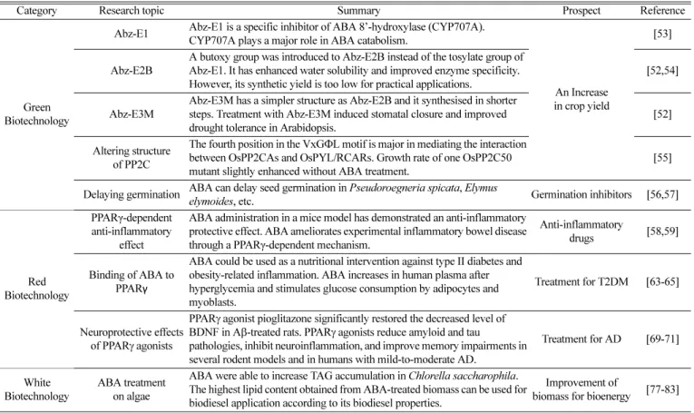
다양한 생명공학 분야에서 앱시스산의 응용 연구가 이루어지고 있으며 Table 1에 생명공학 분야별 앱시스산의 연구와 내용 그리고 전망을 요약하였다. 농생명공학 분야에서는 식물체 내 앱시스산 신 호전달 기작 및 분해 기작에 대한 이해를 바탕으로 작물량 증대의 기반을 마련하고 있다. 앱시스산의 이화작용에서 주요한 역할을 하 는 앱시스산 수산화효소(hydroxylase)를 억제하거나, PP2C의 구조 를 변형하는 등의 방식이다. 한편, 앱시스산은 식물뿐 아니라 동물, 조류(algae) 등에서도 생성되어 다양한 기능을 수행하기 때문에 의 생명공학 및 산업생명공학 분야에서도 이를 활용하기 위한 연구가 이루어지고 있다[48,49].
먼저 의생명공학 분야에서는 포유동물 내 앱시스산의 신호전달을 통한 항염증 작용이 연구되고 있다. 그리고 PPARγ (peroxisome proliferator-activated receptor γ) 작용제로서의 앱시스산의 인슐린 (insulin) 분비 및 시냅스 가소성 개선 기능이 연구되고 있어, 앱시 스산을 활용한 질병 치료 및 개선의 가능성이 제시되고 있다. 이 밖
Table 1. Summary of ABA applications in biotechnology
Category Research topic Summary Prospect Reference
Green Biotechnology
Abz-E1 Abz-E1 is a specific inhibitor of ABA 8’-hydroxylase (CYP707A).
CYP707A plays a major role in ABA catabolism.
An Increase in crop yield
[53]
Abz-E2B
A butoxy group was introduced to Abz-E2B instead of the tosylate group of Abz-E1. It has enhanced water solubility and improved enzyme specificity.
However, its synthetic yield is too low for practical applications.
[52,54]
Abz-E3M
Abz-E3M has a simpler structure as Abz-E2B and it synthesised in shorter steps. Treatment with Abz-E3M induced stomatal closure and improved drought tolerance in Arabidopsis.
[52]
Altering structure of PP2C
The fourth position in the VxG ΦL motif is major in mediating the interaction between OsPP2CAs and OsPYL/RCARs. Growth rate of one OsPP2C50 mutant slightly enhanced without ABA treatment.
[55]
Delaying germination ABA can delay seed germination in Pseudoroegneria spicata, Elymus
elymoides, etc. Germination inhibitors [56,57]
Red Biotechnology
PPARγ-dependent anti-inflammatory
effect
ABA administration in a mice model has demonstrated an anti-inflammatory protective effect. ABA ameliorates experimental inflammatory bowel disease through a PPARγ-dependent mechanism.
Anti-inflammatory
drugs [58,59]
Binding of ABA to PPAR γ
ABA could be used as a nutritional intervention against type II diabetes and obesity-related inflammation. ABA increases in human plasma after hyperglycemia and stimulates glucose consumption by adipocytes and myoblasts.
Treatment for T2DM [63-65]
Neuroprotective effects of PPAR γ agonists
PPAR γ agonist pioglitazone significantly restored the decreased level of BDNF in A β-treated rats. PPARγ agonists reduce amyloid and tau
pathologies, inhibit neuroinflammation, and improve memory impairments in several rodent models and in humans with mild-to-moderate AD.
Treatment for AD [69-71]
White Biotechnology
ABA treatment on algae
ABA were able to increase TAG accumulation in Chlorella saccharophila.
The highest lipid content obtained from ABA-treated biomass can be used for biodiesel application according to its biodiesel properties.
Improvement of
biomass for bioenergy [77-83]
Korean Chem. Eng. Res., Vol. 58, No. 4, November, 2020
에도 산업생명공학 분야에서는 조류의 생장량과 지질 함량을 증가시키는 앱시스산을 활용하여 바이오연료의 생산 효율을 증대시키 는 연구가 제시된 바 있다.
5-1. 농생명공학 분야(Green Biotechnology)
물은 농업 생산성에 있어 제한 요인(limiting factor)이며 식물이 가뭄 환경에서 이를 극복하는 반응은 작물 생산에 매우 중요하다 [50]. 앱시스산은 가뭄의 부정적인 영향을 지연시키거나 잎의 광합 성률(photosynthetic rate)을 증가시키는 것으로 나타났다[50,51].
하지만 농생명공학 분야에서 가뭄 저항성과 관련된 앱시스산의 응 용은 CYP707A (ABA 8'-hydroxylase)에 의한 수산화(hydroxylation)와 같은 요인에 의한 앱시스산의 불활성화 때문에 제한되었다[52]. 이 를 극복하기 위해 Okazaki 외(2011)는 CYP707A를 억제하는 것으 로 잘 알려진 식물생장지연제 uniconazole의 유사체를 설계 및 합 성하여 Abz-E1(Abscinazole-E1)이라는 새로운 CYP707A 억제제 를 개발하였다[53]. 이듬해 Okazaki 외(2012)는 Abz-E1의 tosylate group 대신 butoxy group을 도입한 Abz-E2B를 개발하였는데, 그 결과 용해도가 높아졌으며 효소 특이성도 크게 개선되었다[54]. 하 지만 Abz-E2B를 실제로 적용하기에는 합성 수율이 낮기 때문에 Takeuchi 외(2016)는 단순한 구조와 짧은 합성 단계가 특징인 Abz- E3M을 개발하였으며, 이는 애기장대에서 기공 폐쇄와 가뭄 저항성 을 향상시켰다[52]. 이처럼 기후변화에 대응하기 위해 앱시스산 불 활성화를 극복하고, CYP707A 억제제의 경제적인 합성을 위한 연 구가 지속되고 있다.
또한 비생물적 스트레스 저항성이 증진된 작물을 개발하기 위한 방법으로 앱시스산 인식 관련 효소를 변형시킨 연구가 국내 연구진 에 의해 제시되었다. Lee 외(2017)는 애기장대에서 PYL/RCAR과 의 상호작용을 조절하는 PP2C의 특정 모티프를 제시하였으며, 특 정 변이체에서는 앱시스산 민감도의 변화가 없었고 별도의 앱시스 산 처리 없이도 뿌리 성장이 다소 증가했다. 일반적으로 앱시스산
과민성은 식물의 성장을 저해시키는 것으로 알려져 있지만[55], 해당 변이체는 과민성의 영향을 받지 않으므로 작물량 증대의 가능성을 시사한다. 한편 앱시스산의 발아 억제 기능을 응용한 활용 방안도 제시되고 있다. Richardson 외(2019)는 Pseudoroegneria spicata의 발아를 지연시키기 위해 종자를 앱시스산으로 코팅하였으며, 그 결 과 코팅 비율이 높을수록 발아시기를 더 지연시킬 수 있음이 밝혀 졌다[56]. 이와 유사하게 Elymus elymoides를 대상으로 한 연구에 서도 발아가 지연되었다[57]. 따라서 종자를 보존하거나 발아 시기 의 선택적 조절을 가능하게 하는 발아 지연제로서 앱시스산의 산업 적 응용을 기대해 본다.
5-2. 의생명공학 분야(Red Biotechnology)
인간 그리고 모델 동물에서 앱시스산을 처리한 실험군은 염증성 장 질환(inflammatory bowel disease, IBD), 제2형 당뇨병(type 2 diabetes mellitus, T2DM), 알츠하이머(Alzheimer’s disease, AD), 우울증(depression) 등의 질병 치료에 도움이 되는 것으로 나타났다 [58]. IBD에 대한 앱시스산의 항염증 작용은 동물 내 면역계 세포 에서의 신호전달 과정을 바탕으로 이루어지는데 이는 PPARγ의 활 성화에 달려있다. Fig. 4는 포유동물의 면역계에서 이루어지는, 앱 시스산의 신호전달을 통한 전염증 및 항염증 작용(pro- and anti- inflammatory actions)을 나타낸 것이며, 앱시스산은 염증 반응에서 이중적인 기능을 하는 것으로 보인다[13]. Guri 외(2011)는 앱시스 산이 PPARγ-의존적으로 IBD에 대해 항염증 효과를 나타내지만, T 세포에 PPARγ가 없을 때는 염증을 악화시킨다는 사실을 밝혔다 [59]. 따라서 앱시스산에 반응하는 서로 다른 신호 시스템이 존재할 수 있으며, 이는 대식세포 활성화와 항염증에 관한 앱시스산 연구 사이의 상충된 결과를 설명할 수 있다[58]. 결론적으로 항염증제의 후보 물질로서 앱시스산의 구체적인 작용 기전에 대한 연구가 진행 된다면 IBD를 포함한 염증성 질환의 개선이 가능할 것으로 전망 된다.
Fig. 4. The ABA signaling pathway in mammalian cells of the immune system. LANCL-2, Lanthioninesynthetase C-like protein 2; PLC, Phos-
pholipase C; DAG, Diacylglycerol; IP3, Inositol trisphosphate; AC, Adenylyl cyclase; PKA, protein kinase A; CD38, Cluster of differ-
entiation 38; cADPR, Cyclic ADP; ROS, Reactive oxygen species; NO, Nitric oxide; PPARγ, Peroxisome proliferator-activated
receptor γ.
520 이정호 · 김승희 · 유하영
한편, 전염증 작용과 관련된 앱시스산의 신호전달 과정은 원형질막에서의 앱시스산 인식, cADPR (cyclic ADP ribose)의 과생산, Ca2+의 증가를 수반한다[60]. 앱시스산이 과립구(granulocyte) 등 면역계 세포의 원형질막에 결합하고 PLC (phospholipase C)가 활 성화되어 DAG(diacylglycerol), IP3 (inositol trisphosphate)를 증가 시키는데, DAG는 신호전달 매개 효소를 통해 AC (adenylyl cyclase)를 자극한다. 이 과정으로 활성화된 PKA(protein kinase A)는 CD38 (cluster of differentiation 38)의 인산화를 유도하여 cADPR를 증가 시킨다. 최종적으로 세포질의 Ca2+농도는 증가하여 식세포 작용을 가능하게 하고 ROS와 NO(nitric oxide)가 생성 및 방출된다[61].
Bruzzone 외(2007)는 인간 백혈구에 앱시스산을 농도별로 처리한 실험을 통해 인간 과립구에서 앱시스산이 식세포 작용과 ROS 및 NO 생성량을 자극한다는 사실을 밝혔다[62].
다음으로 앱시스산과 PPARγ의 신호전달을 응용한 T2DM 및 AD의 개선 가능성이 있다. T2DM은 비만과 관련된 질병 중 하나로 고혈압, 동맥 심장 질환 등으로 이어질 수 있는 인슐린 저항성 질병 이다. T2DM에 효과적인 약물 중 하나로 인슐린 민감도를 증가시키는 TZD (thiazolidinedione)가 알려졌으며, 그의 수용체인 PPARγ는 TZD에 의해 선택적으로 작용한다[63]. 하지만 TZD는 체중 증가, 간독성 등의 부작용이 있기 때문에 이를 대체할 치료제가 요구되었 다. Guri 외(2007)는 TZD의 대안으로서 TZD와 구조적으로 유사 하고 PPARγ 리간드 결합 부위에 결합하는 앱시스산의 활용 가능 성을 제시하였다[64]. 앱시스산은 포도당 농도가 높을 때 인간 및 쥐의 β 세포로부터 방출되며 이로 인해 인슐린이 분비된다[61]. 실 제로 앱시스산이 인슐린 분비 및 설치류의 지방세포(adipocyte)와 근원세포(myoblast)에 의한 포도당 흡수를 자극하는 등 포도당 대 사 조절에 관여한다는 사실이 밝혀졌다[65]. 따라서 앱시스산은 T2DM 치료에 있어 TZD를 대체할 후보 물질로 기대된다.
치매는 기억, 언어, 성격, 행동 등 2개 이상의 인지적 영역(cognitive domain)이 점차 퇴화하는 것이 특징이며, AD는 치매의 가장 흔한 원인이다[66]. AD를 유발하는 병리적인 요인으로는 아밀로이드 베 타(amyloid beta, Aβ) 플라크, 타우(tau) 단백질의 엉킴, 신경염증 (neuroinflammation) 등이 있다[67]. 특히, 타우 단백질은 시냅스의 결손과 기억 장애를 일으킨다[68]. 따라서 AD를 치료하기 위한 이 상적인 치료제는 질병 초기의 염증과 질병 후기의 타우 단백질 과 인산화 및 시냅스 결손을 모두 완화시킬 수 있어야 한다. 수상돌기 가시(dendritic spine)의 손실은 시냅스 기능의 상실과 직접적인 관 련이 있으므로 수상돌기가시의 밀도와 시냅스 가소성(synaptic plasticity)을 증가시켜야 하는데, 이러한 기능을 하는 물질로 BDNF (brain derived neurotrophic factor)가 있다[69]. Prakash 외(2014)는 레트(rats)에 Aβ를 처리하여 AD를 유도하였으며, 이후 PPARγ의 작용제인 피오글리타존(pioglitazone)을 처리하였다[70]. 그 결과 떨어졌던 BDNF 수치와 시냅스 가소성이 모두 증가하였는데, 이는 PPARγ 작용제가 BDNF의 발현을 조절하여 시냅스 가소성을 개선 시켰음을 보여주는 결과이다. 또한 쥐에 앱시스산을 처리한 결과 학습 능력과 기억력이 향상된 연구가 보고되었다[71]. 앱시스산이 PPARγ 리간드에 결합한다는 사실과 앞선 연구 결과들을 종합적으로 생각했을 때, 앱시스산 또한 AD를 개선할 가능성이 있을 것으로 추정된다. 따라서 앱시스산에 의한 PPARγ의 활성화 과정 및 AD 완화 기작에 대한 연구가 앞으로 더 진행된다면 AD의 새로운 치료 제 개발이 가능할 것으로 기대된다.
5-3. 산업생명공학 분야(White Biotechnology)
화석연료를 대체할 수 있는 바이오연료의 유망한 공급원으로 미 세조류(micro algae)가 급속히 떠오르고 있다. 미세조류는 성장 속 도가 빠르고 식물의 경작지를 침범하지 않으며 성장 과정에서 이산 화탄소를 흡수하기 때문에 인류의 지속가능한 발전을 위한 대체 자 원으로 주목받고 있다[72]. 물론 식물유래 바이오매스 생산을 위한 에너지 작물 개발 연구들이 수행되고 있지만[73], 월등한 생산성을 가진 미세조류 기반의 바이오연료는 전 세계적인 운송 연료 수요를 충족시킬 수 있는 재생 에너지로 분석되었으며[74], 이러한 결과를 바탕으로 3세대 바이오매스 기반 바이오리파이너리(biorefinery)의 상용화를 목표로 많은 연구들이 수행되고 있다[75-77]. 녹조류인 클로렐라(Chlorella)는 성장 속도가 빠르며 단위세포 무게 당 지질 함량이 높기 때문에 바이오디젤 생산 공정에 적합하다고 알려져 있 으며[78], Contreras-Pool 외(2016)는 클로렐라 사카로필라(Chlorella saccharophila)의 배양에 앱시스산을 첨가한 결과 트리아실글리세 롤(triacylglycerol, TAG)의 함유량이 2배 이상 증가되었음을 확인 하였다[79]. 특히 질소가 결핍된 조건 하에서 앱시스산을 첨가했을 때, 세포 농도와 지질 함유량이 증가되는 것으로 밝혀졌다[79,80].
또한, 녹조류인 Scenedesmus quadricauda[81], Nannochloropsis oceanica[82], Chlorella vulgaris[83]에 앱시스산을 첨가한 결과 이 전 연구들과 유사하게 바이오매스 또는 지질의 수율이 향상된 것으 로 보고되었다. Sivaramakrishnan과 Incharoensakdi (2020)는 클로 렐라 배양에서 앱시스산, 제아틴(zeatin), 인돌아세트산(indole acetic acid), 지베렐린 등 다양한 식물 호르몬의 첨가에 대한 효과를 확인한 결과 앱시스산을 첨가한 실험군에서 오메가-3(Omega-3)와 같은 고급 지방산의 생성에 영향을 주는 것으로 밝혀졌다[84]. 따라 서 지질 생산을 위한 미세조류 배양에서 앱시스산의 잠재력은 매우 높으며 관련 기작에 대한 연구를 통해 바이오리파이너리의 효율이 보다 향상될 것으로 기대한다.
6. 결 론
앱시스산은 식물에서 스트레스 반응에 대한 신호전달 과정을 매 개하는 역할과 잎의 노화, 종자의 휴면 등의 생리작용에 관여하는 것으로 잘 알려져 있었지만, 식물뿐만 아니라 동물, 조류(algae) 등 다른 생물계에서도 특정한 기능이 수행되는 것이 밝혀진 이후 다양 한 생명공학 분야에서 앱시스산을 응용하는 연구들이 수행되고 있 다. 농생명공학 분야에서 CYP707A 억제제 개발을 통해 앱시스산 의 불활성화를 방지하고, PP2C의 특정 모티프를 변형시킴으로써 앱시스산 관련 스트레스 저항성을 향상시켰다. 앞으로 이러한 시도 들이 산업에 적용되면 지구의 기후변화로 인한 작물 생산량 감소를 해결할 수 있을 것으로 기대된다. 의생명공학 분야에서는 앱시스산 을 활용한 다양한 질병 치료의 가능성이 제시되고 있는데, 특히 PPARγ의 작용제로서 앱시스산은 항염증 작용, 인슐린 분비 등의 기능을 나타내므로 염증성 질환 및 당뇨병의 치료제로서 활용할 수 있을 것으로 보인다. 또한 앱시스산이 시냅스 가소성을 개선시켜 학습 능력과 기억력을 개선시키는 것으로 추정되는데, 향후 앱시스 산과 PPARγ 사이의 신호전달 메커니즘이 명확히 밝혀진다면 이를 응용한 질병 치료제 개발이 가능할 것으로 예상된다. 한편 산업생 명공학 분야에서는 앱시스산이 미세조류의 세포 농도와 지질 함량 의 증가에 관여하는 것으로 밝혀졌으며, 향후 응용 연구들은 바이
Korean Chem. Eng. Res., Vol. 58, No. 4, November, 2020
오리파이너리의 효율 개선을 통한 상용화에 큰 기여를 할 것으로기대한다.
감 사
본 연구는 2020학년도 상명대학교 교내연구비를 지원받아 수행 하였음.
References
1. Dangi, A. K., Sharma, B., Khangwal, I. and Shukla, P., “Com- binatorial Interactions of Biotic and Abiotic Stresses in Plants and Their Molecular Mechanisms: Systems Biology Approach,”
Mol. Biotechnol., 60(8), 636-650(2018).
2. Atkinson, N. J. and Urwin, P. E., “The Interaction of Plant Biotic and Abiotic Stresses: From Genes to the Field,” J. Exp. Bot., 63(10), 3523-3543(2012).
3. Addicott, F. T., Lyon J. L., Ohkuma, K., Thiessen, W. E., Carns, H. R., Smith, O. E., Cornforth, J. W., Milborrow, B. V., Ryback, G. and Wareing, P. F., “Abscisic Acid: A New Name for Absci- sin II (Dormin),” Science, 159(3822), 1493(1968).
4. Strausz, S. D., “A Study of the Physiology of Dormancy in the Genus Pyrus,” Ph.D. Dissertation, Oregon State University, Cor- vallis, Oregon(1970).
5. Cornforth, J. W., Milborrow, B. V., Ryback, G., Rothwell, K. and Wain, R. L., “Identification of the Yellow Lupin Growth Inhib- itor as (+)-Abscisin II ((+)-Dormin),” Nature, 211(5050), 742- 743(1966).
6. Zhang, X. L., Jiang, L., Xin, Q., Liu, Y., Tan, J. X. and Chen, Z.
Z., “Structural Basis and Functions of Abscisic Acid Receptors PYLs,” Front. Plant Sci., 6, 88(2015).
7. Sah, S. K., Reddy, K. R. and Li, J., “Abscisic Acid and Abiotic Stress Tolerance in Crop Plants,” Front. Plant Sci., 7, 571(2016).
8. Lawas, L. M. F., Zuther, E., Jagadish, S. K. and Hincha, D. K.,
“Molecular Mechanisms of Combined Heat and Drought Stress Resilience in Cereals,” Curr. Opin. Plant Biol., 45(Part B), 212- 217(2018).
9. Pareek, A., Dhankher, O. P. and Foyer, C. H., “Mitigating the Impact of Climate Change on Plant Productivity and Ecosystem Sustainability,” J. Exp. Bot., 71(2), 451-456(2020).
10. Ray, S., Mondal, W. A. and Choudhuri, M. A., “Regulation of Leaf Senescence, Grain-filling and Yield of Rice by Kinetin and Abscisic Acid,” Physiol. Plant., 59(3), 343-346(1983).
11. Kang, J., Yim, S., Choi, H., Kim, A., Lee, K. P., Lopez-Molina, L., Martinoia, E. and Lee, Y., “Abscisic Acid Transporters Cooper- ate to Control Seed Germination,” Nat. Commun., 6(1), 8113(2015).
12. Kobayashi, Y. and Tanaka, K., “Extraction and Measurement of Abscisic Acid in a Unicellular Red Alga Cyanidioschyzon mero- lae,” Bio Protoc. 6(23): e2033(2016).
13. Baliño, P., Gómez-Cadenas, A., López-Malo, D., Romero, F. J.
and Muriach, M., “Is There A Role for Abscisic Acid, A Proven Anti-Inflammatory Agent, in the Treatment of Ischemic Retinopa- thies?,” Antioxidants, 8(4), 104(2019).
14. Finkelstein, R., “Abscisic Acid Synthesis and Response,” Arabi- dopsis Book, 11, e0166(2013).
15. Xiong, L. and Zhu, J. K., “Regulation of Abscisic Acid Biosyn-
thesis,” Plant Physiol., 133(1), 29-36(2003).
16. Vishwakarma, K., Upadhyay, N., Kumar, N., Yadav, G., Singh, J., Mishra, R. K., Kumar, V., Verma, R., Upadhyay, R. G., Pan- dey, M. and Sharma, S., “Abscisic Acid Signaling and Abiotic Stress Tolerance in Plants: A Review on Current Knowledge and Future Prospects,” Front. Plant Sci., 8, 161(2017).
17. Saroj, K. S., Kambham, R. R. and Jiaxu, L., “Abscisic Acid and Abiotic Stress Tolerance in Crop Plants,” Front. Plant Sci., 7, 571(2016).
18. Wu, F. Q., Xin, Q., Cao, Z., Liu, Z. Q., Du, S. Y., Mei, C., Zhao, C. X., Wang, X. F., Shang, Y., Jiang, T., Zhang, X. F., Yan, L., Zhao, R., Cui, Z. N., Liu, R., Sun, H. L., Yang, X. L., Su, Z. and Zhang, D. P., “The Magnesium-Chelatase H Subunit Binds Abscisic Acid and Functions in Abscisic Acid Signaling: New Evidence in Arabidopsis,” Plant Physiol., 150(4), 1940-1954(2009).
19. Müller, A. H. and Hansson, M., “The Barley Magnesium Che- latase 150-kD Subunit Is Not an Abscisic Acid Receptor,” Plant Physiol., 150(1), 157-166(2009).
20. Tsuzuki, T., Takahashi, K., Inoue, S., Okigaki, Y., Tomiyama, M., Hossain, M. A., Shimazaki, K., Murata, Y. and Kinoshita, T.,
“Mg-chelatase H Subunit Affects ABA Signaling in Stomatal Guard Cells, but Is Not an ABA Receptor in Arabidopsis thali- ana,” J. Plant Res., 124(4), 527-538(2011).
21. Wang, X. F. and Zhang, D. P., “Abscisic Acid Receptors: Mul- tiple Signal-perception Sites,” Ann. Bot., 101(3), 311-317(2008).
22. Pandey, S., Nelson, D. C. and Assmann, S. M., “Two Novel GPCR- Type G Proteins Are Abscisic Acid Receptors in Arabidopsis,”
Cell, 136(18), 136-148(2009).
23. Park, S. Y., Fung, P., Nishimura, N., Jensen, D. R., Fujii, H., Zhao, Y., Lumba, S., Santiago, J., Rodrigues, A., Chow, T. F., Alfred, S. E., Bonetta, D., Finkelstein, R., Provart, N. J., Desveaux, D., Rodriguez, P. L., McCourt, P., Zhu, J. K., Schroeder, J. I., Volkman, B. F. and Cutler, S. R., “Abscisic Acid Inhibits Type 2C Protein Phosphatases via the PYR/PYL Family of START Proteins,”
Science, 324(5930), 1068-1071(2009).
24. Ma, Y., Szostkiewicz, I., Korte, A., Moes, D., Yang, Y., Christmann, A. and Grill, E., “Regulators of PP2C Phosphatase Activity Function as Abscisic Acid Sensors,” Science, 324(5930), 1064-1068(2009).
25. Lim, C. W., Baek, W., Han, S. W. and Lee, S. C., “Arabidopsis PYL8 Plays an Important Role for ABA Signaling and Drought Stress Responses,” Plant Pathol. J., 29(4), 471-476(2013).
26. Qiu, J., Hou, Y., Wang, Y., Li, Z., Zhao, J., Tong, X., Lin, H., Wei, X., Ao, H. and Zhang, J., “A Comprehensive Proteomic Survey of ABA-Induced Protein Phosphorylation in Rice (Oryza sativa L.),” Int. J. Mol. Sci., 18(1), 60(2017).
27. Tischer, S. V., Wunschel, C., Papacek, M., Kleigrewe, K., Hofmann, T., Christmann, A. and Grill, E., “Combinatorial Interaction Net- work of Abscisic Acid Receptors and Coreceptors from Arabi- dopsis thaliana,” Proc. Natl. Acad. Sci. USA, 114(38), 10280-10285 (2017).
28. Khan, Z. H., Kumar, B., Dhatterwal, P., Mehrotra, S. and Meh- rotra, R., “Transcriptional Regulatory Network of Cis-Regulatory Elements (Cres) and Transcription Factors (Tfs) In Plants During Abiotic stress,” Int. J. Plant Biol. Res., 5(2), 1064-1081(2017).
29. Nakashima, K. and Suenaga, K., “Toward the Genetic Improve- ment of Drought Tolerance in Crops,” Jpn. Agric. Res. Q., 51(1), 1-10(2017).
522 이정호 · 김승희 · 유하영
30. Pei, Z. M., Ghassemian, M., Kwak, C. M., McCourt, P. andSchroeder, J. I., “Role of Farnesyltransferase in ABA Regulation of Guard Cell Anion Channels and Plant Water Loss,” Science, 282(5387), 287-290(1998).
31. Zeevaart, J. A. D. and Creelman, R. A., “Metabolism and Phys- iology of Abscisic Acid,” Annu. Rev. Plant Physiol., 39(1), 439- 473(1988).
32. Osakabe, Y., Osakabe, K., Shinozaki, K. and Tran, L. P., “Response of Plants to Water Stress,” Front. Plant Sci., 5, 86(2014).
33. Darwin, F., “Observations on Stomata,” Philos. Trans. Royal Soc., London, 190, 531-621(1898).
34. Bauer, H., Ache, P., Lautner, S., Fromm, J., Hartung, W., Al- Rasheid, K. A., Sonnewald, S., Sonnewald, U., Kneitz, S., Lach- mann, N., Mendel, R. R., Bittner, F., Hetherington, A. M. and Hed- rich, R., “The Stomatal Response to Reduced Relative Humidity Requires Guard Cell-autonomous ABA Synthesis,” Curr. Biol., 23(1), 53-57(2013).
35. Mori, I. C. and Schroeder, J. I., “Reactive Oxygen Species Acti- vation of Plant Ca2+ Channels. A Signaling Mechanism in Polar Growth, Hormone Transduction, Stress Signaling, and Hypotheti- cally Mechanotransduction,” Plant Physiol., 135(2), 702-708(2004).
36. Joshi-Saha, A., Valon, C. and Leung, J., “A Brand New START:
Abscisic Acid Perception and Transduction in the Guard Cell,”
Sci. Signal, 4(201), re4(2011).
37. Munemasa, S., Hauser, F., Park, J., Waadt, R., Brandt, B. and Schroeder, J. I., “Mechanisms of Abscisic Acid-mediated Control of Stomatal Aperture,” Curr. Opin. Plant Biol., 28, 154-162(2015).
38. Lisar, S. Y., Motafakkerazad, R., Hossain, M. M. and Rahman, I.
M. M., in I. M. M. Rahman(Ed.), Water Stress, InTech, Croatia (2012).
39. Van der Graaff, E., Schwacke, R., Schneider, A., Desimone, M., Flugge, U. I. and Kunze, R., “Transcription Analysis of Arabidop- sis Membrane Transporters and Hormone Pathways During Devel- opmental and Induced Leaf Senescence,” Plant Physiol., 141(2), 776-792(2006).
40. Himelblau, E. and Amasino, R. M., “Nutrients Mobilized from Leaves of Arabidopsis thaliana During Leaf Senescence,” J. Plant Physiol., 158(10), 1317-1323(2001).
41. Asad, M. A. U., Zakari, S. A., Zhao, Q., Zhou, L., Ye, Y. and Cheng, F., “Abiotic Stresses Intervene with ABA Signaling to Induce Destructive Metabolic Pathways Leading to Death: Pre- mature Leaf Senescence in Plants,” Int. J. Mol. Sci., 20(2), 256 (2019).
42. Riov, J., Dagan, E., Goren, R. and Yang, S. F., “Characterization of Abscisic Acid-induced Ethylene Production in Citrus Leaf and Tomato Fruit Tissues,” Plant Physiol., 92(1), 48-53(1990).
43. Zhao, Y., Chan, Z., Gao, J., Xing, L., Cao, M., Yu, C., Hu, Y., You, J., Shi, H., Zhu, Y., Gong, Y., Mu, Z., Wang, H., Deng, X., Wang, P., Bressan, R. A. and Zhu, J. K., “ABA Receptor PYL9 Promotes Drought Resistance and Leaf Senescence,” Proc. Natl. Acad. Sci.
USA, 113(7), 1949-54(2016).
44. Zhao, Y., Gao, J., Kim, G. I., Chen, K., Bressan, R. A. and Zhu, J. K., “Control of Plant Water Use by ABA Induction of Senes- cence and Dormancy: An Overlooked Lesson from Evolution,”
Plant Cell Physiol., 58(8), 1319-1327(2017).
45. Huo, H., Dahal, P., Kunusoth, K., McCallum, C. M. and Brad- ford, K. J., “Expression of 9-cis-EPOXYCAROTENOID DIOX-
YGENASE4 Is Essential for Thermoinhibition of Lettuce Seed Germination but Not for Seed Development or Stress Toler- ance,” Plant Cell, 25(3), 884-900(2013).
46. Martínez-Andújar, C., Ordiz, M. I., Huang, Z., Nonogaki, M., Beachy, R. N. and Nonogaki, H., “Induction of 9-cis-epoxycar- otenoid Dioxygenase in Arabidopsis thaliana Seeds Enhances Seed Dormancy,” Proc. Natl. Acad. Sci. USA, 108(41), 17225- 17229(2011).
47. Vishal, B. and Kumar, P. P., “Regulation of Seed Germination and Abiotic Stresses by Gibberellins and Abscisic Acid,” Front.
Plant Sci., 9, 838(2018).
48. Cowan, A. K. and Rose, P. D., “Abscisic Acid Metabolism in Salt-Stressed Cells of Dunaliella salina: Possible Interrelation- ship with β-Carotene Accumulation,” Plant Physiol., 97(2), 798- 803(1991).
49. Lee, K. W., Hong, S., Rahman, M. A., Ji, H. C., Cha, J. Y., Jones, C. S., Son, D. and Lee, S. H. “Ectopic Overexpression of Teff Grass (Eragrostis tef) Phi-class Glutathione S-transferase 1 (EtGSTF1) Enhances Prokaryotic Cell Survivability against Diverse Abiotic Stresses,” Biotechnol. Bioprocess Eng., 24(3), 552-559(2019).
50. Brito, C., Dinis, L. T., Ferreira, H., Moutinho-Pereira, J. and Correia, C. M., “Foliar Pre-Treatment with Abscisic Acid Enhances Olive Tree Drought Adaptability,” Plants, 9(3), 341(2020).
51. He, J., Jin, Y., Palta, J. A., Liu, H.-Y., Chen, Z. and Li, F.-M.,
“Exogenous ABA Induces Osmotic Adjustment, Improves Leaf Water Relations and Water Use Efficiency, But Not Yield in Soy- bean under Water Stress,” Agronomy, 9(7), 395(2019).
52. Takeuchi, J., Okamoto, M., Mega, R., Kanno, Y., Ohnishi, T., Seo, M. and Todoroki, Y., “Abscinazole-E3M, a Practical Inhib- itor of Abscisic Acid 8′-hydroxylase for Improving Drought Tol- erance,” Sci. Rep., 6(1), 37060(2016).
53. Okazaki, M., Nimitkeatkai, H., Muramatsu, T., Aoyama, H., Ueno, K., Mizutani, M., Hirai, N., Kondo, S., Ohnishi, T. and Todoroki, Y., “Abscinazole-E1, a Novel Chemical Tool for Exploring the Role of ABA 8'-hydroxylase CYP707A,” Bioorg. Med. Chem., 19(1), 406-413(2011).
54. Okazaki, M., Kittikorn, M., Ueno, K., Mizutani, M., Hirai, N., Kondo, S., Ohnishi, T. and Todoroki, Y., “Abscinazole-E2B, a Practical and Selective Inhibitor of ABA 8'-hydroxylase CYP707A,”
Bioorg. Med. Chem., 20(10), 3162-3172(2012).
55. Han, S., Min, M. K., Lee, S. Y., Lim, C. W., Bhatnagar, N., Lee, Y., Shin, D., Chung, K. Y., Lee, S. C., Kim, B. G. and Lee, S.,
“Modulation of ABA Signaling by Altering VxGΦL Motif of PP2Cs in Oryza sativa,” Mol. Plant, 10(9), 1190-1205(2017).
56. Richardson, W. C., Badrakh, T., Roundy, B. A., Aanderud, Z. T., Petersen, S. L., Allen, P. S., Whitaker, D. R. and Madsen, M. D.,
“Influence of an Abscisic Acid (ABA) Seed Coating on Seed Germination Rate and Timing of Bluebunch Wheatgrass,” Ecol.
Evol, 9, 7438-7447(2019).
57. Badrakh, T., “Effects of Abscisic Acid (ABA) on Germination Rate of Three Rangeland Species,” MSc Dissertation, Brigham Young University, Provo, Utah(2016).
58. Lievens, L., Pollier, J., Goossens, A., Beyaert, R. and Staal, J.,
“Abscisic Acid as Pathogen Effector and Immune Regulator,”
Front. Plant Sci., 8, 587(2017).
59. Guri, A. J., Evans, N. P., Hontecillas, R. and Bassaganya-Riera, J., “T Cell PPARγ Is Required for the Anti-inflammatory Effi-
Korean Chem. Eng. Res., Vol. 58, No. 4, November, 2020
cacy of Abscisic Acid Against Experimental IBD,” J. Nutr. Bio-chem, 22(9), 812-819(2011).
60. Li, H. H., Hao, R. L., Wu, S. S., Guo, P. C., Chen, C. J., Pan, L.
P. and Ni, H., “Occurrence, Function and Potential Medicinal Applications of the Phytohormone Abscisic Acid in Animals and Humans,” Biochem. Pharmacol, 82(7), 701-712(2011).
61. Zocchi, E., Hontecillas, R., Leber, A., Einerhand, A., Carbo, A., Bruzzone, S., Tubau-Juni, N., Philipson, N., Zoccoli-Rodriguez, V., Sturla, L. and Bassaganya-Riera, J., “Abscisic Acid: A Novel Nutraceutical for Glycemic Control,” Front. Nutr., 4, 24(2017).
62. Bruzzone, S., Moreschi, I., Usai, C., Guida, L., Damonte, G., Salis, A., Scarfì, S., Millo, E., De Flora, A. and Zocchi, E., “Abscisic Acid Is an Endogenous Cytokine in Human Granulocytes with Cyclic ADP-ribose as Second Messenger,” Proc. Natl. Acad. Sci.
USA, 104(14), 5759-5764(2007).
63. Lehmann, J. M,, Moore, L. B., Smith-Oliver, T. A., Wilkison, W.
O., Willson, T. M. and Kliewer, S. A., “An Antidiabetic Thiazo- lidinedione Is a High Affinity Ligand for Peroxisome Prolifera- tor-activated Receptor Gamma (PPAR gamma),” J. Biol. Chem., 270(22), 12953-12956(1995).
64. Guri, A. J., Hontecillas, R., Si, H., Liu, D. and Bassaganya-Riera, J., “Dietary Abscisic Acid Ameliorates Glucose Tolerance and Obesity-related Inflammation in db/db Mice Fed High-fat Diets,”
Clin. Nutr., 26(1), 107-116(2007).
65. Magnone, M., Emionite, L., Guida, L., Vigliarolo, T., Sturla, L., Spinelli, S., Buschiazzo, A., Marini, C., Sambuceti, G., De Flora, A., Orengo, AM., Cossu, V., Ferrando, S., Barbieri, O. and Zoc- chi, E., “Insulin-independent Stimulation of Skeletal Muscle Glucose Uptake by Low-dose Abscisic Acid via AMpK Activa- tion,” Sci. Rep., 10(1), 1454(2020).
66. Weller, J. and Budson, A., “Current Understanding of Alzheimer’s Disease Diagnosis and Treatment,” F1000Res., 7, 1161(2018).
67. Kinney, J. W., Bemiller, S. M., Murtishaw, A. S., Leisgang, A.
M. and Lamb, B. T., “Inflammation as a Central Mechanism in Alzheimer’s Disease,” Alzheimers Dement (NY), 4, 575-590(2018).
68. Biundo, F., Del Prete, D., Zhang, H., Arancio, O. and D'Adamio, L., “A Role for Tau in Learning, Memory and Synaptic Plas- ticity,” Sci. Rep., 8(1), 3184(2018).
69. Govindarajulu, M., Pinky, P. D., Bloemer, J., Ghanei, N., Suppi- ramaniam, V. and Amin, R., “Signaling Mechanisms of Selective PPARγ Modulators in Alzheimer’s Disease,” PPAR Res., 2018, 2010675(2018).
70. Prakash, A. and Kumar, A., “Role of Nuclear Receptor on Reg- ulation of BDNF and Neuroinflammation in Hippocampus of β- amyloid Animal Model of Alzheimer’s Disease,” Neurotox. Res., 25(4), 335-347(2014).
71. Liu, J., Gu, X., Zou, R., Nan, W., Yang, S., Wang, H. L. and Chen, X. T., “Phytohormone Abscisic Acid Improves Spatial Memory and Synaptogenesis Involving NDR1/2 Kinase in Rats,” Front.
Pharmacol., 9, 1141(2018).
72. Lee, K., Lee, Y. J., Chang, H. N. and Jeong, K. J., “Engineering Trichosporon oleaginosus for Enhanced Production of Lipid
from Volatile Fatty Acids as Carbon Source,” Korean J. Chem.
Eng., 36(6), 903-908(2019).
73. Joshi, R., Singla-Pareek, S. L. and Pareek, A., “Engineering Abi- otic Stress Response in Plants for Biomass Production,” J. Biol.
Chem., 293(14), 5035-5043(2018).
74. Rahpeyma, S. S. and Raheb, J., “Microalgae Biodiesel as a Valu- able Alternative to Fossil Fuels,” Bioenergy Res., 12(4), 958-965 (2019).
75. Ju, J. H., Oh, B. R., Ryu, S. K., Heo, S. Y., Kim, S. Y., Hong, W.
K., Kim, C. H. and Seo, J. W., “Production of Lipid Containing High Levels of Docosahexaenoic Acid by Cultivation of Aurantioch- ytrium sp. KRS101 using Jerusalem artichoke extract,” Biotech- nol. Bioprocess Eng., 23(6), 726-732(2018).
76. Choi, Y. Y., Hong, M. E., Chang, W. S. and Sim, S. J., “Auto- trophic Biodiesel Production from the Thermotolerant Microalga Chlorella sorokiniana by Enhancing the Carbon Availability with Temperature Adjustment,” Biotechnol. Bioprocess Eng., 24(1), 223-231(2019).
77. Lee, J. H., Lee, H. U., Lee, J. H., Lee, S. K., Yoo, H. Y., Park, C.
and Kim, S. W., “Continuous Production of Bioethanol Using Microalgal Sugars Extracted from Nannochloropsis gaditana,”
Korean J. Chem. Eng., 36(1), 71-76(2019).
78. Muthuraj, M., Selvaraj, B., Palabhanvi, B., Kumar, V. and Das, D., “Enhanced Lipid Content in Chlorella sp. FC2 IITG via High Energy Irradiation Mutagenesis,” Korean J. Chem. Eng., 36(1), 63-70(2019).
79. Contreras-Pool, P. Y., Peraza-Echeverria1, S., Ku-González, Á.
F. and Herrera-Valencia, V. A., “The Phytohormone Abscisic Acid Increases Triacylglycerol Content in the Green Microalga Chlorella saccharophila (Chlorophyta),” ALGAE, 31(3), 267-276(2016).
80. Sulochana, S. B. and Arumugam, M., “Influence of Abscisic Acid on Growth, Biomass and Lipid Yield of Scenedesmus quadricauda Under Nitrogen Starved Condition,” Bioresour. Technol., 213, 198-203(2016).
81. Sulochana, S. B. and Arumugam, M., “Influence of Abscisic Acid on Growth, Biomass and Lipid Yield of Scenedesmus quadricauda under Nitrogen Starved Condition,” Bioresour. Technol., 213, 198- 203(2016).
82. Lu, Y., Tarkowská, D., Turečková, V., Luo, T., Xin, Y., Li, J., Wang, Q., Jiao, N., Strnad, M. and Xu, J., “Antagonistic Roles of Abscisic Acid and Cytokinin During Response to Nitrogen Depletion in Oleaginous Microalga Nannochloropsis oceanica Expand the Evolutionary Breadth of Phytohormone Function,”
Plant J., 80(1), 52-68(2014).
83. Lin, B., Ahmed, F., Du, H., Li, Z., Yan, Y., Huang, Y., Cui, M., Yin, Y., Li, B., Wang, M., Meng, C. and Gao, Z., “Plant Growth Regulators Promote Lipid and Carotenoid Accumulation in Chlo- rella vulgaris,” J. Appl. Phycol., 30, 1549-1561(2017).
84. Sivaramakrishnan, R. and Incharoensakdi, A., “Plant Hormone Induced Enrichment of Chlorella sp. Omega-3 Fatty Acids,” Bio- technol. Biofuels, 13(7), 1-14(2020).